1790
Simultaneous acquisition of 1H/19F/23Na/31P MR imaging in Phantom at 3T using a quadruple-nuclear RF coil system
Nan Li1,2, Xing Yang1,2, Feng Du1,2, Kang Yan3, Bei Liu3, Yiping Du3, Chunsheng Yang4,5, Zhi Zhang4,5, Li Chen4,5, Fang Chen4,5, Xiaoliang Zhang6, Xin Liu1,2, Hairong Zheng1,2, and Ye Li1,2
1Lauterbur Imaging Research Center, Shenzhen Institutes of Advanced Technology, Chinese Academy of Sciences, Shenzhen, China;, shenzhen, China, 2Key Laboratory for Magnetic Resonance and Multimodality Imaging of Guangdong Province, Shenzhen, China, Shenzhen, China, 3Institute for Medical Imaging Technology, School of Biomedical Engineering, Shanghai Jiao Tong University, Shanghai, China., Shanghai, China, 4State Key Laboratory of Magnetic Resonance and Atomic and Molecular Physics, Wuhan Center for Magnetic Resonance, Innovation Academy for Precision Measurement Science and Technology, the Chinese Academy of Sciences, Wuhan 430071, China;, Wuhan, China, 5University of Chinese Academy of Science, Beijing 100049, China, Wuhan, China, 6Department of Biomedical Engineering, State University of New York at Buffalo, NY, United States, Buffalo, NY, United States
1Lauterbur Imaging Research Center, Shenzhen Institutes of Advanced Technology, Chinese Academy of Sciences, Shenzhen, China;, shenzhen, China, 2Key Laboratory for Magnetic Resonance and Multimodality Imaging of Guangdong Province, Shenzhen, China, Shenzhen, China, 3Institute for Medical Imaging Technology, School of Biomedical Engineering, Shanghai Jiao Tong University, Shanghai, China., Shanghai, China, 4State Key Laboratory of Magnetic Resonance and Atomic and Molecular Physics, Wuhan Center for Magnetic Resonance, Innovation Academy for Precision Measurement Science and Technology, the Chinese Academy of Sciences, Wuhan 430071, China;, Wuhan, China, 5University of Chinese Academy of Science, Beijing 100049, China, Wuhan, China, 6Department of Biomedical Engineering, State University of New York at Buffalo, NY, United States, Buffalo, NY, United States
Synopsis
Affected by the inherent physical properties, it was challenging to obtain the imaging of heteronuclear at 3T, which usually have weak MR signals. Hence, high performance was required of the RF system to excite and receive MR signals. In this study, a range of imaging tests were performed on the phantom by using the developed specific quadruple-nuclear RF coil system. The results demonstrated the feasibility of synchronized 1H/19F/ 23Na/31P MR imaging at 3T. Furthermore, the imaging approach combined the quadruple-nuclear RF coil and triple tuned local 19F/23Na/31P receive coil was also indicated the improved SNR of corresponding nucleus.
Introduction
Simultaneously acquisition of MRI imaging of multiple nucleus can not only provide high-quality anatomical signals, but reflect the information of tumor molecules and ion metabolism, which is expected to employ to the early prediction and evaluation of therapeutic effect of tumor1-3. The detection capability of extremely weak signals depends on the corresponding RF transceivers, which is the core component of RF signal acquisition. Significant SNR enhancement can be achieved with improved radiofrequency (RF) coils3.A four-frequency 1H / 19F / 23Na/ 31P RF coil system was developed in our previous study4. Good tuning, matching and decoupling performance was obtained from the bench tests. In this study, we renew the structure of the RF coil with adding the triple tuned local 19F / 23Na / 31P receive coil to improve the weak nuclei MR imaging quality. Furthermore, the phantom imaging studies were conducted at self-made 3T multinuclear MR system. The phantom experiments at 3T were shown the ability of the RF coil system MR imaging of 1H / 19F / 23Na / 31P imaging. The combination of triple tuned local 19F / 23Na / 31P receive coil also indicated the improved SNR of corresponding nucleus.
Method
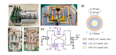
The structure of the quadruple-nuclear RF coil system was improved with adopted three-layer nested structure and was shown in Fig. 1. The outer and inner layer coils worked in 1H/19F and 23Na/31P resonated frequencies, respectively. The new triple-tuned local receive coils was added in the innermost layer to improve the 19F/23Na/31P signal sensitivity. The circuit schematics of the coil elements are shown in Fig.1 (e), which contained a dual-tuned rectangle loop element for 19F/31P Imaging and a butterfly element for 23Na imaging. The size of channels of 19F/31P were each 40 mm × 40 mm and of 23Na were each 70 mm × 55 mm.All on-system studies were scanned using 3T MRI system, the operative mode of the quadruple-nuclear RF coil was controlled by the multi-frequency and multichannel sequential control system. The solution composition of phantoms for multiple nucleus imaging were described as follows: phosphoric acid solution(H3PO4) with a concentration of 85 % was used for 31P imaging, the cylindrical phantom with 300 mmol/L NaCl solution used for 1H imaging and 23Na imaging, C6F6 solution with a concentration of 98 % were used as the 19F phantom. In addition, a specific phantom filled with three solution was customized to evaluate the quality of 1H/19F/23Na/31P imaging simultaneously as shown in Fig.1 (f). The Multinuclear MR imaging was scanned by the self-made GRE(gradient echo)sequence5,which utilized the double gradient echo to achieve the simultaneous acquisition of 1H / 19F / 23Na/ 31P MR imaging. 31P signal was excited by the first echo and the 1H、19F、23 Na was excited by the second one simultaneously. The detailed information of RF pulse was shown in the Fig. 2.
The scan parameters were as follows parameters was used to obtain signals: TE/TR= 5.4/400 ms, Slice thickness=20 mm, Matrix =128×128, Average = 4, Scan time = 3 min 40 s. FOV of the 1H acquisition was 200 mm × 200 mm. Based on the sequence, the corresponding FOV of other non-proton was determined by the frequencies. Complete image reconstruction with the independently developed spectrometer system. SNR distributions were calculated by dividing the signal value by the noise standard deviation for the quantitative evaluation of 1H / 19F / 23Na/ 31P imaging quality.
Result
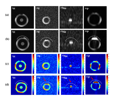
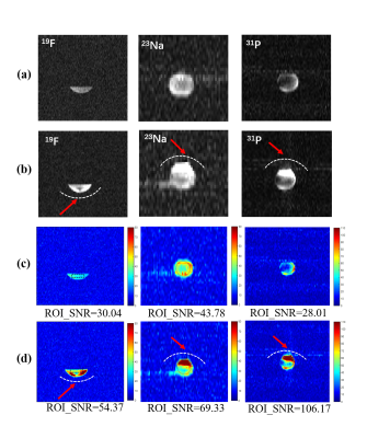
Fig. 3 shows the 1H / 19F / 23Na/ 31P MR measured results obtained with the specific phantom which including the four nuclei signals simultaneously. Fig. 4 shows the MR images obtained with the individual test phantom. All the tests indicated the developed coil system can be used for multi-nuclei MR imaging. SNR comparisons between the approach with and without the local triple-tuned receive RF coils were also shown in Fig. 3 (c)-(d) and Fig. 4 (c)-(d). The position of the local 19F / 23Na/ 31P receive RF coil was indicated by the white dotted-line, the SNR of the region indicated by the red arrow is significantly improved. Especially, in the individual phantom for 19F / 23Na/ 31P imaging, the SNR of the ROIs in 19F / 23Na/ 31P image showed in Fig. 4 (d) was all Increased more than 50 % by adding the local triple-tuned receive RF coils.Discussion/conclusion
In this study, simultaneous 1H/19F/23Na/31P MR images at 3T are successfully acquired in phantom using the developed quadruple-nuclear RF coil with a self-made 3T multinuclear MR RF system. The results demonstrated the feasibility of synchronized 1H/19F/23Na/31P imaging. The improved SNR of 19F/23Na/31P was also achieved by the imaging approach combined the quadruple-nuclear RF coils and triple tuned local receive coils. Future investigation will focus on the animal study, which aimed to obtain the multi-nuclei information of tumor simultaneously in vivo.Acknowledgements
This work was supported in part NSFC under Grant No. 81627901; the Strategic Priority Research Program of Chinese Academy of Sciences (Grant No. XDB25000000); city grant JCYJ20170413161314734; Youth Innovation Promotion Association of CAS No. 2017415.References
- Hong S. M, Choi C.H, Magill A. W, et al. Design of a quadrature 1H/31P coil using bent dipole antenna and 4-channel loop at 3T MRI. IEEE Trans.Med. Imag., 2018; 37(12): 2613–2618.
- Ha Y.H, Choi. C. H, Shah N.J, et al. Development and Implementation of a PIN-Diode Controlled, Quadrature-Enhanced, Double-Tuned RF Coil for Sodium MRI. IEEE Trans. Med. Imaging. 2018; 37(7): 1626–1631.
- Ryan .B, Karthik. L, Guillaume. M, et al, A nested phosphorus and proton coil array for brain magnetic resonance imaging and spectroscopy”. Neuroimage. 2016; 124: 602-611
- Yang. X , Li. N, Du. F. et al, A four-frequency 1H/19F/23Na/31P RF coil system for simultaneous multinuclear MRI and MRS at 3.0T. ISMRM. 2020; 4096.
- Yang. C. S, Liu C.Y, Li. Ye, et al, Multi-nuclear and multi-channel synchronization control system for tumor molecular imaging (under preparation)
Figures

Fig. 1 (a) The new entirety structure of the
quadruple-nuclear RF coil system. (b) The outer layer 1H/19F
dual-tuned coils. (c) The inner layer 23Na/31P RF coils. (d)
The innermost layer of local triple-tuned receive 19F/23Na/31P
RF coils. (e) The circuits of local triple-tuned receive 19F/23Na/31P
RF coils. (f) The specific phantom filled with three kinds of solution
simultaneously.

Fig. 2. The RF pulse of the simultaneous
acquisition for the 1H / 19F / 23Na/ 31P
MR imaging.

Fig. 3.
(a)-(b): The 1H/19F/23Na/31P MR
measured images obtained with the specific phantom. (a) only
scanned by using the quadruple-nuclear RF coil. (b) Combined the
quadruple-nuclear coil and triple tuned local 19F/23Na/31P
receive coil. (c)-(d): SNR maps for the 1H/19F/23Na/31P
imaging. (c) Without the local triple-tuned RF coils. (d) Combined with the local triple-tuned 19F/23Na/31P
receive RF coils. The position of the local
receive RF coil was indicated by the white dotted-line, and the SNR of the
region indicated by the red arrow is significantly improved.

Fig. 4.
(a)-(b): The 1H/19F/23Na/31P MR
measured images obtained with the independent
nuclide test phantom. (a) only
scanned by using the quadruple-nuclear RF coil. (b) Combined the
quadruple-nuclear coil and triple tuned local 19F/23Na/31P
receive coil. (c)-(d): SNR maps for the 1H/19F/23Na/31P
imaging. (c) Without the local triple-tuned RF coils. (d) Combined with the local triple-tuned 19F/23Na/31P
receive RF coils. The position of the local
receive RF coil was indicated by the white dotted-line, and the SNR of the
region indicated by the red arrow is significantly improved.