1765
Artificial Intelligence based smart MRI: Towards development of automated workflow for reduction of repeat and reject of scans
Raghu Prasad, PhD1, Harikrishna Rai, PhD1, and Sanket Mali1
1GE Healthcare, Bangalore, India
1GE Healthcare, Bangalore, India
Synopsis
Majority of the pre-scan errors in MRI radiological workflows are due to a) in-appropriate patient positioning, b) incorrect protocol selection for the anatomy to be scanned, c) operator/technologist negligence. In this work, we propose and develop an AI (Artificial Intelligence) based computer vision solution to correct patient positioning errors and reduce the scan time. Our approach relies on identification of RF coil and anatomy of the patient when occluded with coils using a 3D depth camera. Camera based solution has shown significant improvements in some of the critical MRI based workflow such as auto-landmarking, coil/protocol selection and scan range overlay.
Purpose
To avoid repeat and reject of MRI scans caused by human errors such as improper scanning methods, patient positioning, and incorrect protocol. To improve efficiency of MRI technologist.Introduction
The technologist who is responsible for scanning the patient tends to commit pre-scan errors such as scanning the patient with incorrect orientation, incorrect landmark, inappropriate pose, angle, and direction. Further, when the patients checks-in the scanner room with a prescription of the body part to be scanned, the technologist reviews the prescription, and then prepares the patient for the scan. During this process, the following may lead to pre scan errors: A) the technologist mis-interprets the prescription and instructs the patient towards an in-appropriate pose and orientation. B) The patient mis-interprets the instructions given by technologist, leading to inappropriate pose and orientation. C) The technologist does not pay attention to incorrect pose and orientation of patient and completes the scan. All these positioning errors lead to repeat and reject of scans. This causes a) discomfort to patient b) increases patient wait time c) cognitive stress on technologist and radiologists, and d) reduces throughput of the scanner machine. To address the above pain points, we have developed an AI based Deep Neural Network architecture that can identify the type of RF coil wrapped around the patient, and detect the pose/orientation of the patient through camera video frames and suggest the right protocol based on the coil identified. Proposed solution is capable of correcting errors under various lighting conditions, clothing and patient demographics. A feasibility study was performed on the effects of EMI on camera data and have evaluated the impact of camera on magnetic in-homogeneity.Methods
In our method we mount a depth camera over ceiling of the MRI room such that FoV of camera includes MRI table and bore (Fig.1). We place one camera on ceiling and one inside the bore and then acquire color and depth frames from the camera to identify various anatomical coils, patient orientation, anatomy of the patient occluded under RF coil (Fig.2). To identify anatomical regions, we have developed a Deep Convolutional Neural Network that identifies shape, spatial location and thickness of the coil (Fig.3). This neural network is based on focal loss class imbalance approach to identify and classify various RF anatomical coils. The focal loss is calculated as follows: 𝐹𝐿(𝑝𝑡) = −(1 − 𝑝𝑡)𝛾log(𝑝𝑡), 𝑝𝑡 is a notation for the probability of the class labelled with numeric value 1. The modulating factor (1 − 𝑝𝑡)𝛾 is thus added to the original cross entropy formula. Once RF coils are identified (Fig.4), the probable anatomies along with their orientation (all DICOM orientations) that cover this coil region are also identified through a Generative Adversarial Neural Network. Orientation in our case are typically postures of the patient wrapped with the RF coils. The orientation serves as important clue for protocol selection. Identified coil name and patient orientation is then matched with the protocol that operator selects in the console and in case of a mismatch the operator is alerted. Neural network then identifies the anatomical points and its spatial locations when the patient anatomy is occluded with coil. The landmarking positions are determined based on the key anatomical points and identified coil locations. This automatic identification of landmarking positions through camera serves as an input to scan range overlay which determines the starting and ending location of scan. Now, the anatomy identified, orientation of the patient, the scan range along with the RF coil name identified is compared with the MRI prescription. If found correct, the operator can start the scan, else operator is requested to correct for the errors before start of the scan.Results
The proposed algorithm was able to identify RF coil, anatomy and patient orientation accurately when patient is covered with blanket . The correlation between ground truth of all quantitative parameters and obtained value from the proposed system were significantly high (r=0.92) for coil and anatomy identification. The classification results for orientation identification was found to be 95.33%. This shows that our algorithm has high precision in identification/classification. The operator error was significantly reduced by 62%. The error was measured as the difference of mistakes the operator commits due to cognitive stress during the MRI scan with and without camera-based workflow. Further, the repeat-reject of scans was significantly reduced (p<.001) by adopting this workflow. These statistical results were obtained in lab set up on a GE MRI system (GE 1.5T, SIGNA Creator) using a Spherical chemical phantom.Discussion and Conclusions
Our camera based experimental analysis reveals that using the depth cameras we can improve the pre-scan MR workflow efficiently. This has shown to have significant benefit towards a). Scan time reduction b). Waiting time of patient c). Improvement in image quality. All the factors that contribute to repeat and reject of scans such as improper patient positioning, improper protocol selection, incorrect coil placement can be reduced to a large extent using proposed AI based computer vision workflow. MRI modality can benefit significantly from this camera-based workflow due to the complexity involved in scanning the patients with additional apparatus such as RF coils, MRE driver for elastography, MR guided biopsy.Acknowledgements
The authors would like to thank Tony Linz and Saban Kurucay of GE Healthcare MRI group for their insightful comments and suggestions.References
- Booij, R., van Straten, M., Wimmer, A. et al. Automated patient positioning in CT using a 3D camera for body contour detection: accuracy in pediatric patients. Eur Radiol (2020). https://doi.org/10.1007/s00330-020-07097-w
- Booij R, Budde RPJ, Dijkshoorn ML, van Straten M. Accuracy of automated patient positioning in CT using a 3D camera for body contour detection. Eur Radiol. 2019 Apr;29(4):2079-2088. doi: 10.1007/s00330-018-5745-z. Epub 2018 Oct 10. PMID: 30306328; PMCID: PMC6420476.
Figures
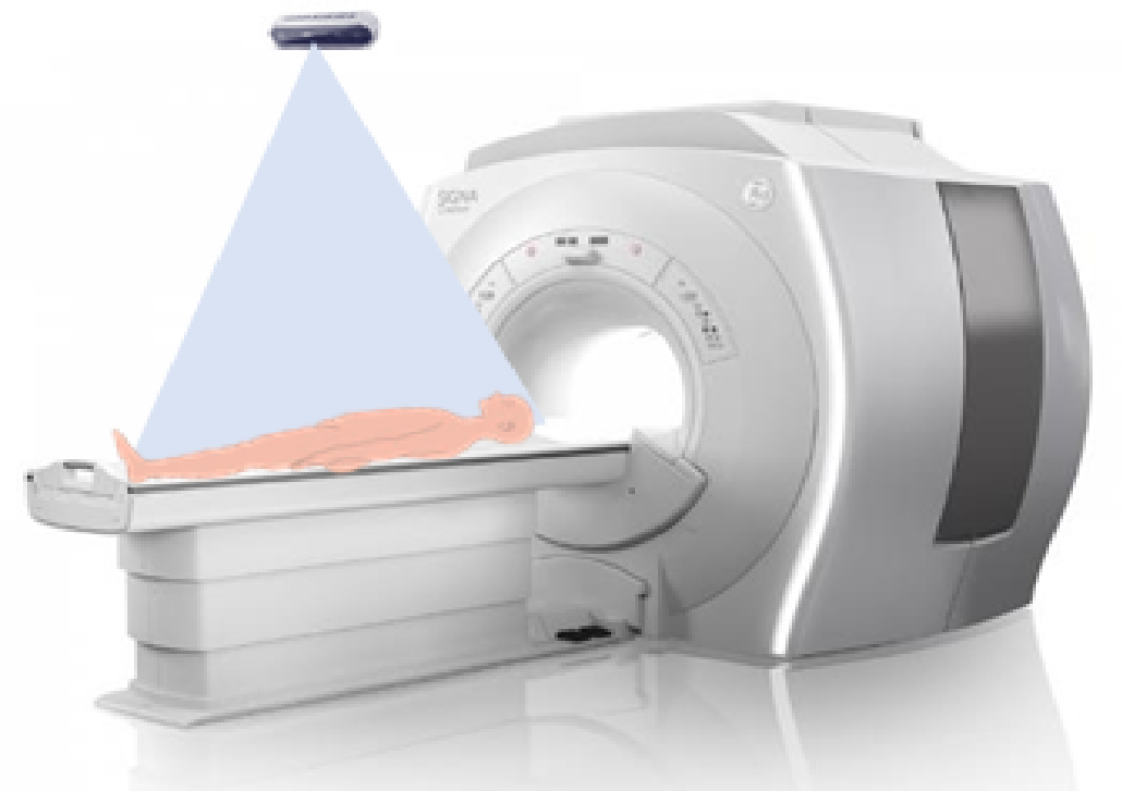
Fig.1 Placement of Camera in MRI scanner room
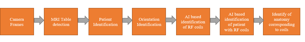
Fig.2 Workflow of smart patient positioning
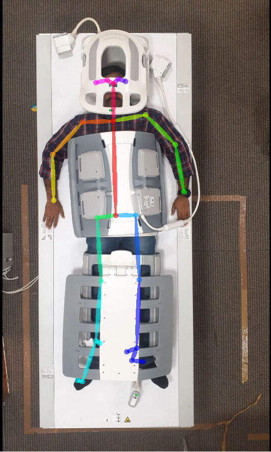
Fig.3 Anatomical points identification under occlusion of coils using deep neural network
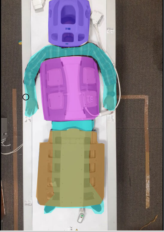
Fig.4 RF coil and patient identification using deep learning based network with shape and texture as priors