1741
Cerebral Microstructural Alteration in Follow-up COVID-19 Patients: A Diffusion Kurtosis Imaging Study1Radiology, Zhongnan Hospital of Wuhan University, Wuhan, China, 2MR Research, GE Healthcare, Beijing, China, 3Neurology, Zhongnan Hospital of Wuhan University, Wuhan, China
Synopsis
This study aims to investigate the cerebral microstructural alteration of follow-up recovered COVID-19 (rCOVID-19) patients and explore the corrections of symptoms and microstructure changes. Decreased axial kurtosis (Kax), radial kurtosis (Krad), mean kurtosis (Kmean), mean kurtosis tensor (KTmean) and increased fractional anisotropy (FA) in several brain regions of rCOVID-19 patients indicated demyelination and increased cell membrane permeability of cerebral cortex. Hyposmia was negatively correlated with Kmean and Krad in right anterior cingulum of rCOVID-19 patients, partly explaining the residual olfactory impairment several months after recovery. DKI metrics can help reveal microstructural changes of CNS for rCOVID-19 patients.
Introduction
The Novel Severe Acute Respiratory Syndrome Coronavirus-2 (SARS-CoV-2) infection can cause series of central nervous system (CNS) symptoms in the acute onset of illness, such as dizziness, headache and hyposmia1. However, little studies were reported about the alteration of symptoms and brain structures of cured patients with COVID-192. Here we used diffusion kurtosis imaging (DKI) to explore long-term cerebral microstructure changes of COVID-19 patients after rehabilitation.Methods
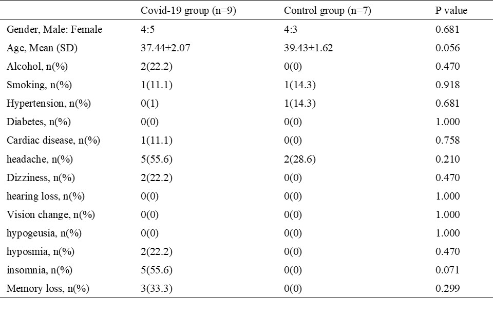
This study was approved by the Institutional Review Board. Nine patients recovered from COVID-19 for 8 to 9 months were recruited. The diagnosis of COVID-19 was confirmed by positive RT-PCR testing for SARS-CoV-2 on pharyngeal swabs. Seven volunteers without any history of COVID-19 were enrolled as control group. Clinical data and DKI imaging were recorded. The DKI imaging was collected on a 3 Tesla MRI scanner (Discovery MR 750w, GE Healthcare) with a 32-channel head coil and using a spin-echo single-shot echo-planar imaging sequence (TR/TE 6000ms/82ms, 60 diffusion direction with b value of 2000 s/mm2). The DKE and SPM12 software were utilized to analysis the whole brain DKI data. The DKI metrics including fractional anisotropy (FA), mean kurtosis (Kmean), axial kurtosis (Kax), radial kurtosis (Krad), and mean kurtosis tensor (KTmean) were obtained using a customized and automated algorithm in the MATLAB software R2019a (Mathworks, Natick, Massachusetts, USA). The demographic data and whole -brain DKI metrics were compared between groups using SPSS (version 22.0; SPSS Inc., Chicago, IL, USA).Results
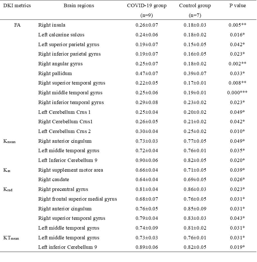
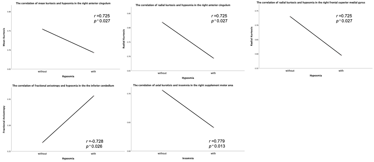
There was no significant difference of gender, age and symptoms (p >0.050,) between groups except insomnia (p = 0.021)(Table 1). Decreased Kax, Krad, Kmean, KTmean and increased FA in COVID-19 group compared to the control group (Table 2). In addition, Hyposmia was negatively correlated with Kmean in right anterior cingulum (r = 0.725, p = 0.027), Krad in right anterior cingulum (r = 0.725, p = 0.027) and right frontal superior medial gyrus (r = 0.725, p = 0.027) and positively correlated with FA in the inferior cerebellum (r = -0.728, p = 0.026) in COVID-19 group. Insomnia was negatively correlated with Kax in the right supplement motor area (r = 0.779, p = 0.013) (Figure 1).Discussion
rCOVID-19 patients showed decreased Kax, Kmean, Krad, KTmean and increased FA in bilateral brain regions, suggesting that COVID-19 can induce cerebral microstructure alterations. Kmean reflects the complexity of neural structures; Kax and Krad provide diffusion information in the parallel and perpendicular directions, respectively. Kax is generally considered to reflect axonal integrity and Krad is believed to reflect myelin integrity3. We suspected that COVID-19 might induce demyelination and increase cell membrane permeability in the cerebral cortex. Hyposmia was related to decreased Krad and Kmean in right anterior cingulum, an important role in the limbic system. As the limbic system is a part of the olfactory circuit, it may partly explain why patients with COVID-19 manifested residual olfactory impairment even lasting for 9 months after recovery4. Insomnia was related to decreased Kax in the right supplementary motor area (SMA). A previous study has shown that insomnia was correlated with the changes of connectivity patterns between the Default Mode Network (DMN) and region of SMA5. We assumed the decreased Kax in SMA might alter the function connectivity between SMA and other brain regions in rCOVID-19 patients with insomnia.Conclusion
DKI could provide evidence for the exploration of intracranial microstructural changes in patients recovering from COVID-19. The patients had decreased Kax, Krad, Kmean, KTmean and increased FA in several brain regions. The symptom hyposmia may be induced by the altered function of anterior cingulum, a part of limbic system in the olfactory circuit.Acknowledgements
No acknowledgement found.References
1.Pennisi M, Lanza G, Falzone L, et al. SARS-CoV-2 and the Nervous System: From Clinical Features to Molecular Mechanisms. Int J Mol Sci. 2020 Jul 31;21(15):5475.
2. Lu Y, Li X, Geng D, et al. Cerebral Micro-Structural Changes in COVID-19 Patients - An MRI-based 3-month Follow-up Study. EClinicalMedicine. 2020 Aug;25:100484.
3. Marrale M, Collura G, Brai M, et al. Physics, Techniques and Review of Neuroradiological Applications of Diffusion Kurtosis Imaging (DKI). Clin Neuroradiol. 2016 Dec;26(4):391-403.
4.Han P, Zang Y, Akshita J, et al. Magnetic Resonance Imaging of Human Olfactory Dysfunction. Brain Topogr. 2019 Nov;32(6):987-997.
5.Santarnecchi E, Del Bianco C, Sicilia I, et al. Age of Insomnia Onset Correlates with a Reversal of Default Mode Network and Supplementary Motor Cortex Connectivity. Neural Plast. 2018 Apr 1;2018:3678534.
Figures