1703
MIITRA atlas: Construction of high resolution T1w and DTI brain templates in a common space, based on 400 older adults1Illinois Institute of Technology, Chicago, IL, United States, 2Rush Alzheimer's Disease Center, Rush University, Chicago, IL, United States
Synopsis
As a critical step to establish the Multichannel Illinois Institute of Technology & Rush university Aging (MIITRA) atlas, the present work aimed to: a) develop high quality 0.5mm resolution T1-weighted (T1w) and diffusion tensor imaging (DTI) templates in a common space using data from a large, diverse, community cohort of non-demented older adults, and b) quantitatively compare the new templates to existing templates in terms of spatial normalization accuracy of external data. The new T1w and DTI templates allowed higher inter-subject and inter-modality spatial normalization of older adult data compared to other templates.
Introduction
An MRI atlas representative of the older adult brain is in high demand. As new methods for multimodal analyses emerge and as more sub-millimeter voxel datasets are collected on older adults, a multimodal older adult brain atlas with high spatial resolution is desirable. The Multichannel Illinois Institute of Technology & Rush university Aging (MIITRA) atlas project aims at addressing these needs. The goal of the present work was twofold: a) to develop high quality 0.5mm resolution T1-weighted (T1w) and diffusion tensor imaging (DTI) templates in a common space using data from a large, diverse, community cohort of non-demented older adults, and b) quantitatively compare the new templates to existing templates in terms of spatial normalization accuracy of external data.Methods
Data:T1w (1mm isotropic) and DTI (2mm isotropic) data were collected on 400 non-demented older adults (50% male; 64.9-98.9 years of age; 54% white, 43% black; 318 with no cognitive impairment and 82 with mild cognitive impairment) participating in longitudinal cohort studies of aging1,2. All data were collected on two 3T MRI scanners.
Template construction:
The template construction process can be divided into 5 steps and combined a recently introduced approach for the development of high quality multimodal templates in a common space3 (steps 1-4) and an approach for the development of high spatial resolution templates based on principles of super-resolution4,5 (step 5). In step 1, ANTs6 registration was driven by T1w data and the resulting transformations were also applied on the DTI data. In step 2, DRTAMAS7 registration was driven by DTI data and the resulting transformations were also applied on the T1w data. Steps 3 and 4 included a second iteration of steps 1 and 2, respectively. The transformations from each step were combined to minimize interpolations. Steps 1-4 were conducted in a 0.5mm isotropic resolution space. The above approach ensured that each step maximized the quality of the corresponding template (steps 1, 3 maximized T1w template quality; steps 2, 4 maximized DTI template quality) and increased the spatial matching between templates. In step 5, the final transformations for the T1w and DTI data were used to map signals from raw space to exact physical locations in final template space, eliminating interpolations that occur in conventional template building methods4,5. The final signal in each voxel in template space was calculated as the weighted average of only those signals contained in that voxel. The weights were derived using a Gaussian kernel with a standard deviation equal to the standard deviation of the signals included in that voxel and centered at the median signal. This approach is less sensitive to residual misregistration4,5. The resulting templates are referred to in the following as MIITRA T1w and MIITRA DTI templates.
Evaluation:
T1w and DTI data from 202 non-demented older adults (50% male, 65-93.2 years of age) participating in ADNI38 were used for evaluation.
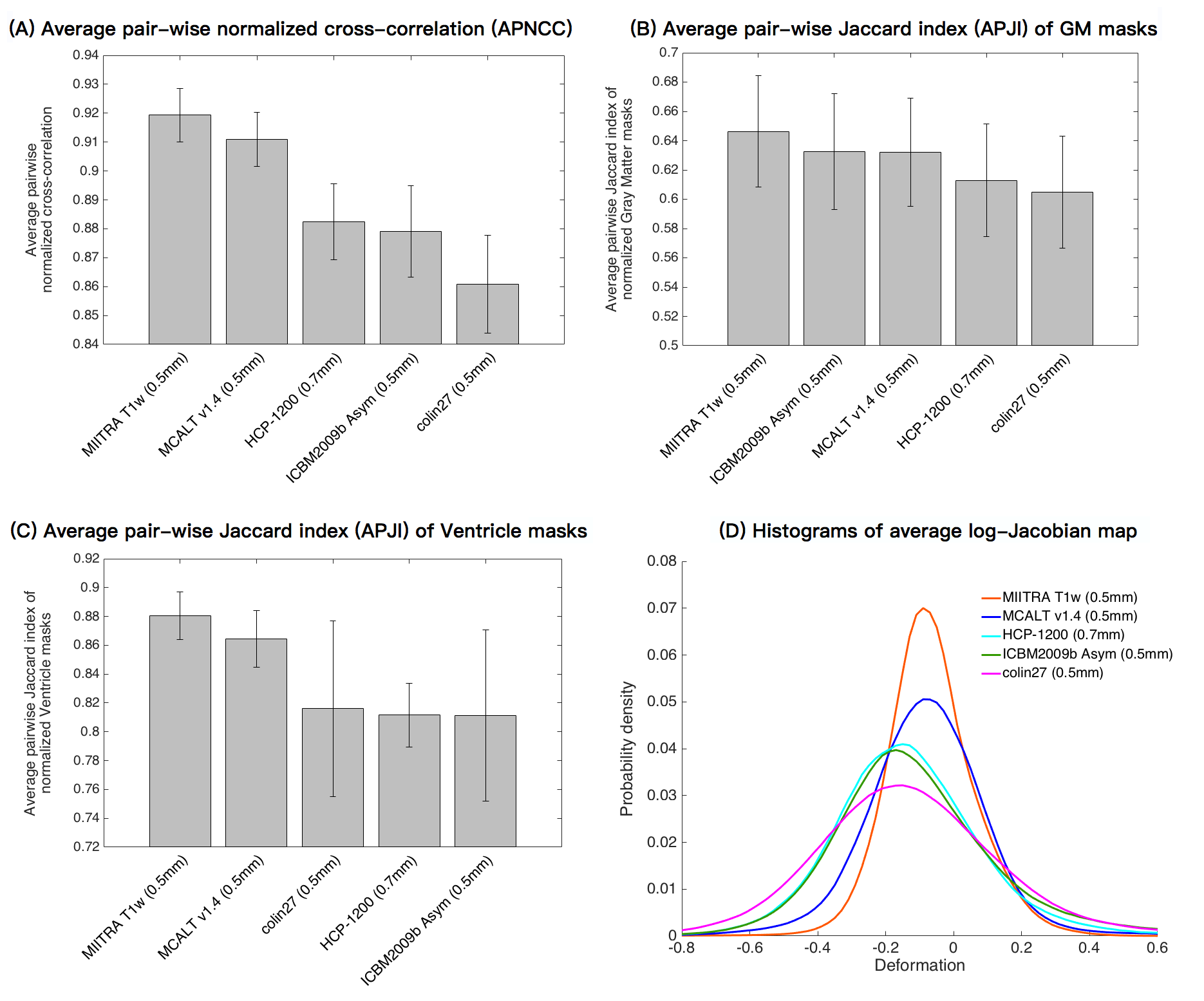
The MIITRA T1w template was compared to other high resolution T1w templates (MCALTv1.4_0.5mm9, ICBM2009b_Asym_0.5mm10,11, colin27_0.5mm12, HCP1200_0.7mm13) in terms of the accuracy of inter-subject spatial normalization of ADNI3 data achieved when each template was used as a reference (ANTs registration). The average pair-wise normalized cross-correlation (APNCC) of normalized T1w images, average pair-wise Jaccard index (APJI) of normalized masks of gray matter and ventricles, and average log-Jacobian determinant of deformations were compared across T1w templates.
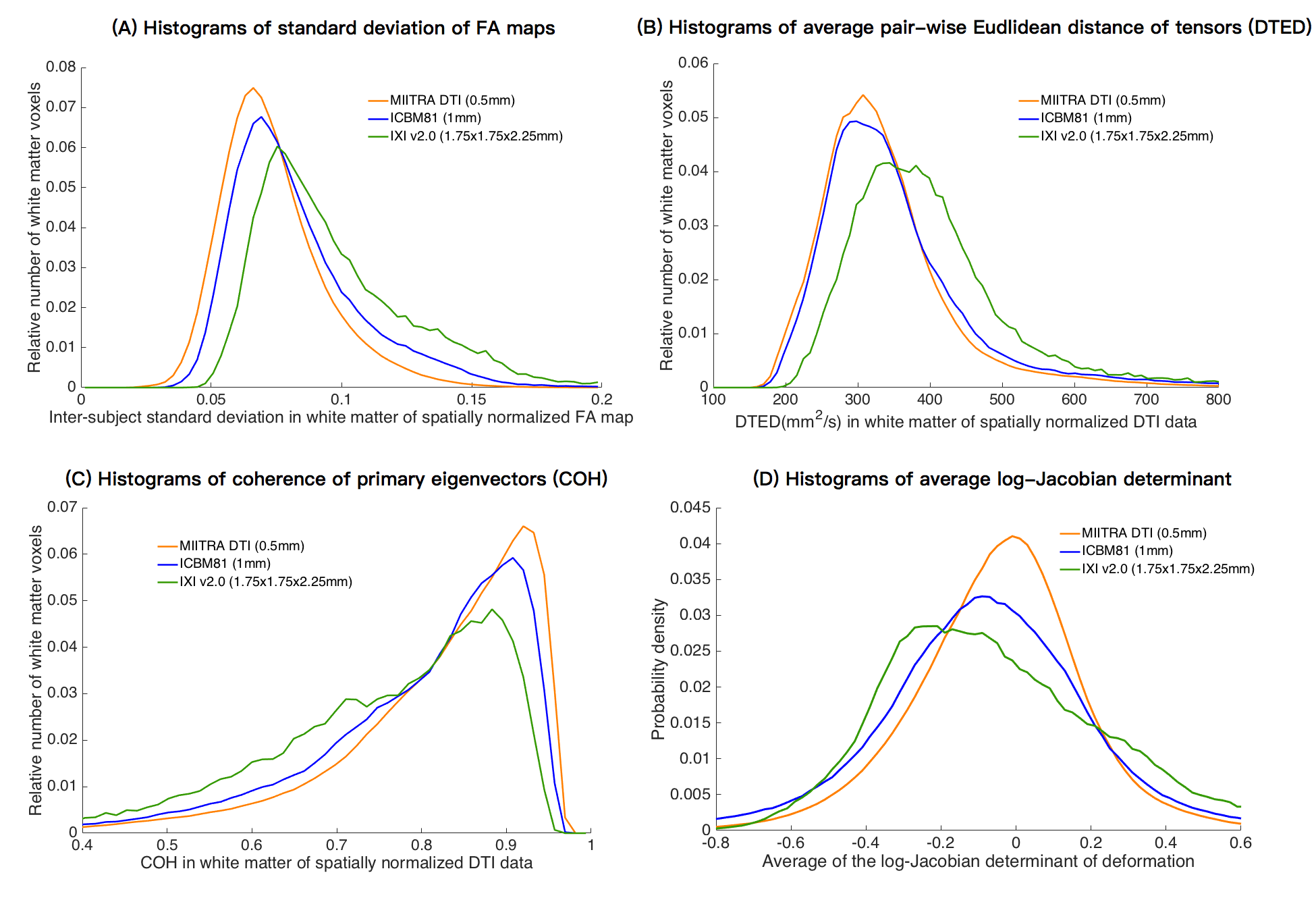
The MIITRA DTI template was compared to other DTI templates that were constructed using at least some data on older adults (IXI v2.014, ICBM8115) in terms of the accuracy of inter-subject spatial normalization of ADNI3 data achieved when each template was used as a reference (DRTAMAS registration). The standard deviation of normalized FA maps, average pair-wise Euclidean distance of normalized tensors (DTED)16, coherence of primary eigenvectors (COH)16 of normalized tensors, and average log-Jacobian determinant of deformations were compared across DTI templates.
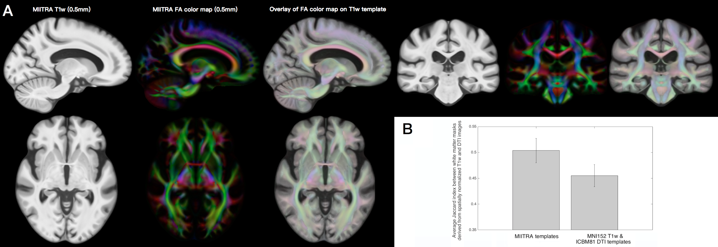
The inter-modality spatial matching achieved with the MIITRA templates was compared to that achieved with the MNI152-T1w17 and ICBM81-DTI15 templates by means of the average Jaccard index between white matter masks generated from T1w images and FA maps (k-means clustering) of spatially normalized ADNI3 data.
Results and Discussion
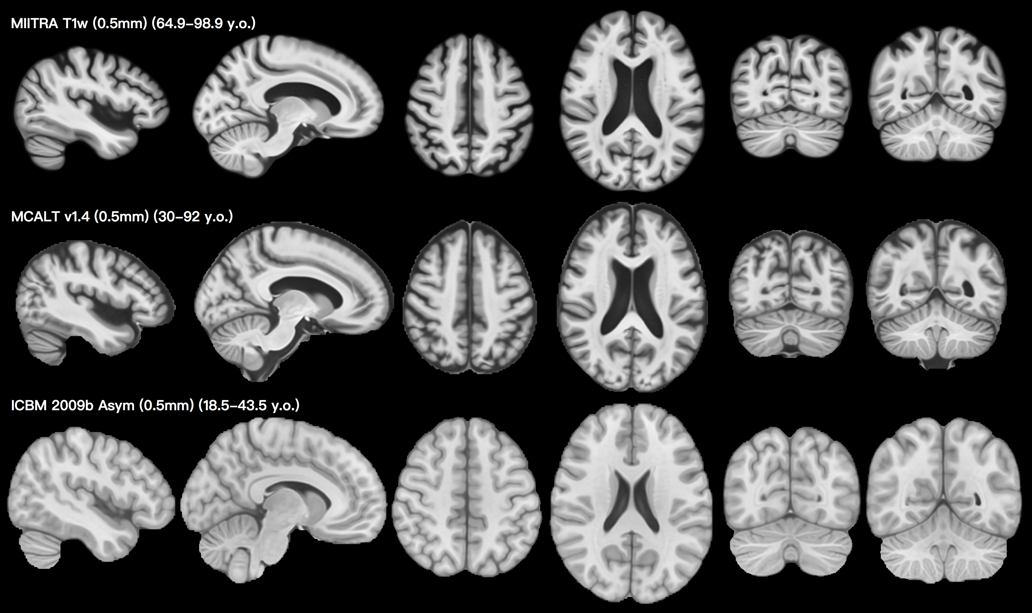
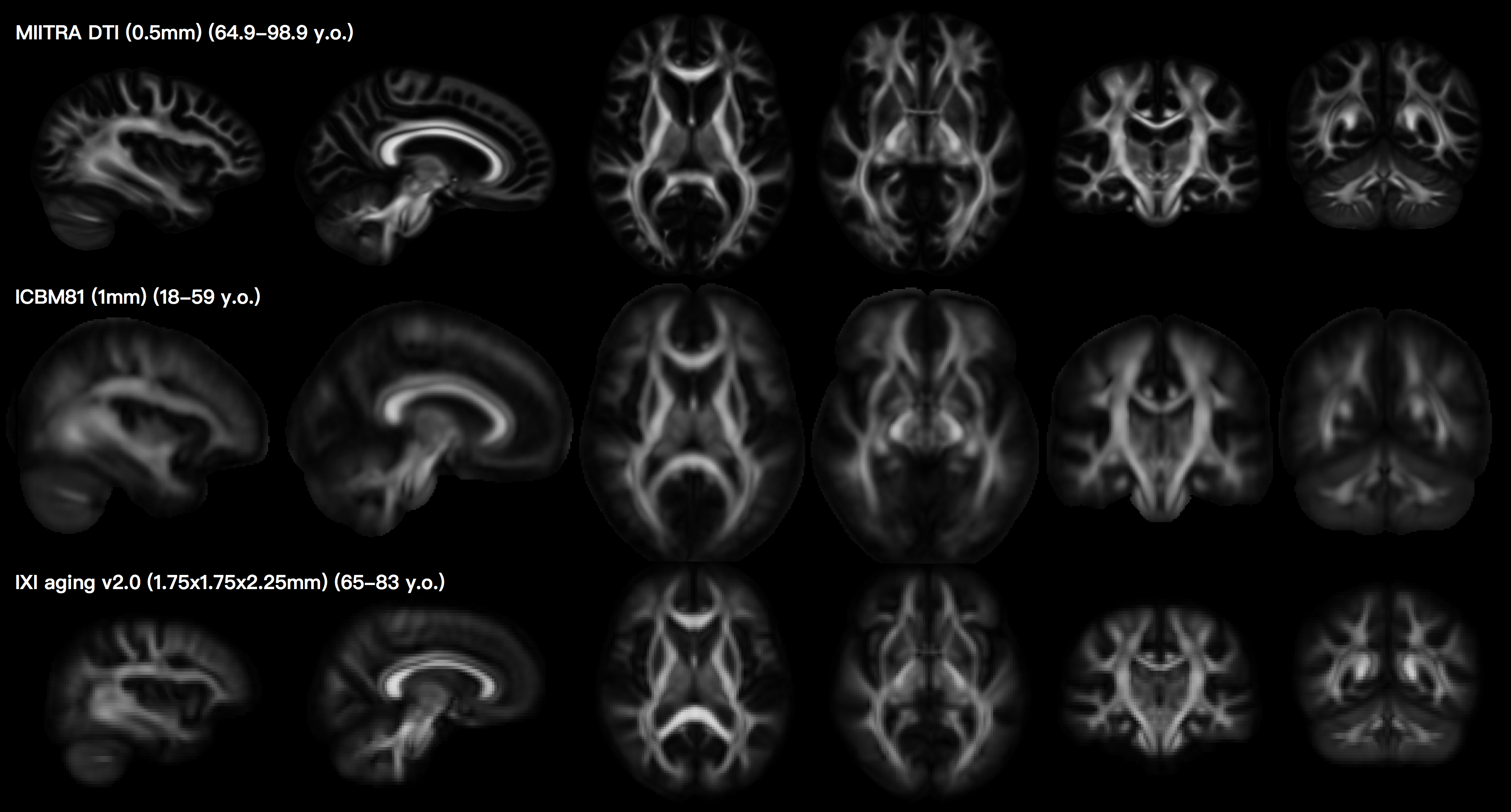
Visual inspection shows that the MIITRA T1w (Fig.1) and DTI (Fig.2) templates with 0.5mm isotropic voxels exhibit high sharpness and are free of artifacts. MIITRA T1w allowed higher APNCC and APJI and less deformation for spatial normalization of ADNI3 data compared to other T1w templates (Fig.3). The MIITRA DTI template allowed lower standard deviation of FA maps, lower DTED, higher COH and less deformation for spatial normalization of ADNI3 data compared to other DTI templates (Fig.4). These results indicate that the MIITRA templates provided higher inter-subject spatial normalization accuracy of older adult data and required less deformation than other templates. Finally, the MIITRA T1w and DTI templates exhibited high spatial matching to each other (Fig.5A) and allowed higher inter-modality spatial matching of normalized older adult data (Fig.5B).Conclusion
This work developed high quality 0.5mm resolution T1w and DTI templates for the MIITRA atlas using data from a large, diverse, community cohort of non-demented older adults, and demonstrated that the new templates allowed higher inter-subject and inter-modality spatial normalization of older adult data compared to other templates.Acknowledgements
This study was supported by:
National Institute on Aging (NIA) R01AG052200
National Institute on Aging (NIA) P30AG010161
National Institute on Aging (NIA) R01AG017917
National Institute on Aging (NIA) RF1AG022018
National Institute on Aging (NIA) R01AG056405
References
1. Bennett DA, Buchman AS, Boyle PA, Barnes LL, Wilson RS, Schneider JA. Religious Orders Study and Rush Memory and Aging Project. J Alzheimers Dis. 2018;64(s1):S161-S189.
2. Barnes LL, Shah RC, Aggarwal NT, Bennett DA, Schneider JA. The Minority Aging Research Study: Ongoing Efforts to Obtain Brain Donation in African Americans without Dementia. Curr Alzheimer Res. 2012;9(6):734-745.
3. Wu Y, et al. Development of High Quality T1w and DTI Templates of the Older Adult Brain in a Common Space. Proc. Intl. Soc. Mag. Reson. Med. 2019;27.
4. Niaz, M.R., et al. Development and evaluation of a 0.5mm isotropic resolution structural template of the older adult brain. Proc. Intl. Soc. Mag. Reson. Med. 2019;27.
5. Niaz, M.R., et al. Development and evaluation of a high spatial resolution diffusion tensor template of the older adult human brain. Proc. Intl. Soc. Mag. Reson. Med. 2020;28.
6. Avants et al. ANTS: Open-Source Tools for Normalization And Neuroanatomy.
7. Irfanoglu M. O., Nayak, A., Jenkins, J., Hutchinson, E. B., Sadeghi, N., Thomas, C. P., et al. DR-TAMAS: diffeomorphic registration for tensor accurate alignment of anatomical structures. Neuroimage 2016;132:439–454.
8. http://adni.loni.usc.edu/
9. Schwarz CG, et al. THE MAYO CLINIC ADULT LIFE SPAN TEMPLATE: BETTER QUANTIFICATION ACROSS THE LIFE SPAN. Alzheimer's & Dementia: The Journal of the Alzheimer's Association. 2017;13(7):P93-4.
10. Fonov V, Evans AC, Botteron K, et al. Brain Development Cooperative Group. Unbiased average age-appropriate atlases for pediatric studies. Neuroimage. 2011;54(1):313-27.
11. Fonov VS, Evans AC, McKinstry RC, et al. Unbiased nonlinear average age-appropriate brain templates from birth to adulthood. NeuroImage. 2009(47):S102.
12. Holmes CJ, Hoge R, Collins L, et al. Enhancement of MR images using registration for signal averaging. Journal of computer assisted tomography. 1998;22(2):324-33.
13. Glasser, M. F., Sotiropoulos, S. N., Wilson, J. A., et al. The minimal preprocessing pipelines for the Human Connectome Project. Neuroimage, 2013;80:105-124.
14. Zhang, H., et al. A computational white matter atlas for aging with surface-based representation of fasciculi. Biomedical Image Registration. WBIR 2010. Lecture Notes in Computer Science, vol 6204.
15. Mori S, et al. Stereotaxic white matter atlas based on diffusion tensor imaging in an ICBM template. Neuroimage 2008;40(2):570-582.
16. Zhang S., et al. Enhanced ICBM diffusion tensor template of the human brain. Neuroimage. 2011;54(2):974-84.
17. G. Grabner, A. L. Janke, M. M. Budge, et al. Symmetric atlasing and model based segmentation: an application to the hippocampus in older adults. Med Image Comput Comput Assist Interv Int Conf Med Image Comput Comput Assist Interv. 2006;9:58–66.
Figures