1693
Connectometry analysis of diffusion MRI in adolescents with sports-related concussion
Hon J Yu1, Mark Fisher2, and Min-Ying Su1
1Radiological Sciences, University of California, Irvine, CA, United States, 2Neurology, University of California, Irvine, Orange, CA, United States
1Radiological Sciences, University of California, Irvine, CA, United States, 2Neurology, University of California, Irvine, Orange, CA, United States
Synopsis
This study evaluates a feasibility of diffusion MRI based connectometry in capturing sports-related concussion (SRC) induced effects in adolescents using high school football players with and without concussion history as cohorts. The results revealed that although there were some common tract bundles whose segments showed significant correlation with concussion history independent of the diffusion metrics used (QA, MD, or FA), there were also different tract bundles that showed either positive or negative correlation with concussion depending on the diffusion metric used. A further study would be necessary to fully examine clinical relevance of this study’s findings in adolescents with SRC.
Introduction
In a recent estimate,1 more than 5 million youth and high school athletes participate in organized American football in the United States with increased risks to repeated head impacts, and sports-related concussion (SRC) continues to be the most common form of mild traumatic brain injury (mTBI) in adolescents.2 MRI-based techniques such as resting-state functional MRI (rs-fMRI) and diffusion tensor imaging (DTI) have been widely used for TBI study in research setting. DTI, especially, has increasingly been touted as a method that offers great potential in both research and clinical settings, with a growing body of literature suggesting certain diffusion metric may serve as a biomarker for microstructural white matter changes in mTBI.3,4 Connectome based on diffusion MRI (dMRI) gathered much interest recently as a non-invasive means to map and measure the structural connections between parcellated gray matter targets using a fiber tracking algorithm, and the subsequent development of connectometry concept introduced a new analytical approach based on local connectomes possible for study of mTBI.5,6 In this study, whole-brain based differences in high school football players between those with history of concussion and those without were explored using DTI and connectometry.Methods
The study protocol was approved by the institutional review board and all subjects gave written informed consent. Fourteen male high school football players (mean-age: 16.9 ± 1.6; range: 15-20 yrs.) were included in the study, 7 with history of concussion and 7 without. Two subjects from each subject cohort also had a repeated MRI 1-4 yrs. after their initial study, resulting in 18 MRI datasets for the study. All MR studies were performed on a 3T scanner (Vantage Galan, Canon Medical Systems, Japan) using a 32-channel, dedicated receive-only head coil. Following T1w scan in sagittal orientation (3D-GRE; 1-mm3 voxel-size; TR/TE/TI=7.3/3.2/900 [ms]), 30-direction DTI scan was performed based on SE-EPI sequence in axial orientation (2 x 2 x 2 mm3 voxel-size; TR/TE=10,800/90 [ms]; b=1000 s/mm2). The analyses to assess differences in DTI data between the players with concussion and those without were conducted based on connectometry,5 using DSI Studio (http://dsi-studio.labsolver.org). Briefly, after motion correction, each of the datasets was reconstructed into the Montreal Neurological Institute (MNI) template space using Q-Space Diffeomorphic Reconstruction (QSDR),5 after which a connectometry database can be created based on different diffusion metric, such as quantitative anisotropy (QA), mean diffusivity (MD), fractional anisotropy (FA), etc. The local connectomes that express positive or negative association with history of concussion using a multiple regression model (age and career in years were included as covariates) were then tracked along a common pathway to reveal the segments of the fascicles that have significant association. A T-score threshold of 2.5 was assigned and tracked using a deterministic fiber tracking algorithm7 to obtain correlational tractography. A length threshold of 40 mm was used to select tracks and a total of 4000 randomized permutations were applied to the group label to obtain the null distribution of the track length and estimate the false discovery rate (FDR). Group connectometry analyses based on 3 different diffusion metrics: QA, MD, and FA, were investigated in this exploratory study.Results
Only the tracks with FDR < 0.05 were shown in this study. Fig. 1 shows the tracks with QA positively correlated with concussion history in mosaic fashion both in axial and coronal orientation as well as in 3-D representation using glass brain. The tracks with positive correlation included: middle Cerebellar Penduncle, Vermis, left/right Cerebellum, left/right Dentatorubrothalamic Tract, left/right Corticospinal Tract, left Medial Lemniscus, left/right Inferior Cerebellar Peduncle, and right Reticulospinal Tract. The FDR was estimated to be 0.0438. There were no tracks with QA negatively correlated with concussion history with significance, however. Fig. 2 shows the tracks with MD positively correlated with concussion history in mosaic fashion both in axial and coronal orientation as well as in 3-D representation using glass brain. The tracks with positive correlation included: left/right Cerebellum, left/right Fornix, Middle Cerebellar Penduncle, left/right Medial Lemniscus, right Thalamic Radiation Superior, Corpus Callosum Body, and right Corticopontine Tract Parietal. The FDR was estimated to be 0.0033. There were no tracks with MD negatively correlated with concussion history with FDR < 0.05, however. Fig. 3 shows the tracks with FA negatiely correlated with concussion history in mosaic fashion both in axial and coronal orientation as well as in 3-D representation using glass brain. The tracks with negative correlation included: left/right Cerebellum, Vermis, Middle Cerebellar Peduncle, and left Arcuate Fasciculus. The FDR was estimated to be 0.0089. There were no tracks with FA positively correlated with concussion history with FDR < 0.05, however.Discussion
Although direct comparison to previously reported results of dMRI on mTBI is difficult to make due to different study design, analytical methodology and/or diffusion metrics used, some of the fascicles with segments that are found to have statistically significant correlation with history of concussion in this study have also been noted to be associated with mTBI or SRC in literature, including the Corticospinal Tract, Arcuate Fasciculus, and Corpus Callosum. Despite small sample size, this study clearly demonstrates a feasibility of connectometry based approach in study of SRC in adolescents and warrants a further study.Acknowledgements
No acknowledgement found.References
1. Daniel RW, Rowson S, Duma SM. Head impact exposure in youth football. Ann Biomed Eng. 2012;40:976-981; 2. Bryan MA, Rowhani-Rahbar A, Comstock RD, et al. Sports-and recreational-related concussions in US youth. Pediatrics 2016;138(1):e20154635; 3. Niogi SN, Mukherjee P. Diffusion tensor imaging of mild traumatic brain injury. J Head Trauma Rehabil. 2010;25:241-255; 4. Zappala G, Thiebaut de Schotten M, Eslinger PJ. Traumatic brain injury and the frontal lobes: What can we gain with diffusion tensor imaging? Coretx 2012;48:156-165; 5. Yeh FC, Badre D, Verstynen T. Connectometry: A statistical approach harnessing the analytical potential of the local connectome. NeuroImage 2016;125:162-171; 6. Murdaugh DL, King TZ, Sun Binjian, et al. Longitudinal Changes in Resting State Connectivity and White Matter Integrity in Adolescents With Sports-Related Concussion. J Int Neuropsychol. 2018;24:781-792; 7. Yeh FC, Verstynen TD, Wang Y, et al. Deterministic diffusion fiber tracking improved by quantitative anisotropy. PLoS One 2013;8:e80713.Figures
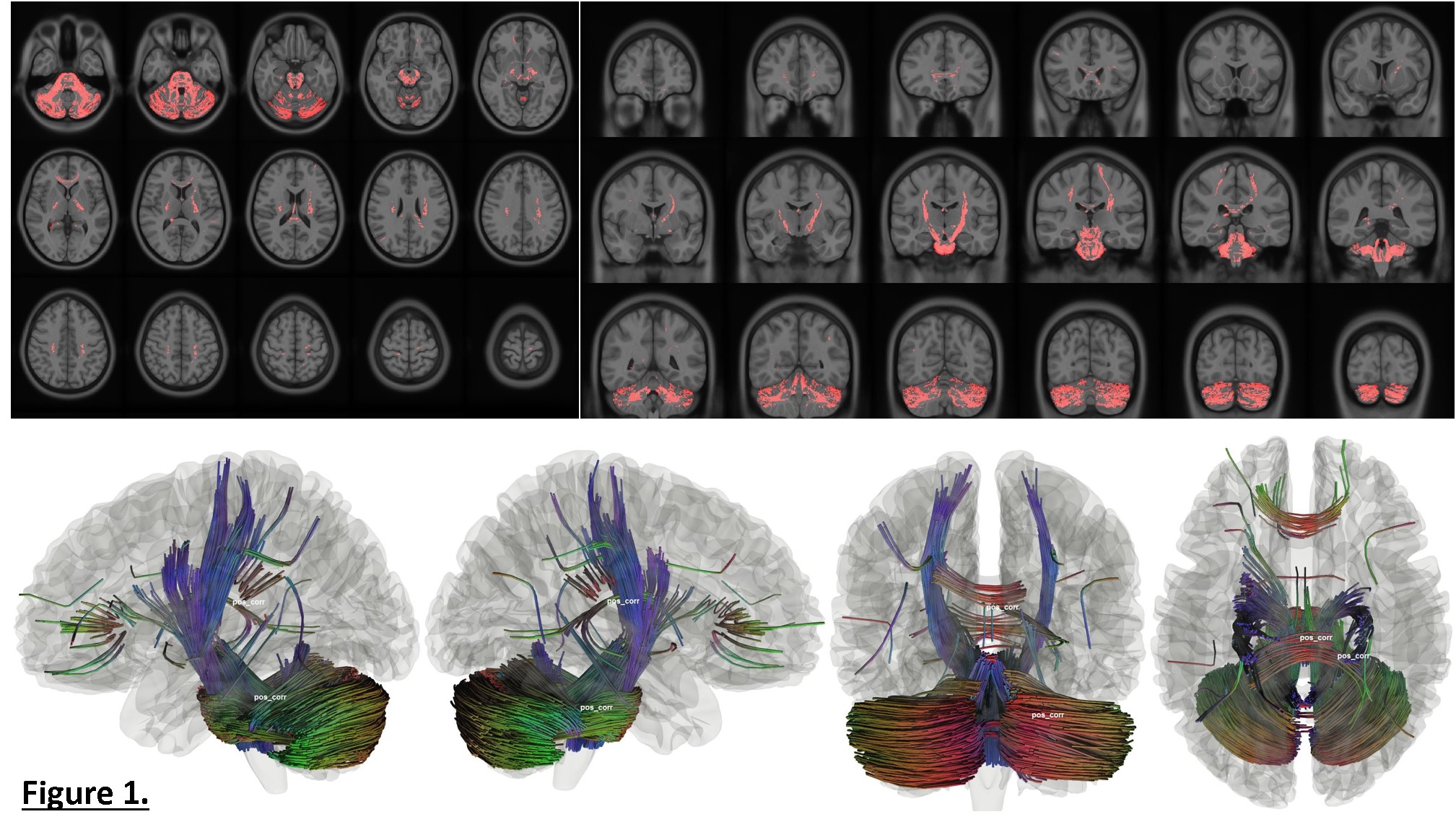
Figure 1. The tracks with segments whose QA
(quantitative anisotropy) values positively correlated with concussion history
shown in mosaic format in axial (top-left) and coronal (top-right) orientation
as well as 3D visualization in glass brain (bottom).
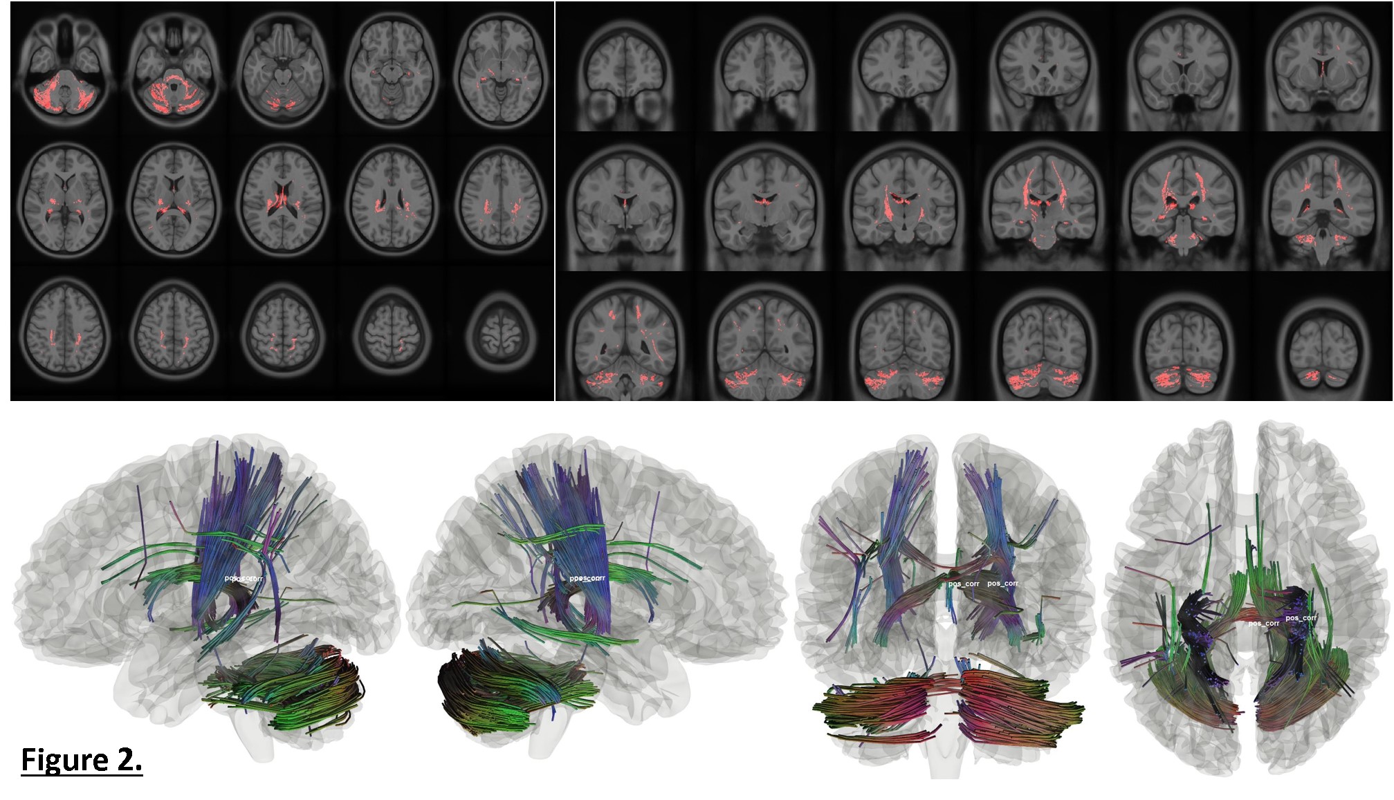
Figure 2. The tracks with segments whose MD
(mean diffusivity) values positively correlated with concussion history shown
in mosaic format in axial (top-left) and coronal (top-right) orientation as
well as 3D visualization in glass brain (bottom).
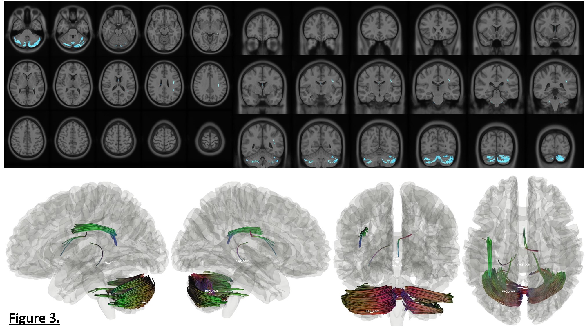
Figure
3. The
tracks with segments whose FA (fractional anisotropy) values negatively
correlated with concussion history shown in mosaic format in axial (top-left) and
coronal (top-right) orientation as well as 3D visualization in glass brain
(bottom).