1689
Resting-state functional connectivity and brain network abnormalities in depressive patients with suicidal ideation1Department of Medical Imaging and Radiological Sciences, and Bachelor Program in Artificial Intelligence, Chang Gung University, Taoyuan, Taiwan, 2Medical Imaging Research Center, Institute for Radiological Research, Chang Gung University and Chang Gung Memorial Hospital at Linkou, Taoyuan, Taiwan, 3Department of Psychiatry, Chang Gung Memorial Hospital, Chiayi, Taiwan, 4Department of Radiology, Taichung Veterans General Hospital, Taichung, Taiwan, 5School of Medicine, Chang Gung University, Taoyuan, Taiwan, 6Department of Diagnostic Radiology, Chang Gung Memorial Hospital, Chiayi, Taiwan
Synopsis
Our study aimed to investigate whether changes in brain function measured with functional magnetic resonance imaging (fMRI) can be detected among individuals with depressive disorders and suicidal ideation, including amplitude of low-frequency fluctuation (ALFF), regional homogeneity (ReHo), and graph theoretical analysis (GTA). We results suggest that brain functional connectivity may be affected in depressive patients with suicidal ideation. The findings from present study can serve as the basis for further algorithm studies by machine learning method to stratify the risk population.
Introduction
Suicide is an important and serious public health problem worldwide, and its effects are observed throughout all regions, countries, and cultures 1. Our study aimed to investigate whether changes in brain function measured with functional magnetic resonance imaging (fMRI) can be detected among individuals with depressive disorders and suicidal ideation, including amplitude of low-frequency fluctuation (ALFF), regional homogeneity (ReHo), and graph theoretical analysis (GTA).Methods
We recruited three groups of 111 participants: 35 depressive patients with suicidal ideation (SI) (age 21–60 years, mean = 41.11 years, SD = 11.78 years), 32 depressive patients without suicidal thoughts (NS) (age 21–60 years, mean = 47.88 years, SD = 8.99 years), and 44 healthy controls (HCs) (age 20–58 years, mean = 41.98 years, SD = 9.27 years). All participants were scanned using a 3T MRI (Verio, SIEMENS, Erlangen, Germany) system. For all participants, the following EPI sequence was used: repetition time (TR)/ echo time (TE) = 2000/30 ms, in-plane resolution (pixel size) = 3.4 x 3.4 mm2, thickness = 4 mm, number of repetitions = 300, scan time for functional imaging = 10 min, and 31 axial slices to cover the whole cerebrum.We obtained mean fractional amplitude of low frequency fluctuation (mfALFF), mean regional homogeneity (mReHo), and graph theoretical topology from rs-fMRI to portray functional changes among three groups. Analysis of variance (ANOVA) and post-hoc two-sample t tests were implemented to compare mfALFF and mReHo among groups using SPM. The functional connectivity toolbox, CONN, simplified the connectivity matrix generation procedure. We first segmented the whole brain of each subject into 90 regions based on the Automated Anatomical Labeling atlas, with each region considered a node. The connection between each node was viewed as an edge. The 90 × 90 connectivity matrix was then computed through Pearson correlations for each participant. Graph Analysis Toolbox (GAT) accepted the output connectivity matrix of each group from CONN 2. GAT was used to obtain the topological parameters, and the area under the curve (AUC) within chosen ranges for each topology index and comparing them between each group. Topological parameters were conducted in densities between 0.2 and 0.5, with an increment of 0.01. The density represents the ratio of existing connections to possible connections. A two-sample t test and nonparametric permutation test was performed by GAT for statistical comparison.
Results
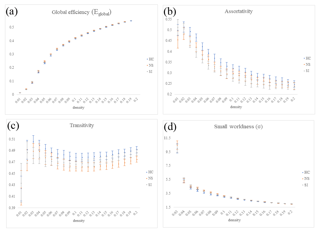
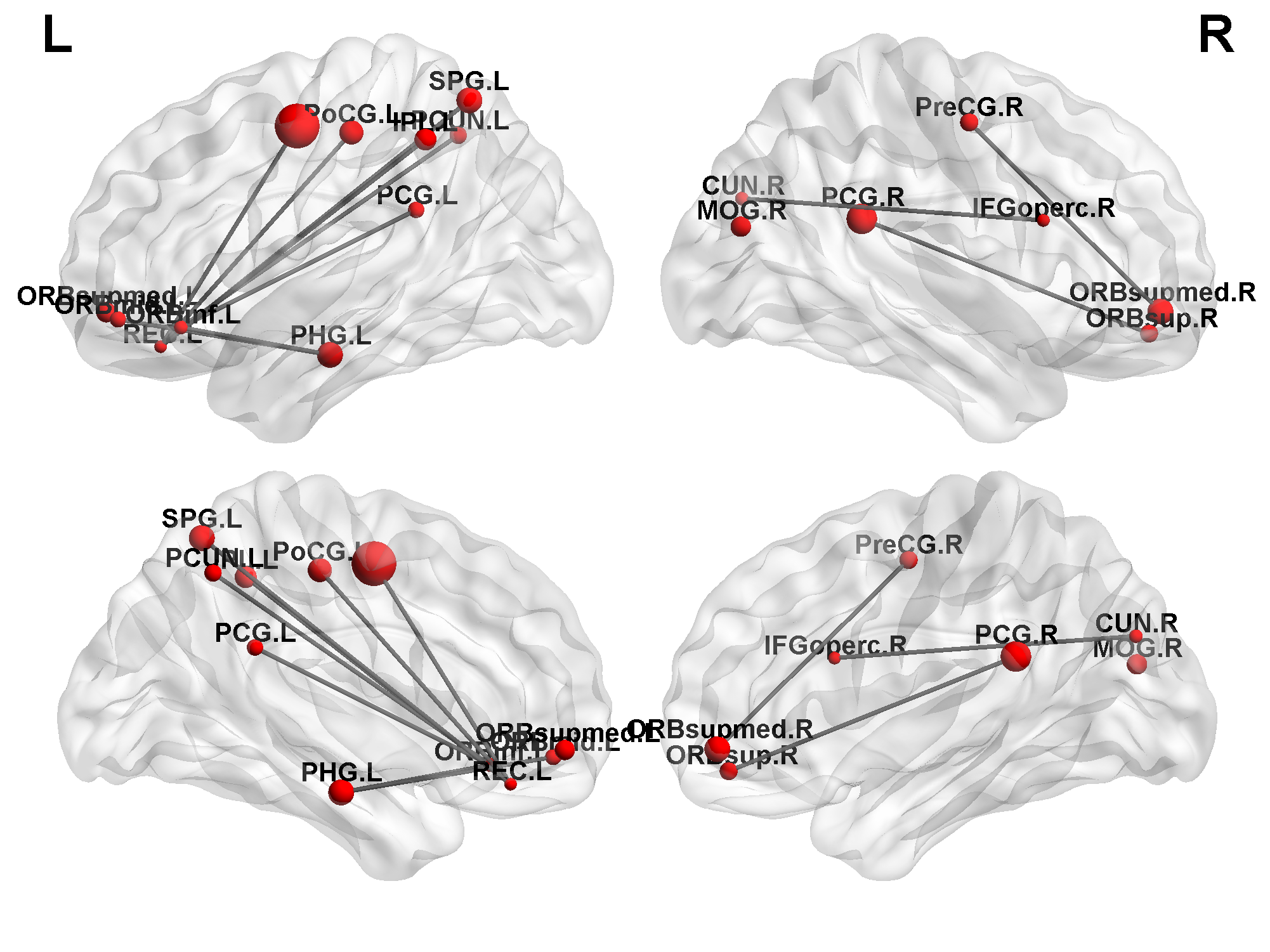
In the VBA of mfALFF between the SI and the NS groups (Fig. 1a, NS > SI), we found lower mfALFF activation of the left cuneus in the SI group compared with the NS group (Fig. 1b) and higher mfALFF activation of the right middle temporal pole gyrus (Fig. 1c) in the SI group compared with the NS group. In the VBA of mReHo between the SI and the NS groups (Fig. 1d, NS > SI), we found lower regional homogeneity of the right cuneus in the SI group compared with the NS group (Fig. 1e). We also found higher regional homogeneity of the left middle temporal gyrus (MTG) (Fig. 1f) in the SI group compared with the NS group (all corrected p < 0.05).In the GTA of the three groups, we found a significant tendency (NS > SI > HC) in the global efficiency (Fig. 2a, corrected p < 0.05). However, no significant tendency was found in assortativity (Fig. 2b), transitivity (Fig. 2c), and small-worldness index (σ) (Fig. 2d). Although all participants maintained a small-worldness functional brain network according to the σ calculation, the network was more like a random network in the SI group. For the NBS analysis, one subnetwork showed more edges in the NS group compared with the SI group (Fig. 3, p < 0.05).
Discussion
Our results demonstrated that widespread and disrupted network changes in brain functional networks and their interconnectivity was associated in depressive patients with suicidal ideation. A graph theoretical analysis (GTA) and network-based statistical (NBS) analysis revealed different topological organization and slightly better local segregation of the brain network in healthy participants compared with those in depressive patients with suicidal ideation. Our results suggest that the neural basis underlying the psychopathology in depressive patients with suicidal ideation involves multiple brain functional networks and their interaction.Conclusion
We concluded that the brain function of depressive disorders with suicidal ideation was different than that in depressive disorders without suicidal ideation and in healthy participants. The findings from present study can serve as the basis for further algorithm studies by machine learning method to stratify the risk population.Acknowledgements
This study was supported by the research programs, MOST106-2314-B-182-040-MY3 and MOST109-2314-B-182-047-MY3, which were sponsored by the Ministry of Science and Technology, Taipei, Taiwan.References
1. Sveticic, J. and D. De Leo, The hypothesis of a continuum in suicidality: a discussion on its validity and practical implications. Ment Illn, 2012. 4(2): p. e15.
2. Hosseini, S.M., F. Hoeft, and S.R. Kesler, GAT: a graph-theoretical analysis toolbox for analyzing between-group differences in large-scale structural and functional brain networks. PLoS One, 2012. 7(7): p. e40709.
Figures