1687
Limbic System Lateralization of Amide Proton Transfer Weighted Signals in Young Healthy Subjects1the First Affiliated Hospital of Dalian Medical University, Dalian, China, 2Philips Healthcare, Beijing, China
Synopsis
Amide proton transfer weighted (APTw) imaging is a novel molecular imaging technique to acquire the proton exchanged signals from the amide in proteins or peptides to water. Previous studies discover lateralization in the brain structure and function, including limbic system. However, it is not clear whether APTw also has lateral advantage. In this prospective study, we applied the automatic brain segmentation method to quantify the APTw signal values of limbic system in young healthy subjects. Significant differences of APTw signals between left and right brain hemisphere were found, which may suppose related with right-handedness.
Introduction
Amide proton transfer weighted (APTw) MR imaging is a novel non-invasive molecular imaging technique. The signals in APTw are based on the amide protons content in endogenous proteins or peptides. Previous studies have shown that APTw imaging is capable of diagnosis in ischemic stroke, tumors and neurodegenerative diseases[1][2][3]. Although one study on the APTw signal values of different anatomical regions in young and healthy normal brain parenchyma had been published[4]. However, the manual ROI delineation method in this study may led to certain measurement errors. Furthermore, it is not clear whether APTw also has lateral advantage. In this study, we aim to use an automatic segmentation method of brain tissue to quantify the APTw signal intensity in the limbic system in healthy young volunteers.Methods
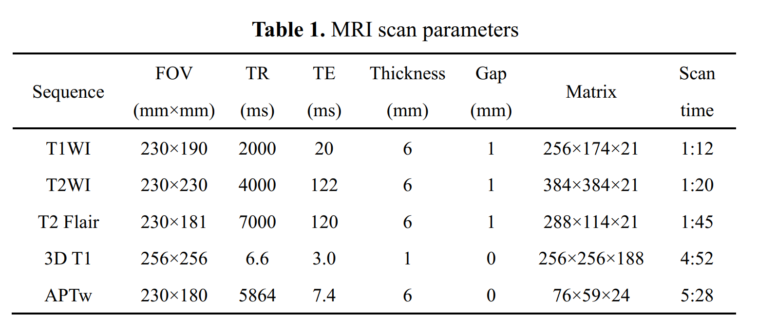
Nineteen healthy volunteers (mean age: 25.37 ± 1.49, range from 22 to 29 years; 10 females, 9 males) were recruited and all were right-handed. Informed consent was acquired from each subject. All volunteers were performed MR examination on a 3.0 T MR scanner (Ingenia CX, Philips Healthcare, the Netherlands) with a 32-channel receive-only head coil. MR protocols were performed in this study including axial T1 sequence, axial T2 sequence, axial T2 FLAIR, sagittal 3D T1 TFE and 3D TSE APTw sequence. Scan parameters were shown in Table 1.All of the images were analysis using SPM 8 and REST plus software package on MATLAB (2013 b) platform. After converting of the original DICOM images into 3D NIFTI format images with dcm2niigui software, the APTw images and 3D T1 structure images were registered to the MNI standard space. The Gaussian smoothing kernel size was set to 6 mm. With the template in the Montreal Neurological Institute (MNI) space, the APTw images of each region of the cerebral hemisphere were extracted. The APTw signals of the left and the right hemisphere are separately measured. The paired-sample t test was used to analyze the APTw signals of bilateral hemispheres in the limbic system. All statistical analyses were performed using the SPSS 22.0 software package.
Results
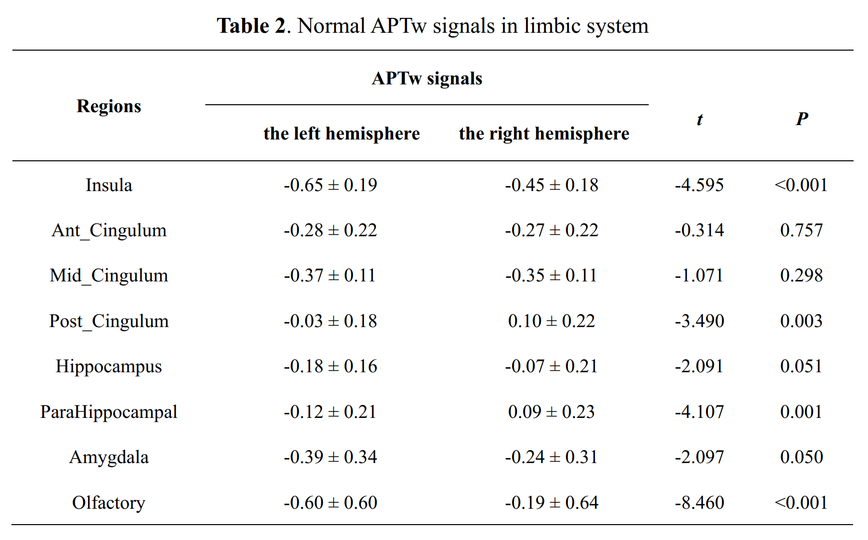
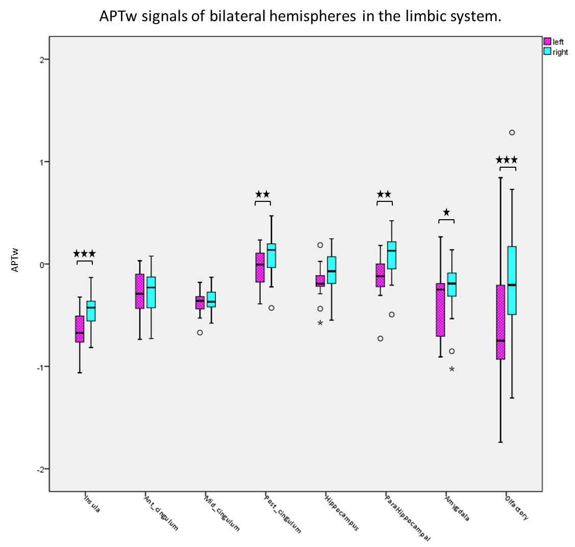
In our data, there were no apparent motion artifacts in all subjects. The averaged APTw signal intensity and standard deviation signals in different brain regions of limbic system are presented in Table 2 and Figure 1-2. Significant elevated APTw signals in the limbic system in the right side were found than those in the left side, especially in the insula, post cingulum, para hippocampus, amygdala and olfactory (P < 0.05). In addition, there are significant differences of APTw signals in different brain regions (P < 0.001). Furthermore, higher differences between APTw signals of bilateral brain were found in the insula (difference: 0.2%) and olfactory (difference: 0.41%).Discussion
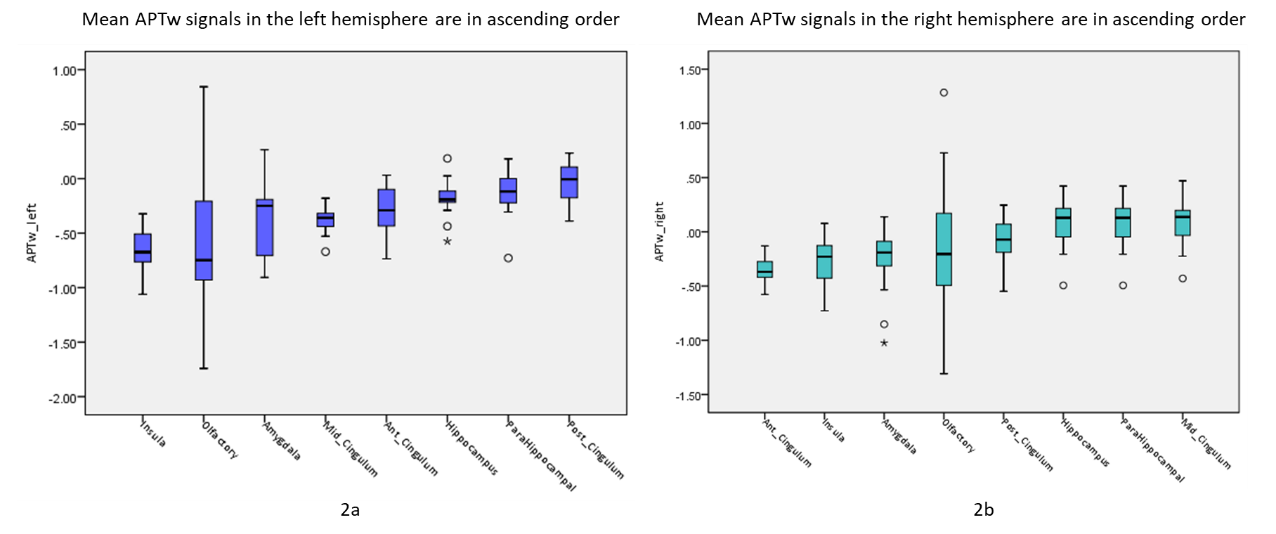
In this study, the averaged APTw signal values in different brain structures in the limbic system were measured in healthy young subjects. We believed that the accuracy of brain region segment is improved with the previous manual ROI delineation results[4]. The method will help for study the protein distribution in cerebral diseases and pathophysiological mechanisms with APT and other chemical exchange saturation transfer (CEST) imaging method[5]. The limbic system is internally interconnected and has extensive connections with other parts of the nervous system. It participates in the regulation of sensory and visceral activities, and is closely related to mental activities such as emotion, behavior, learning and memory[6]. In this study, we found that there are statistical differences of the averaged APTw signal intensity of limbic system structures between bilateral brain structures, and the right side is higher than the left side. We suspect that this difference is related to right-handedness. A recent study has been confirmed that hemispheric lateralisation determines an individual's emotional processing[7]. Meanwhile, we found that the APTw values in different regions of unilateral cerebral hemisphere were also different. We speculate that this is caused by differences in metabolite changes in different regions of the cerebral hemispheres on both sides. But this still needs further research to confirm.Conclusion
The baseline of APTw signal values in different anatomical localizations of limbic system were measured in young healthy subjects. There are differences of APTw signal values in both of unilateral and bilateral cerebral hemisphere, which may relate to right-handedness.Acknowledgements
No acknowledgement found.References
[1] Yu H, Lou H, Zou T, Wang X, Jiang S, Huang Z, Du Y, Jiang C, Ma L, Zhu J, He W, Rui Q, Zhou J, Wen Z. Applying protein-based amide proton transfer MR imaging to distinguish solitary brain metastases from glioblastoma. Eur Radiol. 2017 Nov;27(11):4516-4524. doi: 10.1007/s00330-017-4867-z.
[2] Msayib Y, Harston GWJ, Tee YK, Sheerin F, Blockley NP, Okell TW, Jezzard P, Kennedy J, Chappell MA. Quantitative CEST imaging of amide proton transfer in acute ischaemic stroke. Neuroimage Clin. 2019; 23:101833. doi: 10.1016/j.nicl.2019.101833. Epub 2019 Apr 23. PMID: 31063943; PMCID: PMC6503165.
[3] Li S, Chan P, Li C, et al. Changes of Amide Proton Transfer Imaging in Multiple System Atrophy Parkinsonism Type. Front Aging Neurosci. 2020; 12:572421. Published 2020 Sep 30. doi:10.3389/fnagi.2020.572421
[4] Sartoretti T, Sartoretti E, Wyss M, et al. Amide Proton Transfer Contrast Distribution in Different Brain Regions in Young Healthy Subjects. Front Neurosci. 2019; 13:520. Published 2019 May 22. doi:10.3389/fnins.2019.00520
[5] Ward KM, Aletras AH, Balaban RS. A new class of contrast agents for MRI based on proton chemical exchange dependent saturation transfer (CEST). J Magn Reson. 2000 Mar;143(1):79-87. doi: 10.1006/jmre.1999.1956.
[6] Lopez-Franco A, Alanis AY, Lopez-Franco C, Arana-Daniel N, Lopez-Franco M. Emotional system in complex cognitive activities of working memory: A literature review of its role. J Integr Neurosci. 2018;17(3-4):679-693. doi:10.3233/JIN-180095
[7] Bednarczuk NF, Casanovas Ortega M, Fluri AS, Arshad Q. Vestibulo-cortical hemispheric dominance: The link between anxiety and the vestibular system? Eur J Neurosci. 2018;47(12):1517-1524. doi:10.1111/ejn.13948
Figures