1683
Preliminary Assessment of Intravoxel Incoherent Motion Diffusion-Weighted MRI Metrics in Acute Carbon Monoxide Poisoning1Yan 'an People's Hospital, Yan 'an, China, 2First Affiliated Hospital of Xi 'an Jiaotong University, Xi 'an, China
Synopsis
The purpuse of this study was to assess microvascular perfusion and microstructural integrity using Intravoxel incoherent motion (IVIM) imaging in patients with acute CO poisoning.Thirteen patients with acute CO poisoning and 15 healthy subjects were enrolled. IVIM MRI was collected using a 3.0-T scanner.The ROIs analysis was perforemed. The IVIM perfusion fraction was significantly reduced in multiple brain regions and IVIM diffusion metirc was significantly decreased in centrum semiovale and multiple subcortical gray matter nucleis in CO poisoning. This study shows that IVIM-DWI may be a promising method to assess brain perfusion and injury in acute CO poisoning.
INTRODUCTION
Carbon monoxide (CO) poisoning is one of the most usual kinds of poisoning worldwide.1,2 CO might harm numerous organ systems, particularly systems with high oxygen usage such as the central nervous system (CNS).1 Acute CO poisoning can lead to diverse symptoms, from headache, chest pain, dyspnea, and mental confusion, to coma or death.3,4Magnetic resonance imaging(MRI) is an easily accessible, minimally invasive procedure for the objective evaluation of brain injury of CO poisoning.5 Diffusion tensor imaging and diffusion kurtosis imaging studies found reduced microstructural integrity of the white matter and damage of subcortical gray matter nuclei in patients with CO poisoning compared with healthy controls.6-9 In addition to microstructural changes, several research studies revealed decreased cerebral perfusion in patients with CO poisoning.10-12 Most of these studies examined perfusion differences by single-photon emission computed tomography (SPECT) methods. However, studies that evaluated both diffusion and perfusion in patients with CO poisoning are scarce.
Intravoxel incoherent motion (IVIM) imaging is a diffusion-weighted magnetic resonance imaging (MRI) method with multiple b values developed to identify microvascular perfusion and microstructural integrity concurrently.13,14 IVIM allows quantification of three parameters, which are D, D*, and f, with a biexponential fit. D indicates the pure diffusion coefficient, which can reflect microstructural integrity. D* represents the pseudodiffusion coefficient, and f is a surrogate for microvascular perfusion.13,15 This technique has formerly been used in the brain to characterize perfusion and diffusion of Alzheimer's Disease,16 as well as cerebral ischemia.17,18 According to our literature review, no research with IVIM has been applied to the study of CO poisoning.
Therefore, the purpose of this study was to assess microvascular perfusion and microstructural integrity using IVIM MRI in patients with acute CO poisoning.
METHODS
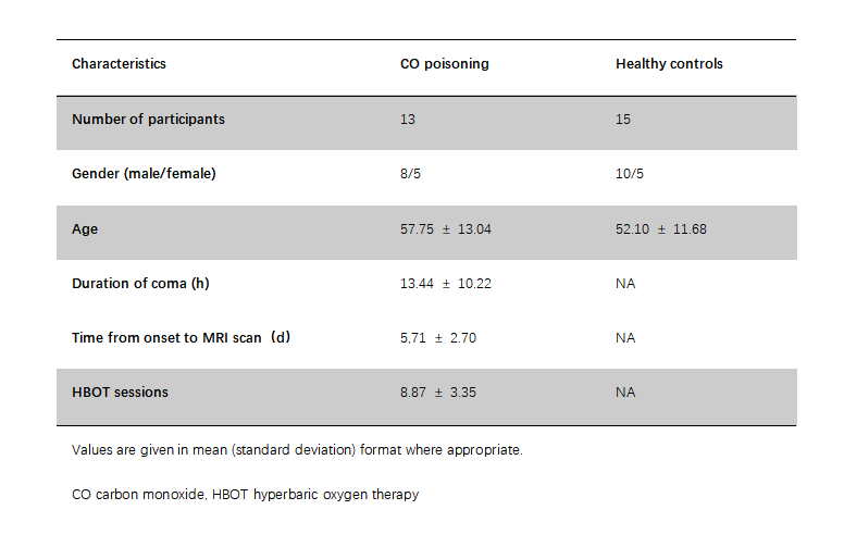
Thirteen patients with acute CO poisoning were included from the Yan 'an People's Hospital, Shan Xi province, China. All of them were received MRI between 2 and 10 days after CO poisoning. Patients satisfying the following diagnostic criteria were chosen: a clear history of recent CO exposure; age ≥ 20 years; there was no traumatic brain injury, cerebral hemorrhage, cerebral infarction, or other intracranial lesions. Meanwhile, 15 healthy subjects were enrolled as controls for the study. All study protocols were approved by the Ethics Committee at Yan'an People's Hospital, and all participants or family members gave written informed consent before study inclusion.MRI was performed using a 3.0-T scanner (Siemens Prisma, Erlangen, Germany) with a twenty-four channel head coil. MRI sequences included T1-weighted image (T1WI), T2WI, and IVIM-DWI. IVIM-DWI was performed using eight b-values (50, 100,150, 200, 400, 600, 800, and 1000 s/mm2 with the following acquisition parameters: repetition time 3,700 ms, echo time 80 ms, field of view 256 × 256 mm2, acquisition matrix 128 × 128, slice thickness 5 mm. Image analysis was performed automatically by the workstation (Advantage Workstation, Siemens Healthcare). Maps of D, D*, f were obtained.
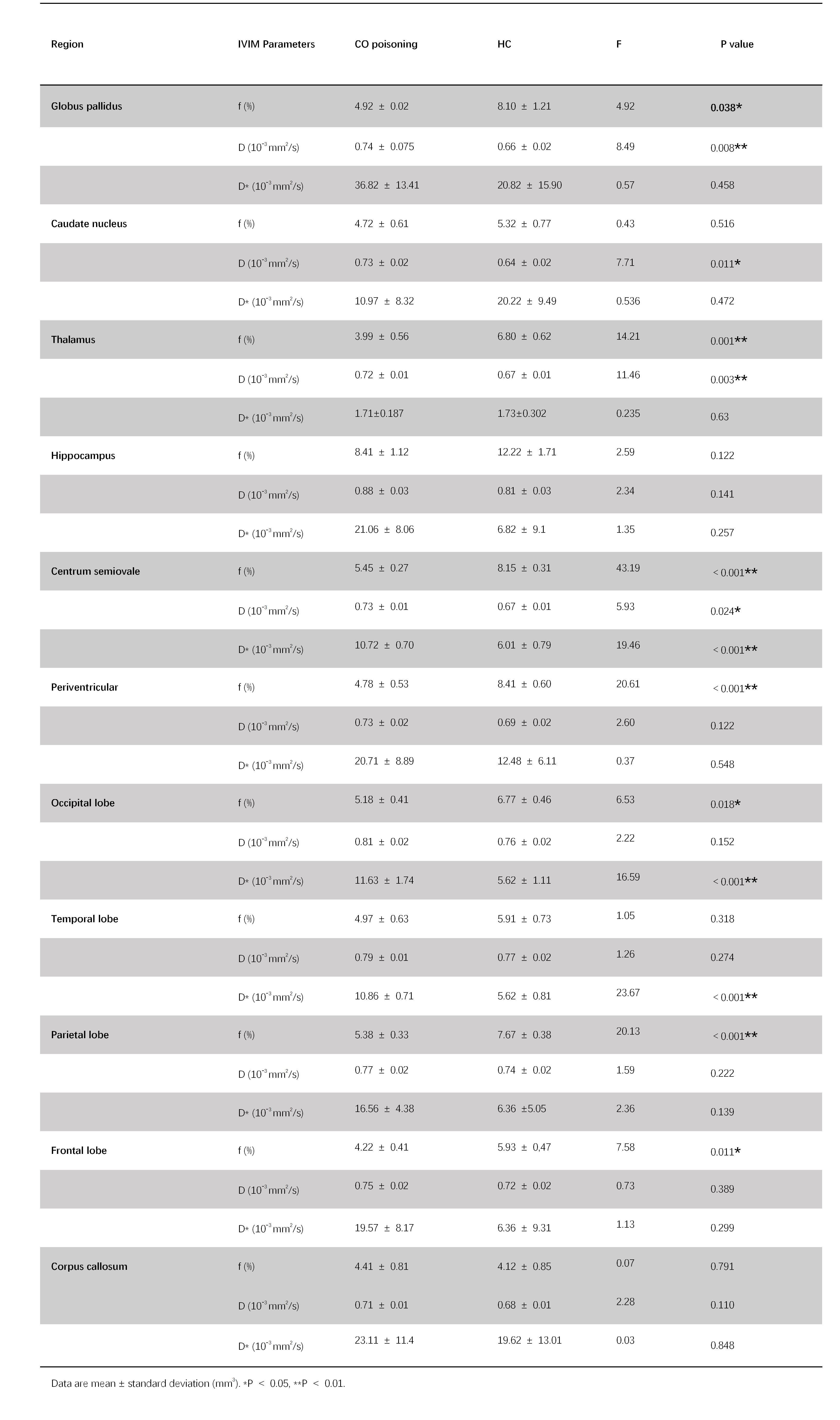
The ROIs analysis on the parametric maps was performed by two radiologists. According to previous literature, ten ROIs were chosen, which were the globus pallidus (GP), caudate nucleus, thalamus, hippocampus, centrum semiovale (CS), periventricular, corpus callosum (CC), frontal lobe, occipital lobe, temporal lobe, and parietal lobe. Except for the CC genu, body, and splenium, other ROIs were placed bilaterally, and the mean value was extracted.
Statistical analysis was performed using SPSS 20. A normality test (Kolmogorov-Smirnov) and homogeneity of variance test (Levene’s test) were performed. Between-group differences were analyzed using analysis of covariance (ANCOVA), controlling for age and sex.
RESULTS
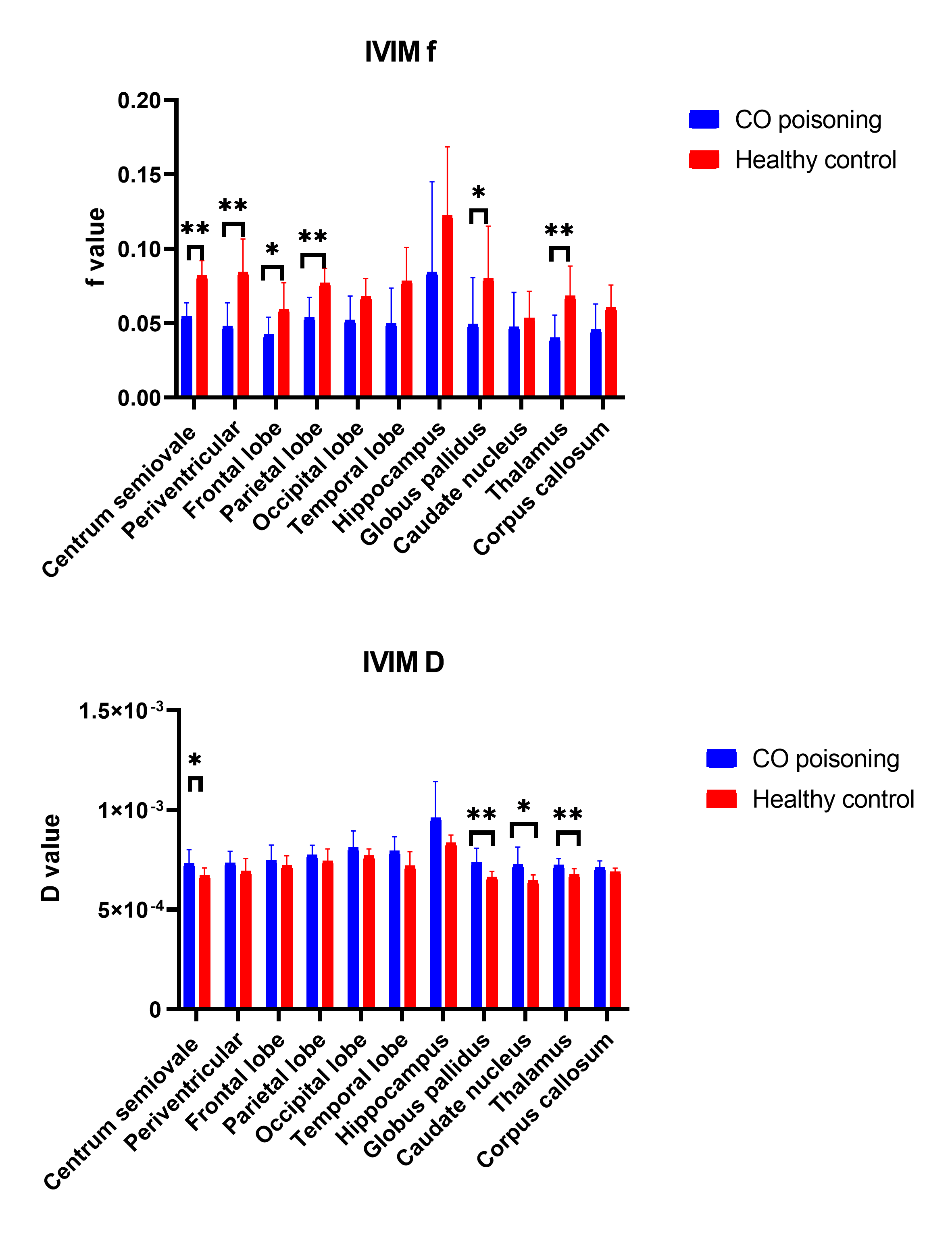
The subject demographics and clinical information are shown in Table 1.As shown in Table 2 and Figure 1, IVIM f was significantly decreased in patients with acute CO poisoning in globus pallidus, thalamus, centrum semiovale, periventricular, parietal lobe, frontal lobe. In the acute CO poisoning, IVIM D was significantly lower in globus pallidus, caudate nucleus, thalamus, and centrum semiovale compared to the HC.
DISCUSSION
In the present study, we found that the IVIM perfusion fraction was significantly reduced in multiple gray matter cortical regions, subcortical white matter regions, and subcortical gray matter nuclei in patients with acute CO poisoning. Meanwhile, we revealed that the IVIM diffusion metric was significantly decreased mainly in centrum semiovale and multiple subcortical gray matter nuclei in CO poisoning.In patients with acute CO poisoning, the lower IVIM perfusion indices in the globus pallidus and thalamus were in parallel with the higher IVIM diffusion metrics. A higher D indicates less restricted water diffusion, which is a surrogate for decreased microstructural integrity. A higher f* is a surrogate for higher microvascular perfusion.13-15 This indicates the vulnerability of this particular brain region in CO poisoning. The predilection of globus pallidus and thalamus involvement in CO poisoning has been frequently reported in MRI analysis and SPECT research.7,9,11,12,19 The reasons for selective damage to globus pallidus may be that this region is easily affected by the hypoxic-hypotension process because of a poor anastomotic blood supply.5,20 The additional affected regions from our IVIM analysis suggested hypoperfusion in occipital, temporal, and frontal regions. This is consistent with previous SPECT studies.10-12
CONCLUSION
This study shows that IVIM-DWI may be a promising method to quantitatively assess brain perfusion and brain injury in acute CO poisoning.Acknowledgements
No acknowledgement found.References
1. Rose JJ, Wang L, Xu Q, et al. Carbon Monoxide Poisoning: Pathogenesis, Management, and Future Directions of Therapy. AM J RESP CRIT CARE 2017;195(5):596-606
2. Hampson NB, Piantadosi CA, Thom SR, Weaver LK. Practice Recommendations in the Diagnosis, Management, and Prevention of Carbon Monoxide Poisoning. AM J RESP CRIT CARE 2012;186(11):1095-1101
3. Ashcroft J, Fraser E, Krishnamoorthy S, Westwood-Ruttledge S. Carbon monoxide poisoning. BMJ 2019:l2299
4. Weaver LK. Clinical practice. Carbon monoxide poisoning. N Engl J Med 2009;360(12):1217-1225
5. Beppu T. The Role of MR Imaging in Assessment of Brain Damage from Carbon Monoxide Poisoning: A Review of the Literature. AM J NEURORADIOL 2014;35(4):625-631
6. Zhang Y, Wang T, Lei J, et al. Cerebral Damage after Carbon Monoxide Poisoning: A Longitudinal Diffusional Kurtosis Imaging Study. AM J NEURORADIOL 2019
7. Chou MC, Li JY, Lai PH. Longitudinal White Matter Changes following Carbon Monoxide Poisoning: A 9-Month Follow-Up Voxelwise Diffusional Kurtosis Imaging Study. AM J NEURORADIOL 2019
8. Tsai P, Chou M, Chiang S, et al. Early white matter injuries in patients with acute carbon monoxide intoxication. MEDICINE 2017;96(5):e5982
9. Fujiwara S, Beppu T, Nishimoto H, et al. Detecting damaged regions of cerebral white matter in the subacute phase after carbon monoxide poisoning using voxel-based analysis with diffusion tensor imaging. NEURORADIOLOGY 2012;54(7):681-689
10. Tsai C, Yip P, Chen S, et al. The impacts of acute carbon monoxide poisoning on the brain: Longitudinal clinical and 99mTc ethyl cysteinate brain SPECT characterization of patients with persistent and delayed neurological sequelae. CLIN NEUROL NEUROSUR 2014;119:21-27
11. Chen NC, Chang WN, Lui CC, et al. Detection of gray matter damage using brain MRI and SPECT in carbon monoxide intoxication: a comparison study with neuropsychological correlation. CLIN NUCL MED 2013;38(2):e53-e59
12. Lu Y, Tsai S, Kao C, Lin W. Regional cerebral blood flow in patients with carbon monoxide intoxication. ANN NUCL MED 2012;26(10):771-776
13. Federau C, Maeder P, O'Brien K, et al. Quantitative measurement of brain perfusion with intravoxel incoherent motion MR imaging. RADIOLOGY 2012;265(3):874-881
14. Le Bihan D, Breton E, Lallemand D, et al. MR imaging of intravoxel incoherent motions: application to diffusion and perfusion in neurologic disorders. RADIOLOGY 1986;161(2):401-407
15. Conklin J, Heyn C, Roux M, et al. A Simplified Model for Intravoxel Incoherent Motion Perfusion Imaging of the Brain. AM J NEURORADIOL 2016;37(12):2251-2257
16. Bergamino M, Nespodzany A, Baxter LC, et al. Preliminary Assessment of Intravoxel Incoherent MotionDiffusion‐Weighted MRI (IVIM‐DWI) Metrics in Alzheimer's Disease. J MAGN RESON IMAGING 2020;52(6):1811-1826
17. Federau C, Wintermark M, Christensen S, et al. Collateral blood flow measurement with intravoxel incoherent motion perfusion imaging in hyperacute brain stroke. NEUROLOGY 2019;92(21):e2462-e2471 18. Heit JJ, Wintermark M, Martin BW, et al. Reduced Intravoxel Incoherent Motion Microvascular Perfusion Predicts Delayed Cerebral Ischemia and Vasospasm After Aneurysm Rupture. STROKE 2018;49(3):741-745
19. Chou M, Lai P, Li J. Early white matter injuries associated with dopamine transporter dysfunction in patients with acute CO intoxication: A diffusion kurtosis imaging and Tc-99m TRODAT-1 SPECT study. EUR RADIOL 2019;29(3):1375-1383
20. Bianco F, Floris R. MRI appearances consistent with haemorrhagic infarction as an early manifestation of carbon monoxide poisoning. NEURORADIOLOGY 1996;38 Suppl 1:S70-S72
Figures