1682
Application of quantitative susceptibility mapping of brain iron content in children with autism1Children's Hospital of Chongqing Medical University, Chongqing, China, 2GE Healthcare, MR Research China, Beijing, Beijing, China
Synopsis
ASD is caused by multiple factors,some of the pathogenic factors or possible pathogenic factors have not been clearly identified. For example, the detection of trace elements in some children with ASD found that iron content was lower than normal. Whether the lower-than-normal level of trace iron causes the lower-than-normal level of brain iron remains to be further confirmed, as does the direction of causality, i.e., whether the abnormal level of brain iron leads to the occurrence of the disease or whether the disease leads to the abnormal level of brain iron .
Purpose
In this study, the brain iron content of normal children and children with autism was compared to determine the differences, providing a new objective basis for the early diagnosis of children with autism.Material and Methods
For the control group, 40 normal children aged 2-3, 3-4, 4-5, and 5-6 years were prospectively selected from June 2018 to December 2018, with equal numbers of males and females in each age group. For the study group, 40 children with autism aged 2-3, 3-4, 4-5, and 5-6 years were prospectively selected from January 2019 to October 2019; once again, there were equal numbers of males and females in each age group. All children received routine head MRI scans and enhanced T2*-weighted angiography (ESWAN) sequence scans, and the ESWAN sequence images were processed by software to obtain magnetic susceptibility maps. The regions of interest (ROI) of the frontal white matter, frontal gray matter, thalamus, red nucleus, substantia nigra, dentate nucleus, globus pallidus, putamen nucleus, caudate nucleus, pons, and splenium of corpus callosum were selected, and the magnetic susceptibility values were measured. The differences in magnetic susceptibility between the two groups were compared in children at the same age.Results
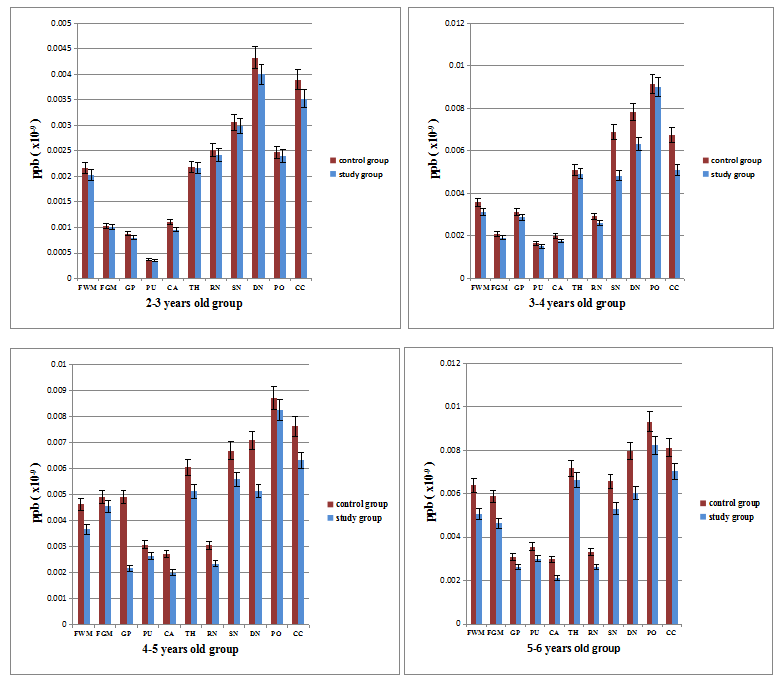
For the children aged 2-3 years, the magnetic susceptibility values in the caudate nucleus, dentate nucleus and splenium of the corpus callosum in the study group were lower than those in the control group (P<0.05). For the children aged 3-4, 4-5, and 5-6 years, the magnetic susceptibility values in the frontal white matter, caudate nucleus, red nucleus, substantia nigra, dentate nucleus, and splenium of the corpus callosum in the study group were lower than those in the control group (P<0.05).Discussion
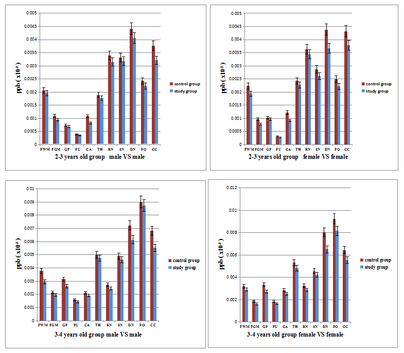
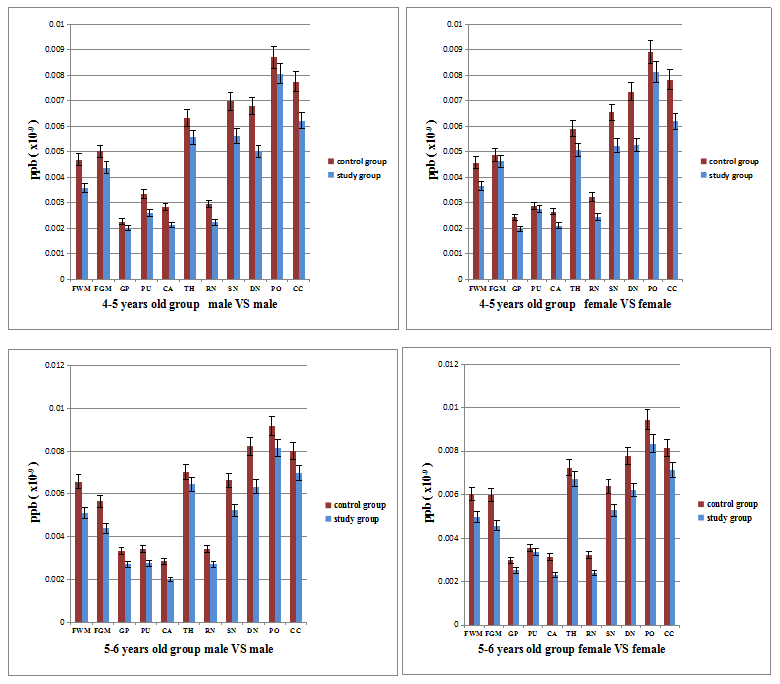
As seen from the error bars in the chart in Figure 1, the differences in magnetic sensitivity values in the substantia nigra and dentate nucleus between the children with autism and normal children in the groups aged 3-6 years was significantly greater than that in the group of children aged 2-3 years. From these results, it can be inferred that the brain iron content of the substantia nigra and dentate nucleus of children with autism may significantly decrease with age. The dentate nucleus plays an important role in the fields of cognition, emotion and movement. It is part of the anatomical circuit connecting the cerebellar cortex, thalamus, cerebral cortex and basal ganglia. Whether the reduction of iron content affects the function of the dentate nucleus remains to be further studied in the future studies .According to the statistical results in Figure 2 and 3, the region of the brain where iron content was reduced in children with autism aged 2-4 years was not affected by sex; the decrease in brain iron content in the children with autism aged 4-6 years was affected by sex; the magnetic susceptibility values in the frontal white matter, putamen nucleus, caudate nucleus, red nucleus, substantia nigra, dentate nucleus, and splenium of the corpus callosum of male children in the study group were lower than those in the control group; the magnetic susceptibility values in the frontal white matter, caudate nucleus, substantia nigra, dentate nucleus, and splenium of the corpus callosum of female children in the study group were lower than those in the controls ; it can be inferred that with increases in age, male children with autism may have more brain regions with iron deficiency than female children with autism, which needs to be further verified in future studies.
Conclusion
This study examined the magnetic susceptibility values in various brain regions of normal children aged 2-6 years in this region and established a reference range for magnetic susceptibility values in various brain regions of normal children in this geographical area, providing a reliable and objective standard for the diagnosis and treatment of some brain diseases in children. The results of this study indicate that the brain magnetic susceptibility values of preschool children with autism is lower than that of normal preschool children.Acknowledgements
No acknowledgement found.References
[1] Gabrielsen TP1, Anderson JS2, Stephenson KG, et al(2018) Functional MRI connectivity of children with autism and low verbal and cognitive performance.Mol Autism. 27(9):67-75.
[2] Cox AD, Virues-Ortega J, Julio F, et al(2017) Establishing motion control in children with autism and intellectual disability: Applications for anatomical and functional MRI. J Appl Behav Anal. 50(1):8-26.
[3] Peterson BS, Zargarian A, Peterson JB, et al(2019)Hyperperfusion of Frontal White and Subcortical Gray Matter in Autism Spectrum Disorder.Biol Psychiatry. 85(7):584-595.
Figures