1681
Correlations between microstructural changes of anterior cingulum cortex and cerebral small vascular disease induced depression : A DKI Study1Department of Radiology, Beijing Chao-Yang Hospital, Beijing, China, 2Department of Neurology, Beijing Chao-Yang Hospital, Beijing, China, 3MR Scientific Marketing, Diagnosis Imaging, Siemens Healthineers China, Beijing, China
Synopsis
Diffusion kurtosis imaging (DKI) is an advanced diffusion model based on an extender b value which characterizes water diffusion process as non-gaussian distribution, accordingly parameters derived from DKI are highly sensitive to changes in the microstructural tissue organization and the complexity of anisotropic environments. This study aimed to investigate the correlation between the microstructural changes of anterior cingulum cortex (ACC) and depression in patients with cerebral small vascular disease (CSVD) by applying diffusion Kurtosis imaging.
Synopsis
Diffusion kurtosis imaging (DKI) is an advanced diffusion model based on an extender b value which characterizes water diffusion process as non-gaussian distribution, accordingly parameters derived from DKI are highly sensitive to changes in the microstructural tissue organization and the complexity of anisotropic environments. This study aimed to investigate the correlation between the microstructural changes of anterior cingulum cortex (ACC) and depression in patients with cerebral small vascular disease (CSVD) by applying diffusion Kurtosis imaging.Introduction
Cerebral small vessel disease (CSVD) is a disease of the small perforating arteries and capillaries commonly seen in the elderly, which leads to barrier of cerebral microcirculation and predominantly affects deep white matter and grey matter[1]. Most elderly people can be found having neuroimaging evidence of CSVD, and some of them does not have clinical manifestations. A high burden of CSVD is associated with mood dysfunction, cognitive and physical problems.[2] and it was also shown that CSVD was strongly associated with late-life depression.[3] At present, the diagnosis of depression is mainly assessed by the neuropsychological assessment scale, which is subjective, and the sensitivity of depression is low in the early diagnosis. Diffusion kurtosis imaging (DKI) is an advanced diffusion model based on an extender b value which characterizes water diffusion process as non-gaussian distribution, accordingly parameters derived from DKI are highly sensitive to changes in the microstructural tissue organization and the compatibility of anisotropic environments. [4] This study aimed to investigate the correlation between the microstructural changes of anterior cingulum cortex (ACC) and depression in patients with cerebral small vascular disease (CSVD) by applying diffusion Kurtosis imaging.Method
A total of 71 CSVD patients (median age: 64 (60,69) years, 41 males, 30 females) were prospectively enrolled in this study. The HAMD of 17 stems were performed to assess the severity of depressive disorders. The subjects were divided into depression group (CSVD-D, n=39, HAMD score ≥ 7 points) and non-depression group(CSVD-ND, n=32, HAMD score < 7 points). All patients underwent MRI scan on a 3T MR scanner (MAGNETOM Skyra, Siemens Healthcare, Erlangen, Germany). T1 weighted images (T1WI) were captured by three-dimensional magnetization prepared rapid acquisition gradient echo (T1WI 3D MPRAGE). The diffusion imaging based on spin-echo echo planar imaging (SE-EPI) was obtained along 30 directions, with 3 b values (0, 1000, 2000 mm2/s) for each direction. The sequence parameters were as follows: TR= 7700ms, TE= 89ms, PAT= 2, FOV= 222 × 222mm, matrix= 74 × 74; slice thickness:3mm; number of slices= 50; acquisition time 588s. The parameters derived from DKI were calculated by DKE software (http://www.nitrc.org/proje cts/dke, version 2.5.1). The DKI parameters include kurtosis fractional anisotropy (KFA), mean kurtosis (Kmean) and mean diffusivity (Dmean). T1WI 3D MPRAGE and DKI maps were registered into MNI space, and the anterior cingulum cortex were automatically segmented by applying SPM8 (http://www.fil.ion.ucl.ac.uk/spm) and the mean value of the parameters were calculated based on the Anatomical Automatic Labeling (AAL) template. Neuropsychological assessments are Hamilton depression rating scale (HAMD). The DKI parameters were calculated in regions of bilateral anterior cingulum cortex (ACC). The intergroup differences were evaluated appropriately with Student’s t test, the Mann–Whitney U-test or χ2 -tests. Spearman rank correlation analysis was used to determine the association between pairs of variables for non-parametric data. P values below 0.05 (two-tailed) were considered statistically significant.Results
Compared with CSVD-ND group, CSVD-D group patients showed significantly higher Dmean in the left ACC (p=0.000). Furthermore, there is a positive correlation between parameter Dmean and HAMD score (The left ACC: R2= 0.126; The right ACC: R2= 0.095, respectively) and there was no correlation between KFA and HAMD score in the left or right ACC.Discussion and Conclusion
In this study, we studied the microstructure changes of ACC, which is an important emotional processing structure in limbic system, in patients with CSVD-induced MMD by DKI. The results showed that the Dmean in DKI maps might be used for early diagnosis of CSVD-induced depression. The microstructure of left ACC changed in patients with CSVD-induced MMD. The DKI parameters of ACC are potentially useful parameters for the quantitative evaluation the degree of CSVD-induced depression.Acknowledgements
No acknowledgement found.References
1. Wardlaw JM, Smith C, Dichgans M. Mechanisms of sporadic cerebral small vessel disease: insights from neuroimaging[J]. The Lancet. Neurology, 2013, 12(5): 483-497.
2. Silbert LC, Nelson C, Howieson DB, et al. Impact of white matter hyperintensity volume progression on rate of cognitive and motor decline[J]. Neurology, 2008, 71(2): 108-113.
3. Taylor WD, Aizenstein HJ, Alexopoulos GS. The vascular depression hypothesis: mechanisms linking vascular disease with depression[J]. Mol Psychiatry, 2013, 18(9): 963-974.
4. Lätt J, Nilsson M, Wirestam R, et al. Regional values of diffusional kurtosis estimates in the healthy brain[J]. Journal of magnetic resonance imaging: JMRI, 2013, 37(3): 610-618.
Figures
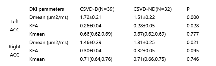
Table 1. A comparison of DKI derived parameters in ACC between two groups.
Note: Values are presented as mean ± SD or median (interquartile range). Abbreviations: Dmean, mean diffusivity; KFA, kurtosis fractional anisotropy; Kmean, mean kurtosis.
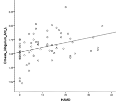
Figure 1. The correlation between the DKI parameter Dmean of the left ACC with HAMD score. Abbreviations: Dmean_cingulum_Ant_L, the mean diffusivity of the left anterior cingulum cortex; HAMD, Hamilton Depression rating scale.
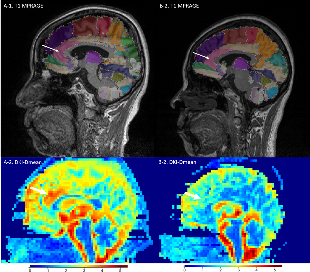
Figure 2. Images of CVSD-D patient (A-1, T1WI 3D MPRAGE, A-2, DKI-Dmean map) and CVSD-ND patient (B-1, T1WI 3D MPRAGE, B-2, DKI-Dmean map). Compared with non-depression CVDS patient, the Dmean map of CVSD-D patient have more warm pixels in region of ACC. White arrow indicate left ACC.