1680
Fibre-specific white matter changes in neonates born to women prescribed methadone in pregnancy1MRC Centre for Reproductive Health, University of Edinburgh, Edinburgh, United Kingdom, 2Developmental Imaging, Murdoch Children's Research Institute, Melbourne, Australia, 3Department of Radiology, Royal Hospital for Sick Children, Edinburgh, United Kingdom, 4Edinburgh Imaging, University of Edinburgh, Edinburgh, United Kingdom
Synopsis
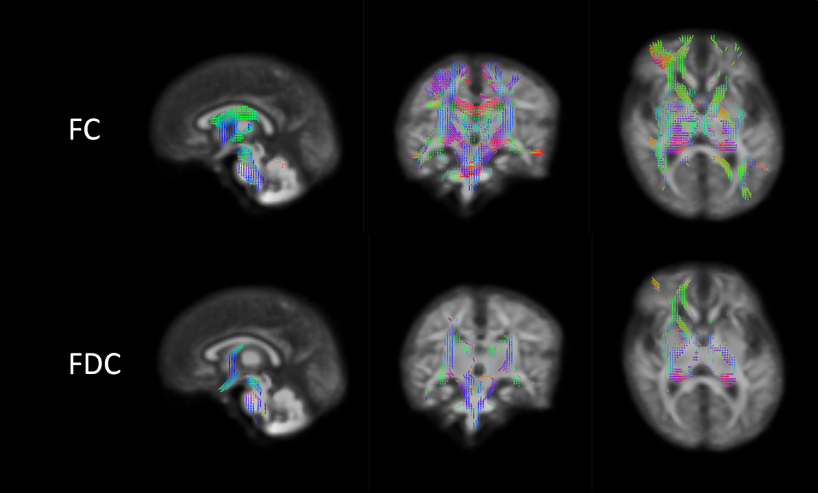
Methadone is often used for medication-assisted treatment of heroin addiction during pregnancy. Children with prenatal exposure to the drug are at increased risk of neurodevelopmental and behavioural impairment. We did fixel-based analysis with a group of 20 term born infants whose mothers had been prescribed methadone during pregnancy for the treatment of heroin addiction and a control group of 20 control infants. There was significant widespread reduction across the WM in fiber-bundle cross-section and fiber density and cross-section in the exposed group, this suggests that affected fibre bundles are less developed, similar to WM structures in preterm born babies.
Introduction
Methadone is often used for medication-assisted treatment of heroin addiction during pregnancy. Children with prenatal exposure to the drug are at increased risk of neurodevelopmental and behavioural impairment [1]. We recently reported that prenatal exposure to methadone is associated with microstructural alterations in major white matter (WM) tracts, assessed by a reduction in fractional anisotropy (FA) apparent soon after birth [2]. This suggests prenatal exposure is linked to abnormal white matter development but inferences about specific microstructural changes are limited when using voxel averaged methods. Here, we apply fixel-based analysis to the same dataset to test the hypothesis that prenatal exposure to methadone is associated with alterations to one or more of the following: fiber density (FD), fiber-bundle cross-section (FC), and fiber density and cross-section (FDC) [3,4].Methods
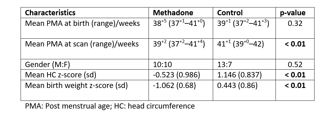
Demographics: 20 term born infants whose mothers had been prescribed methadone during pregnancy for the treatment of heroin addiction and a control group of 20 gestational age matched control infants whose mothers did not use opioids (Table 1). Details of the study group and ethical approvals are provided in [2].Maternal information: The mean methadone dose prescribed at pregnancy booking was 55 mg/day and the mean dose at delivery was 70 mg/day. 95% of the women prescribed methadone smoked tobacco, one reported drinking excessive alcohol (4 units/day at booking). A detailed description of the cohort has been reported previously [2].
Acquisition: MRI was performed on a Siemens Magnetom Verio 3T system (Siemens Healthineers, Erlangen, Germany) using a 12-channel matrix phased array head coil. Diffusion MRI (dMRI) data were acquired using 64 diffusion encoding directions (b = 750 s/mm2) with 2 mm isotropic voxels, TE = 106 ms and TR = 7300 ms, as well as 11 b=0 volumes.
MRI preprocessing: dMRI data were denoised [5], corrected for Gibbs ringing artifacts [6], eddy currents and head movement [7] and bias fields [8]. Fixel-based analysis: The WM fibre orientation distributions (FODs) were calculated using single-shell 3-tissue constrained spherical deconvolution (SS3T-CSD) algorithm [9] (https://3tissue.github.io/) using the average of the response functions for each tissue (WM, GM and CSF) [10]. Then, the standard multi-tissue pipeline for fixel-based analysis was employed to obtain FD, FC and FDC metrics [3,4].
Statistical analysis: For the fibre-specific (fixel-wise) metrics, statistical comparison between groups with and without exposure to methadone during pregnancy was performed using MRtrix3’s fixel-wise statistics with connectivity-based fixel enhancement (CBFE) [11,12]. PMA at scan was used as a covariate of non-interest.
Results
There were no statistically significant differences in fixel-wise FD. However, there were significant widespread differences across the WM in FC and FDC (Figure 1).Discussion
We found that babies whose mothers had been prescribed methadone during pregnancy have reduced FC and FDC widely distributed throughout WM. This suggests that affected fibre bundles are less developed, similar to WM structures in preterm born babies [13,14]. In the original work [2], tract-based spatial statistics (TBSS) [15] was used to assess differences in FA in WM. The comparison between TBSS and fixel-based analysis is challenging as there are technical differences between the analysis frameworks, as well as the limited voxel-wise FA metric compared to the fibre-specific metrics used in fixel-based analyses [3]. For example, it is important to consider that in the TBSS analysis FA values are projected on a mean FA template “skeleton”. In the fixel-based analysis however, all fixels in the WM are considered individually and independently. Despite this we observed substantial overlap between the affected regions in this work and those reported in [2], but the TBSS results appeared less widespread.More work needs to be undertaken to explore the effects of other covariates to the fixel-based derived metrics and investigate other measures of WM microstructure that could help us to gain more insights into the effects of prenatal methadone exposure in the neonatal brain.
Conclusions
We applied the SS3T-CSD algorithm to obtain fibre-specific metrics of WM and studied those in a fixel-based analysis in neonates. We used it to improve understanding of the effects of prenatal methadone exposure on the developing brain and we found widespread reductions of FC and FDC throughout the WM.Acknowledgements
We thank the families that participated in this research; the specialist substance misuse midwives; and the radiographers at the Edinburgh Imaging QMRI facility, Edinburgh, UK. We thank Thorsten Feiweier at Siemens Healthineers for collaborating with dMRI acquisitions (Works-in-Progress Package for Advanced EPI Diffusion Imaging). This work was supported by Theirworld (www.theirworld.org) and was undertaken in the MRC Centre for Reproductive Health, which is funded by MRC Centre Grant (MRC G1002033). The study was sponsored by the University of Edinburgh.References
1 Monnelly et al. Childhood neurodevelopment after prescription of maintenance methadone for opioid dependency in pregnancy: a systematic review and meta‐analysis. Developmental Medicine & Child Neurology. 2019.
2 Monnelly et al. Prenatal methadone exposure is associated with altered neonatal brain development. NeuroImage: Clinical. 2018.
3 Raffelt et al. Investigating white matter fibre density and morphology using fixel-based analysis. NeuroImage. 2017.
4 Dhollander et al. Fixel-based Analysis of Diffusion MRI: Methods, Applications, Challenges and Opportunities. OSFpreprints. 2020.
5 Veraart et al. Denoising of diffusion MRI using random matrix theory. Neuroimage. 2016.
6 Kellner et al. Gibbs‐ringing artifact removal based on local subvoxel‐shifts. Magnetic Resonance in Medicine. 2015.
7 Andersson et al. An integrated approach to correction for off-resonance effects and subject movement in diffusion MR imaging. Neuroimage. 2016
8 Tustison et al. N4ITK: improved N3 bias correction. IEEE Trans Med Imaging. 2010.
9 Dhollander et al. A novel iterative approach to reap the benefits of multi-tissue CSD from just single-shell (+b=0) diffusion MRI data. Proc Intl Soc Mag Reson Med. 2016
10 Dhollander et al. Improved white matter response function estimation for 3-tissue constrained spherical. Proc Intl Soc Mag Reson Med. 2019.
11 Raffelt et al. Connectivity-based fixel enhancement: Whole-brain statistical analysis of diffusion MRI measures in the presence of crossing fibres. Neuroimage. 2015.
12 Tournier et al. MRtrix3: A fast, flexible and open software framework for medical image processing and visualisation. NeuroImage. 2019.
13 Pannek et al. Fixel-based analysis reveals alterations is brain microstructure and macrostructure of preterm-born infants at term equivalent age. NeuroImage: Clinical. 2018.
14 Kelly et al. Long-term development of white matter fibre density and morphology up to 13 years after preterm birth: A fixel-based analysis. NeuroImage. 2020.
15 Smith et al. Tract-based spatial statistics: Voxelwise analysis of multi-subject diffusion data. Neuroimage. 2006.
Figures