1634
The Current Status of Black-Blood Thrombus Imaging in Assessment of Cerebral Venous Thrombosis in a Real-word Clinical Practice1Chaoyang Hospital, Beijing, China, 2Chaoyang Hospitao, Beijing, China, 3Cedars Sinai Medical Center, Los Angeles, CA, United States, 4Xuanwu Hospital, Beijing, China
Synopsis
We evaluated diagnostic and staging performance of BTI in patients with suspected CVST compared with conventional imaging methods in a real-world study. Our results verified high diagnostic value of BTI in a large patient cohort and BTI provided another advantage of accurate staging (acute, subacute and chronic), achieving more thrombosed segments of definite stage, which is important for clinical strategy.
Purpose
Magnetic resonance black-blood thrombus imaging (BTI) has been developed for the detection of cerebral venous thrombosis (CVT). In this study, we have evaluated BTI in the diagnosis and staging of CVT with 5-year real-world clinical experience.Method
Consecutive patients who were suspected of CVT between April 2014 and December 2019 from Xuanwu Hospital were recruited into the current study. Randomized imaging examination were performed on enrolled patients, including CT, MRI, SWI, DSA or BTI (classified into group 1 with BTI and group 2 without BTI). The result of diagnosis and staging from report system between two groups were recorded. In a subgroup analysis, the diagnostic performance of BTI was assessed on a per-patient and per-segment basis, respectively. Thrombus stage were compared between BTI and conventional imaging techniques.Result
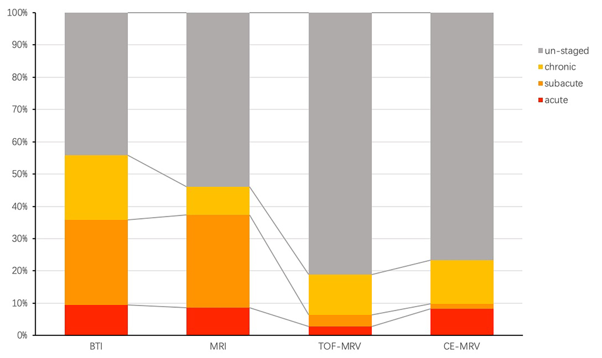
In this real-world study, a total of 308 patients suspected with CVT were enrolled (133 in group 1 and 175 in group 2). We found that 81.6% patients with BTI while only 43.3% patients without BTI got the clear and definite diagnosis. And nearly 100% of patients could obtain the result of staging on the image report while only 29.0% of patients without BTI could be staged. In the subgroup analysis, BTI showed a sensitivity of 96.4% and a specificity of 98.3% in the detection of CVT with an area-under-the-curve (AUC) of 0.995 on the per-patient level. The sensitivity and specificity were 87.9% and 96.8%, respectively, with an AUC of 0.951 on the per-segment level. The rate of matching in segment-level thrombus staging between BTI and clinical staging was 55.8% (148/265). BTI reclassified 97.8% (88/90) of otherwise un-staged thrombus, leading to a reduction in the percentage of un-staged segments from 34.0% to 2.3% (P < 0.001).Conlusion
In this real-world study, BTI aid to make more definite diagnosis. It allowed direct visualization of thrombus with high sensitivity and specificity to enhance confidence of diagnosing CVT and enabled more staged thrombi which is suitable for serving as a first line tool in clinical practice.Acknowledgements
We thank the patients involved in the study and appreciate the contributionof all investigators for our study.References
Ferro JM, Aguiar de Sousa D. Cerebral venous thrombosis: An update. Curr Neurol Neurosci Rep. 2019;19:742.
Dmytriw AA, Song JSA, Yu E, Poon CS. Cerebral venous thrombosis: State of the art diagnosis and management. Neuroradiology. 2018;60:669-6853.
Ferro JM, Canhao P, Aguiar de Sousa D. Cerebral venous thrombosis. Presse medicale (Paris, France : 1983). 2016;45:e429-e4504.
Alami B, Boujraf S, Quenum L, Oudrhiri A, Alaoui Lamrani MY, Haloua M, et al. [cerebral venous thrombosis: Clinical and radiological features, about 62 cases]. Journal de medecine vasculaire. 2019;44:387-3995.
van Dam LF, van Walderveen MAA, Kroft LJM, Kruyt ND, Wermer MJH, van Osch MJP, et al. Current imaging modalities for diagnosing cerebral vein thrombosis - a critical review. Thromb Res. 2020;189:132-1396.
Leach JL, Fortuna RB, Jones BV, Gaskill-Shipley MF. Imaging of cerebral venous thrombosis: Current techniques, spectrum of findings, and diagnostic pitfalls. Radiographics : a review publication of the Radiological Society of North America, Inc. 2006;26 Suppl 1:S19-41; discussion S42-137.
Cai H, Ye X, Zheng W, Ma L, Hu X, Jin X. Pitfalls in the diagnosis and initial management of acute cerebral venous thrombosis. Reviews in cardiovascular medicine. 2018;19:129-1338.
Chiewvit P, Piyapittayanan S, Poungvarin N. Cerebral venous thrombosis: Diagnosis dilemma. Neurology international. 2011;3:e13
Figures