1619
Non-contrast-enhanced MR venography of lower extremity using balanced-TFE mDIXON with robust water-fat swap correction1Philips Japan, Tokyo, Japan, 2Department of Radiological Services, Tokyo Women’s Medical University, Tokyo, Japan, 3Department of Diagnostic Imaging and Nuclear Medicine, Tokyo Women’s Medical University, Tokyo, Japan, 4Philips Healthcare, Best, Netherlands
Synopsis
Magnetic resonance venography (MRV) is often used to assess venous anatomy and pathology. In this study, we developed a 2D dual-echo balanced-turbo field-echo (TFE) mDIXON (bTFE DIXON) sequence to rapidly acquire high SNR MRV of lower extremities, without using cardiac gating and respiratory compensation. We also proposed a new post-processing technique, histogram similarity-based swap correction (HISSCO), to detect and correct water-fat slice swap artifact associated with the mDIXON technique. Our results showed that combined with HISSCO, the bTFE DIXON sequence can potentially be a clinically useful sequence for assessing the venous pathology of the lower extremities.
Introduction
Venous thromboembolic (VTE) disease has incidence rates ranging from 0.75 to 2.69 per 1000 individuals of the general population across the world1. The main cause of the VTE is deep vein thrombosis (DVT), which is usually formed in lower extremities. Therefore, visualizing venous anatomy and pathology in the lower extremities is clinically important. Magnetic resonance venography (MRV) is often used to assess venous anatomy and pathology of the lower extremities. Traditionally, the 2D time-of-flight (TOF) technique has been the gold standard for the MRV2,3. However, the 2D TOF method typically suffers from insufficient SNR and long scan time. Sometimes the long acquisition time can cause respiratory motion artifact and thus phase encoded artifact reduction (PEAR) respiratory compensation technique is required. Previously, the combination of balanced-SSFP acquisition with DIXON-based fat suppression demonstrated SNR efficient and high resolution MR angiography of the lower extremities and heart4–8. To the best of our knowledge, however, this sequence has not been applied for the MRV of the lower extremities. In the present study, we developed a 2D dual-echo balanced-turbo field-echo (TFE) mDIXON (bTFE DIXON) sequence to rapidly acquire high SNR MRV of the lower extremities, without using cardiac gating and respiratory compensation. The purpose of this study was to compare the image quality and acquisition time of the bTFE DIXON sequence with the conventional spectral inversion recovery (SPIR) T1TFE sequence. A new post-processing technique, histogram similarity-based swap correction (HISSCO) was also proposed to correct the water-fat slice swap artifact associated with the mDIXON technique.Methods
Experiments: A total of 3 volunteers were examined on a 3.0T whole-body clinical system (Ingenia, Philips Healthcare). The study was approved by the local IRB, and written informed consent was obtained from all subjects. Non-contrast-enhanced TOF images of the lower extremities were acquired as a series of 2D axial sections using two imaging methods: SPIR T1TFE and bTFE DIXON. Both imaging was performed in each of 4 stations from the lower abdomen to the distal calf. The MRV images were finally visualized by computing maximum intensity projection (MIP).The bTFE DIXON sequence was acquired with FOV=400×219×271mm, voxel size=0.78×0.78×4mm, C-SENSE factor=3, TR/TE1/TE2 =4.1/1.49/2.6, FA=90, NSA=1, total scan time= 12:40min. The SPIR T1TFE sequence was acquired with FOV=400×220×271mm, voxel size=1.25×1.25×4mm, SENSE factor=2.5, TR/TE =13/10.36, FA=25, NSA=1, total scan time=15:18min.
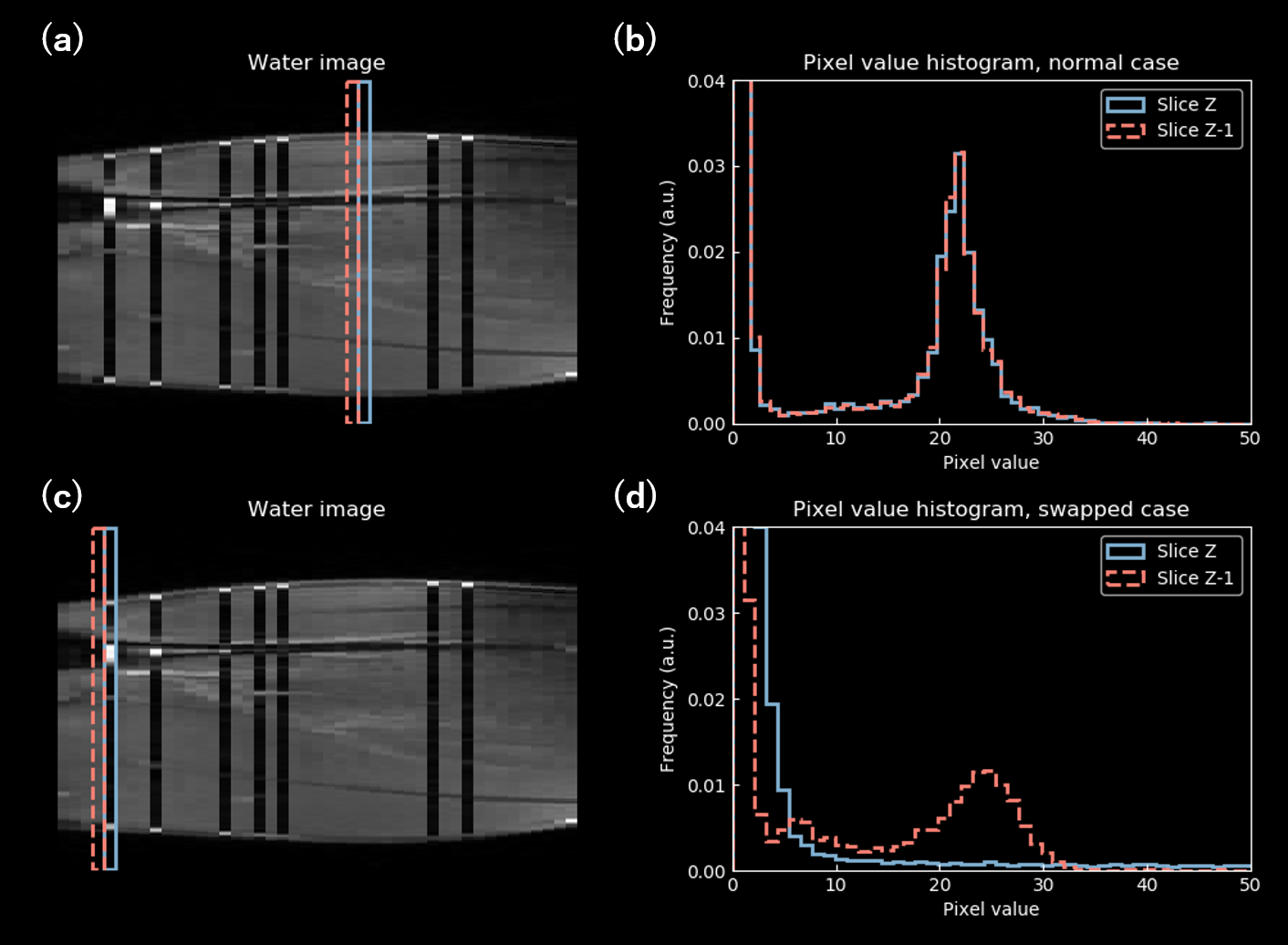
Artifact correction: The proposed slice swap artifact correction algorithm, HISSCO, consists of the following steps (Figure 1). First, two neighboring axial slices were compared across the entire image and the histograms correspond to those slices were obtained. When there is no swap in both slices of interest (Fig. 1a), the two histograms are alike (Fig. 1b). However, when either of the slices is swapped (Fig. 1c, blue box), the histogram shapes become vastly different (Fig. 1d). The similarity of the two histograms was quantified by calculating the root mean squared (RMS) error and the slice with low similarity to the adjacent slice was considered as the potentially swapped slice. This procedure was run separately on right and left legs for each of the water and fat images. If the same slices were detected on both water and fat images, the slice swap was confirmed, and those slices were exchanged between water and fat images. To evaluate the performance of HISSCO, the bTFE DIXON images with and without HISSCO application were qualitatively compared with the conventional SPIR T1TFE.
Results and Discussions:
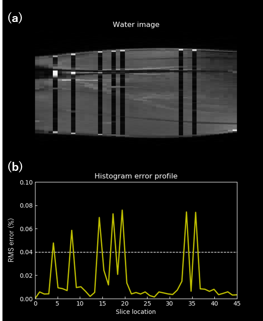
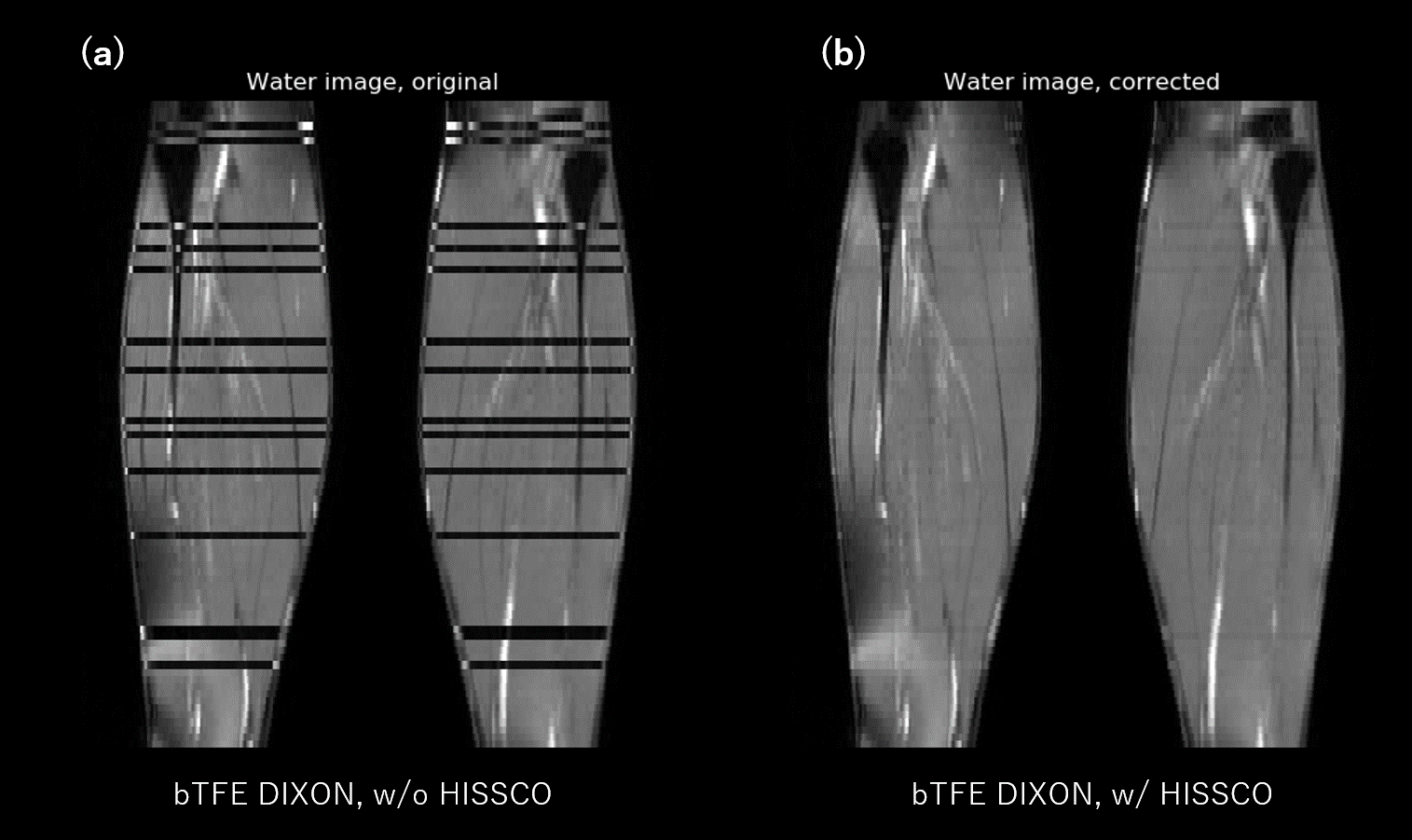
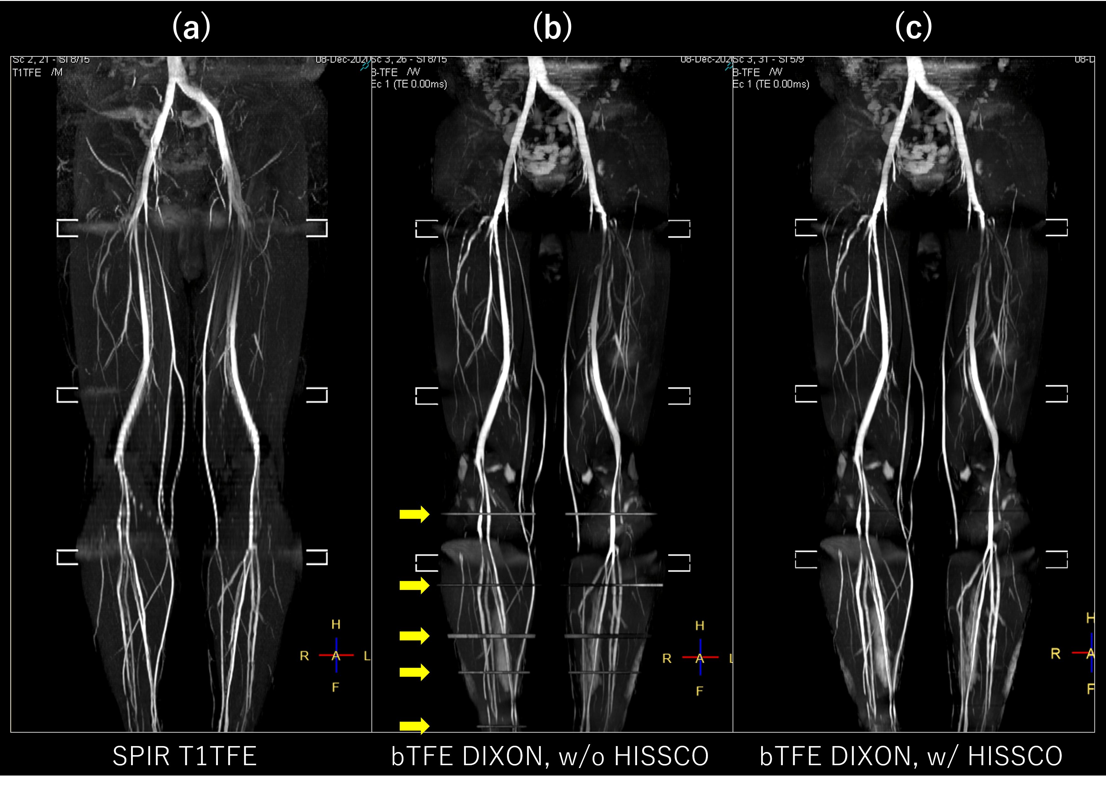
Figure 2 demonstrates the water-fat slice swap artifact detection by HISSCO, using the same image presented in the figure 1a. Slices that showed a marked increase of histogram RMS error (i.e. histogram dissimilarity) in figure 2b exactly matched the location of the slice swaps observed in the original bTFE DIXON water image (Fig. 2a). Figure 3 shows the comparison of the bTFE DIXON water images with and without the application of HISSCO. The water-fat slice swaps observed in the original bTFE DIXON were well corrected by applying the HISSCO, and the zebra pattern due to the swap became negligible. Among all slice swaps observed, the HISSCO was able to detect 94.0% of them correctly. Figure 4 compares MIP images of the SPIR T1TFE, bTFE DIXON without HISSCO, and bTFE DIXON with HISSCO applied. Overall, the SNR of the bTFE DIXON was higher than that of the SPIR T1TFE (Fig. 4a), and the depiction of the venous anatomy clearly improved for all subjects. The slice swaps observed below the knee in the original bTFE DIXON (Fig. 4b, arrow) were successfully corrected even when the swap occurred on one side of the leg (Fig. 4c). The total acquisition time of the bTFE DIXON was 12:40 min and was shorter than that of the conventional SPIR T1TFE. These results indicate that the bTFE DIXON with water-fat slice swap correction using HISSCO might be the preferred approach over the conventional SPIR T1TFE for detecting the DVT of the lower extremities.Conclusion
We demonstrated that bTFE DIXON can rapidly acquire high SNR MRV of the lower extremities, without using cardiac gating and respiratory compensation. Combined with the robust water-fat swap artifact correction technique we developed, bTFE DIXON can be a clinically useful sequence for detecting the DVT of the lower extremities.Acknowledgements
No acknowledgement found.References
1. Raskob GE, Angchaisuksiri P, Blanco AN, et al. Thrombosis: A major contributor to global disease burden. Arterioscler Thromb Vasc Biol. 2014;34(11):2363-2371.
2. Lanzer P, Gross GM, Keller FS, Pohost GM. Sequential 2D inflow venography: Initial clinical observations. Magn Reson Med. 1991;19(2):470-476.
3. Carpenter JP, Holland GA, Baum RA, Owen RS, Carpenter JT, Cope C. Magnetic resonance venography for the detection of deep venous thrombosis: Comparison with contrast venography and duplex Doppler ultrasonography. J Vasc Surg. 1993;18(5):734-741.
4. Brittain JH, Reeder SB, Shimakawa A, et al. Non-contrast-enhanced angiography at 3T using SSFP and “Dixon” fat-water separation. In: Proceedings of the 12th Annual Meeting of ISMRM. ; 2004.
5. Çukur T, Shimakawa A, Yu H, et al. Magnetization-prepared IDEAL bSSFP: A flow-independent technique for noncontrast-enhanced peripheral angiography. J Magn Reson Imaging. 2011;33(4):931-939.
6. Hamatani Y, Abe K, Goto Y, et al. Improved visualization of non-contrast-enhanced MRA of the foot using REACT with balanced SSFP-DIXON (bREACT) at 1.5T. In: Proceedings of the 28th Annual Meeting of ISMRM. ; 2020.
7. Kodaira K, Nagao M, Yoneyama M, et al. Whole heart coronary MRA with 3D non-selective bSSFP-DIXON : comparison with conventional methods. In: Proceedings of the 28th Annual Meeting of ISMRM. ; 2020.
8. Yoneyama M, Zhang S, Hu HH, et al. Free-breathing non-contrast-enhanced flow-independent MR angiography using magnetization-prepared 3D non-balanced dual-echo Dixon method: A feasibility study at 3 Tesla. Magn Reson Imaging. 2019;63(August):137-146.
Figures