1615
Optimization of Ultrashort TE protocol QUTE-CE with ferumoxytol for abdominal MRA1Department of Physics, Northeastern University, Boston, MA, United States, 2Northeastern University Biomedical Imaging Center, Boston, MA, United States, 3Department of Radiology, Massachusetts General Hospital, Boston, MA, United States, 4Siemens Medical Solutions, Boston, MA, United States
Synopsis
A contrast-enhanced MRA protocol with Ultrashort Time-to-Echo (UTE) was optimized on phantoms covering a range of ferumoxytol concentrations for abdominal imaging. This approach produces images with high SNR (~ 4166) at clinical concentrations. The protocol was applied in vivo to human subjects for abdominal vascular imaging. Acquired human images demonstrated great delineation of central and peripheral vessels in the liver, kidneys and other abdominal organs. The combination of an optimized UTE sequence and the blood-pool kinetics of ferumoxytol enables high-resolution vascular imaging across the entire abdomen with maximized image quality. This technique could potentially aid in characterization of vascular diseases.
Introduction
Magnetic resonance angiography (MRA) is an imaging modality for visualizing blood vessels, with the advantage of being noninvasive, lack of radiation exposure and iodinated contrast agent administration [1]. Contrast-enhanced MRA involves administration of contrast agent that shortens the longitudinal relaxation time (T1) and provides bright blood enhancement on T1-weighted images [2].Ferumoxytol is a superparamagnetic iron oxide nanoparticle (SPION) which was approved by FDA as iron replacement therapy for iron-deficiency anemia and currently is used as an off-label MRI contrast agent. Given its relatively large particle size (~30nm) [3], it remains intravascular for about 12 hours [4], allowing for a longer temporal window for data acquisition. This enables repetitive scans and free-breathing scans, beneficial for abdominal MRA where image quality is often degraded by motion artifacts.
The recent development of MRI techniques using ultrashort-TE sequences with ferumoxytol minimizes the negative contrast T2 and T2* effects and provides strong positive contrast in vascular compartment [5]. In 2017, Gharagouzloo et al. [6] demonstrated the Quantitative Ultra-short-Time-to-Echo Contrast Enhanced (QUTE-CE) MRA protocol with ferumoxytol in animal models and showed advantages to angiographic quality and quantitative measurements. In the current study, we focus on further optimizing the QUTE-CE MRA protocol for abdominal imaging and applying this protocol in vivo with human subjects. This results in images with very high vascular SNR and CNR even with high clinical concentrations of ferumoxytol.
Methods
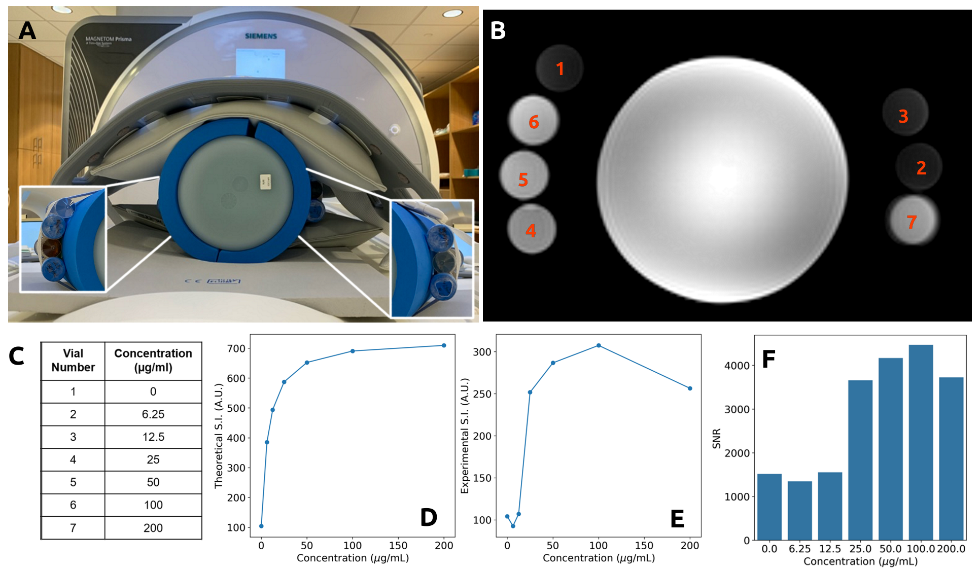
MRI phantoms were prepared by diluting ferumoxytol in 50mL tubes of 0.9% saline at seven concentrations (0 µg/mL, 6.25 µg/mL, 12.5 µg/mL, 25 µg/mL, 50 µg/mL, 100 µg/mL, 200 µg/mL). The phantoms were taped around the large cylindrical “bullet” phantom. Then the whole setup was wrapped by paddings and scanned at Northeastern University Biomedical Imaging Center (Figure 1A).Subjects were recruited at Massachusetts General Hospital and scanned at the Martinos Center for Biomedical Imaging. The subjects were prescribed for 510mg ferumoxytol infusion (Feraheme; AMAG Pharmaceuticals, Waltham, MA) as part of their regular care and consented to imaging about 2 hours after infusion. All procedures were conducted in accordance with the Partners IRB.
Both MR imaging of phantoms and human subjects were performed with the 3T scanner (MAGNETOM Prisma; Siemen, Erlangen, Germany) with spine and body array coils at two different sites. Images were acquired with a prototype 3D stack of spirals UTE VIBE sequence (Phantom experiment: TE=0.05 ms, TR=3.12 ms, FA=5°, resolution=1.56 mm isotropic; Clinical experiment: TE=0.05 ms, TR=2.91 ms, FA=13.5°, resolution=1.56 mm isotropic).
Theoretical signal intensities (with arbitrary unit) for the phantom concentrations were calculated using steady-state spoiled-gradient-echo (SPGR) equation and compared with experimental values [7].
Results
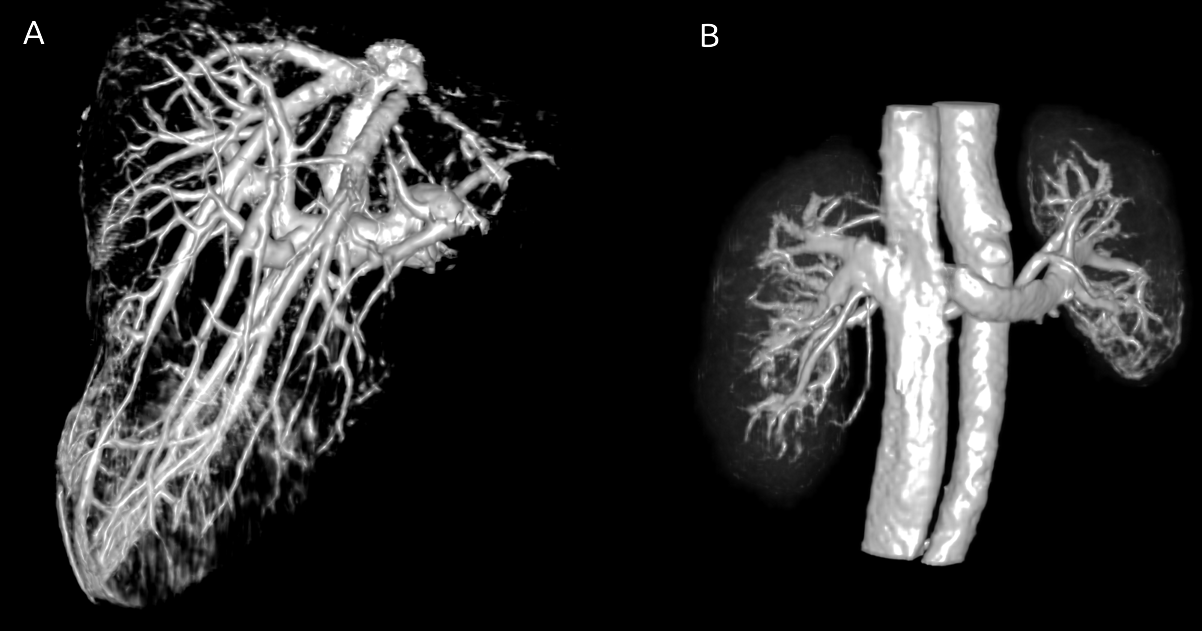
Ferumoxytol concentration MRI phantoms were placed around the large cylindrical phantom and QUTE-CE images were successfully acquired (Figure 1A, 1B) on a 3T clinical machine. Theoretical signal intensity for phantom samples was calculated based on intrinsic values of the sample (T1, T2*, proton density) and acquisition parameters (repetition time, echo time, flip angle) and as a function of ferumoxytol concentration (Figure 1D). The measured intensity versus concentration of the phantoms was presented in Figure 1E. The SNRs were measured 1472 ± 91 for samples at low concentration (0~25µg/mL) and 4317 ± 151 for those at high concentration (50~200µg/mL).Representative CEMRA of one subject rendered in 3D is shown in Figure 2 with annotated abdominal vascular anatomy. The image captured both arteries and veins within the field of view in the abdominal cavity. The liver (Figure 3A) and the kidneys (Figure 3B) were manually cropped from the image. The blood vessels in these organs were visualized with bright contrast with detailed delineation.
Discussion
Ferumoxytol demonstrates high r1 relaxivity of 10 s-1mM-1 and high r2 relaxivity of 62.3 s-1mM-1 at 3T [8], where R1=R10 + r1*[C]. Therefore, it can cause bright contrast on T1-weighted images and strong susceptibility effect. In the phantom experiment, the significant signal loss at high concentration of ferumoxytol is likely due to the strong T2* effect. However, for a 70kg human with an approximate 5L blood volume, the 510mg of elemental iron in ferumoxytol (clinical dose) produces 102µg/mL of intravascular concentration, at which there is no significant signal loss observed in the phantom experiment. Additionally, sampling data at UTE limits the spin dephasing within relatively short time thus reduces susceptibility effects.Images acquired on human shows excellent delineation of large vessels (i.e. inferior vena cava, aorta, portal vein) and small vessels (i.e. renal arteries). By taking advantage of the blood-pool kinetics of ferumoxytol, signal is enhanced in both arteries and veins with high SNR and CNR.
Conclusion
This study demonstrated the potential of UTE sequence with ferumoxytol to produce detailed abdominal vascular images. The QUTE-CE technique enables imaging for central arterial and venous anatomy at a single scan. Given the extremely high SNR and CNR, it has the potential to assess peripheral vessels with high-resolution and accelerated imaging. Further development of the technique in both breath-hold and free-breathing abdominal CEMRA is ongoing. Future research may focus on techniques for comprehensive analysis of the organs and vascular structures captured in these high positive contrast images.Acknowledgements
This work was partially supported by National Institutes of Health (Grant numbers 1R21DK118449-01) awarded to Dr. Sridhar.References
1. Mukundan, S., et al., Ferumoxytol-Enhanced Magnetic Resonance Imaging in Late-Stage CKD. Am J Kidney Dis, 2016. 67(6): p. 984-8.
2. Bashir, M.R., et al., Emerging applications for ferumoxytol as a contrast agent in MRI. J Magn Reson Imaging, 2015. 41(4): p. 884-98.
3. Bullivant, J.P., et al., Materials characterization of Feraheme/ferumoxytol and preliminary evaluation of its potential for magnetic fluid hyperthermia. Int J Mol Sci, 2013. 14(9): p. 17501-10.
4. Wells, S.A., et al., Pharmacokinetics of Ferumoxytol in the Abdomen and Pelvis: A Dosing Study with 1.5- and 3.0-T MRI Relaxometry. Radiology, 2019: p. 190489.
5. Knobloch, G., et al., Comparison of gadolinium-enhanced and ferumoxytol-enhanced conventional and UTE-MRA for the depiction of the pulmonary vasculature. Magn Reson Med, 2019. 82(5): p. 1660-1670.
6. Gharagouzloo, C.A., et al., Quantitative vascular neuroimaging of the rat brain using superparamagnetic nanoparticles: New insights on vascular organization and brain function. Neuroimage, 2017. 163: p. 24-33.
7. Gharagouzloo, C.A., P.N. McMahon, and S. Sridhar, Quantitative contrast-enhanced MRI with superparamagnetic nanoparticles using ultrashort time-to-echo pulse sequences. Magn Reson Med, 2015. 74(2): p. 431-41.
8. Knobloch, G., et al., Relaxivity of Ferumoxytol at 1.5 T and 3.0 T. Invest Radiol, 2018. 53(5): p. 257-263.
Figures