1601
Novel antenna array for ultra-high field MR-PET1INM-4, Forschungszentrum Juelich, Juelich, Germany, 2JARA-BRAIN-Translational Medicine, Aachen, Germany, 3Department of Neurology, RWTH Aachen University, Aachen, Germany, 4INM-11, Forschungszentrum Juelich, Juelich, Germany
Synopsis
Hybrid MR-PET is a powerful imaging technique that capitalised on the advantages of both modalities. However, to optimise these capabilities, the MRI antenna, which is located inside the effective PET detection area, needs to be operated without compromising any aspects of system performance or image quality compared to the stand-alone instrumentation. Here, we report a novel J-pole antenna array concept which is gamma radiation transparent. Furthermore, this unique feature provides advantages in MR-only applications by reducing the coupling effect between cables and the antenna elements and by lowering the potential specific absorption rate burden.
INTRODUCTION
In a simultaneous MR-PET acquisition, the MRI measurements require an RF coil or antenna to be positioned inside the PET imaging region. However, as MRI antennas are generally only optimised for MRI requirements, they often contain high amounts of gamma-ray absorbing materials, e.g. capacitors, massive support structures or co-axial cables, which degrade the quality of the PET images. Therefore, any potential attenuation of photons needs to be avoided to ensure optimal PET images while maintaining the best MR quality. This problem is further exacerbated in ultra-high field, as an RF wavelength is shorter in tissue than in an imaging object and thus requires an entirely different design approach. In this work, we introduce a new design which is, namely, a J-pole or J-shape antenna array.1 This novel concept to building a multi-channel RF antenna array fulfils the optimal condition for concurrently-operating hybrid systems as well as for stand-alone ultra-high field MRI systems.MATERIAL AND METHODS
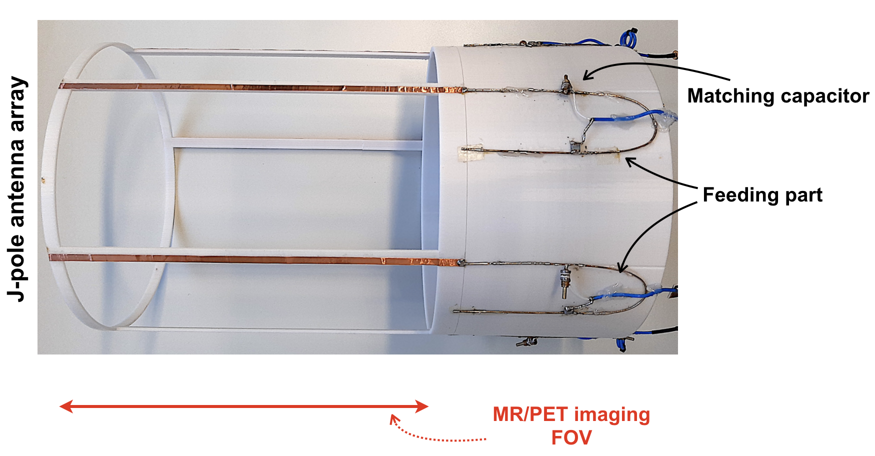
The proposed antenna design, shown in Fig.1, is based on J-pole antennas. Each J-pole antenna comprises a gamma radiation transparent radiating segment and a feeding unit. The element part within the imaging FOV does not include any highly attenuating components, and the antenna pattern is made of thin copper strips. Consequently, the PET acquisition is not degraded and attenuation and scattering artefacts are negligible. A matching network is placed in the feeding unit, which is outside of the MR or PET FOV and at one side of the antenna assembly to avoid any unnecessary interference.In order to evaluate the performance of the newly developed J-pole antenna array, we have also built a standard dipole antenna array. Both antenna arrays were tuned to 297 MHz and using CST simulations, and their characteristics were initially compared. All MR experiments were performed on a 7 T Terra scanner using a 2D FLASH sequence, and a 2-litre spherical water phantom was used. In order to verify the effect in the transmission performance of the J-pole antenna array, transmission scans with rotating 511 keV sources were carried out on a stand-alone PET scanner.
RESULTS
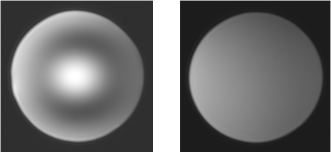
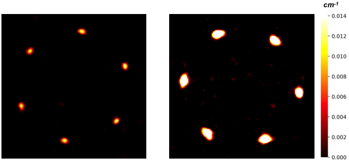
The SNR result from the whole volume, obtained using both antenna arrays, is almost identical. This is in agreement with the simulation results. Since the acquisition was carried out using a circularly-polarised mode, the central brightening2, a known effect at ultra-high field, is seen in the water phantom in Fig. 2. However, images with a uniform B1 were observed with the oil phantom.Fig. 3 shows the reconstructed attenuation maps in one of the various positions of both antenna arrays. Since, importantly, the proposed J-pole antenna array has only thin copper trace and the plastic coil former in the PET sensitive FOV, the map evidently shows very low attenuation for the proposed array. In contrast, relatively high attenuation was detected in the conventional dipole antenna array at several locations - in which the capacitors, inductors and co-axial cables are placed.
DISCUSSION AND CONCLUSIONS
Although multi-modal imaging provides valuable information, it is always challenging to optimise the systems for simultaneous use and often results in performance degradation of at least one modality. In this study, we have introduced and presented a new, gamma radiation transparent, RF antenna array for hybrid multi-modal systems. The MR performance of the new design has been demonstrated on a 7 T whole-body MRI scanner and the evaluation of potential gamma photon attenuation was measured using a 511 keV transmission scan. The novel J-pole antenna array is a unique MRI RF probe with exceptionally low gamma attenuation, allowing simultaneous MR-PET and MR-SPECT experiments to be conducted without compromising any aspects of system performance and image quality compared to the stand-alone instrumentation. The use of this new concept is not restricted to 7 T hybrid multi-modal imaging applications and can be optimised for even higher field strengths and for clinically relevant field strengths.Acknowledgements
No acknowledgement found.References
1. Choi C-H, et al. Dipolantennen-array für hybride MR-PET und MR-SPECT Tomographen sowie dessen Verwendung und MR-PET oder MR-SPECT Tomograph mit einem Dipolantennen-Array, German patent application number 102019003949.1.
2. Adriany G. et al. Transmit and Receive Transmission Line Arrays for7 Tesla Parallel Imaging. Magn. Reson. Med. 2005;53:434–445.
Figures