1572
In-vivo human brain imaging at 5 T using a 48 channel Tx-Rx array1Lauterbur Imaging Research Center, Shenzhen Institutes of Advanced Technology, Chinese Academy of Sciences, Shenzhen, China, 2Key Laboratory for Magnetic Resonance and Multimodality Imaging of Guangdong Province, Shenzhen, China, 3United Imaging Healthcare, Shanghai, China, 4Department of Biomedical Engineering, State University of New York at Buffalo, NY, NY, United States
Synopsis
Intermediate field strength between 3 T and 7 T may provide significant signal-to-noise ratio improvement but with less radio frequency challenges. In this work in-vivo human brain images including anatomic images, angiography images and susceptibility weighted images have been acquired at a prototype 5 T whole body scanner using a 48 channel Tx-Rx array, and compared with those at 3 T commercial scanner. Significant signal-to-noise ratio improvement of 5 T has been shown while image homogeneity is still acceptable.
Introduction
High and ultra-high field (>3 T) MR system provides high signal-to-noise ratio and susceptibility contrast, which leads to high image resolution and better image quality for clinical diagnosis and brain research. Thus, the field strength keeps increasing from 4 T to 10.5 T [1-3]. However, the transmit homogeneity and specific absorption rate (SAR) are two main challenges at ultra-high field, which restricts many applications. Therefore, an intermediate field strength between 3 T and 7 T, such as 5 T, may provide significant signal-to-noise ratio (SNR) improvement but with less critical radio-frequency challenges as what we encountered at 7T [4]. In this work, in-vivo human brain images have been acquired at a novel whole body 5T MRI system using a 48 channel Tx-Rx array. Anatomic images, angiography images and susceptibility weighted images have been acquired and compared with those at 3 T commercial scanner. Image quality at 5 T such as signal-to-noise ratio and image homogeneity were evaluated. The high quality images acquired demonstrate the feasibility and also the benefits of 5T MRI.Methods
In-vivo human brain images were acquired at a novel whole body 5T MRI system (United Imaging Healthcare, Shanghai, China). The diameter of patient bore was 60 cm. Second order shimming was applied to achieve good B0 homogeneity. The maximum gradient strength and slew rate were 120 mT/m and 200 T/m/s respectively. A 48 channel Tx-Rx array was fabricated, which consisted of a shielded quadrature birdcage with 30 cm inner diameter for local transmit and 48-channel helmet-shaped head array for receiving. Firstly, a fast spin echo (FSE) sequences was utilized for T2-weighted anatomic images with the parameters: TR = 3500 ms, TE = 98.56 ms, FOV = 180 × 180 mm2, image matrix = 448 × 448, slice thickness = 3 mm, number of slices = 15, number of average = 4, bandwidth = 150 Hz/pixel. Secondly, a time of flight (TOF) sequence was used for magnetic resonance angiography images (MRA) with the parameters: TR = 18 ms, TE = 3.5 ms, flip angle = 15 degree, FOV = 200 × 180 mm2, image matrix = 336 × 302, slice thickness = 0.6 mm, bandwidth = 220 Hz/pixel. Finally, susceptibility weighted images (SWI) were acquired with the parameters: TR = 30.2 ms, TE = 16 ms, flip angle = 20 degree, in-plane resolution = 0.35 × 0.35 mm2, slice thickness = 1.5 mm, number of slices = 52, number of average = 2, bandwidth = 130 Hz/pixel. SAR monitoring were carried out during the scans to guarantee the safety of volunteer. In order to compare the image quality with 3 T, these sequences with the same parameters were performed at a commercial 3 T human system (uMR 790, United Imaging Healthcare, Shanghai, China). The 24-channel head neck coil was used for comparison.Results
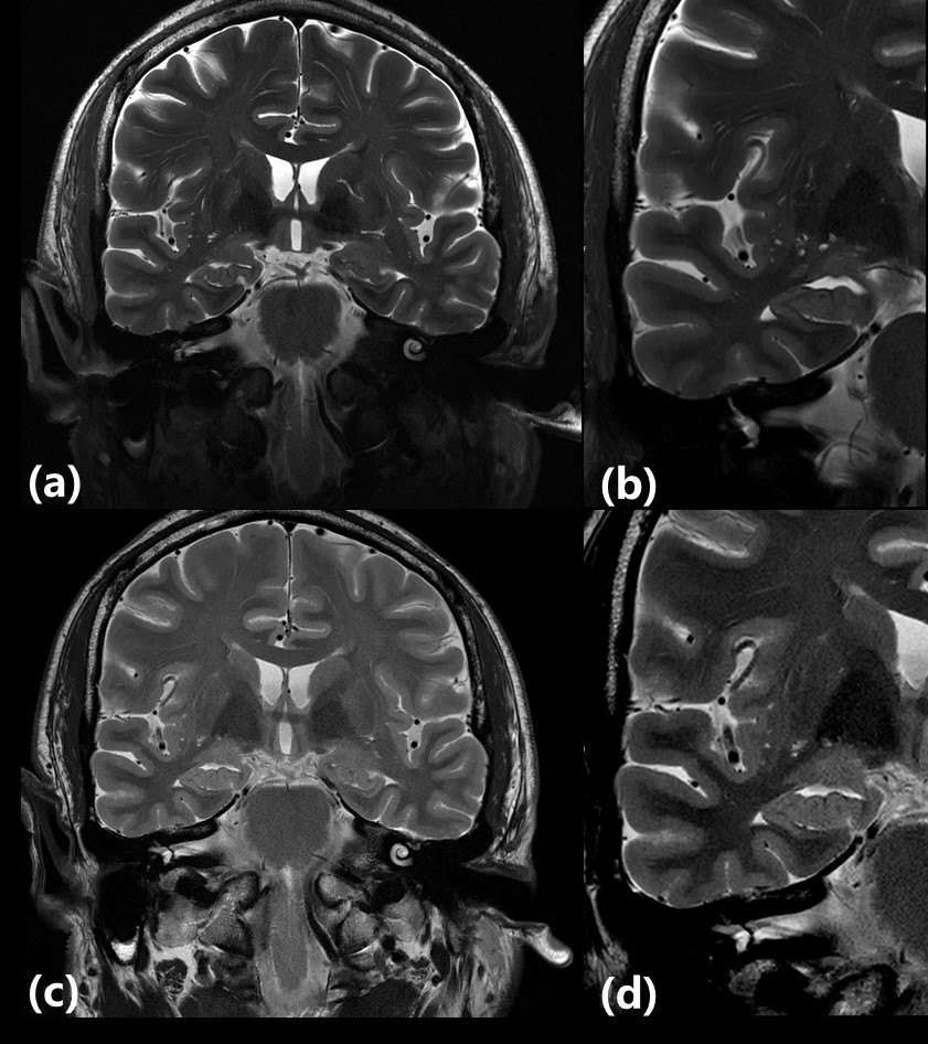
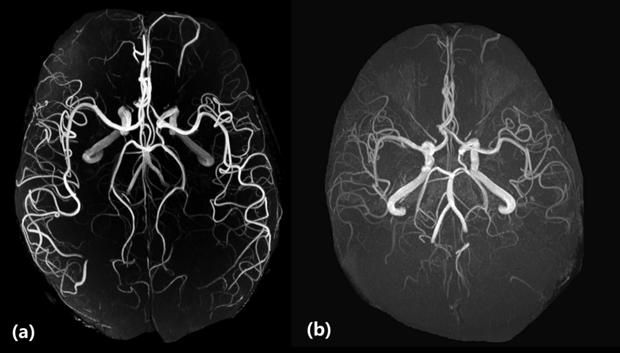
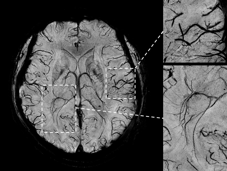
Figure 1 showed T2-weighted FSE brain images with 0.4*0.4*3 mm3 resolution. (a) and (c) were acquired by the local quadrature birdcage transmit and 48-channel receive coil at 5 T and the commercial 24-channel head neck coil at 3 T respectively. (b) and (d) were zoomed-in images of (a) and (c) respectively. Higher signal-to-noise ratio (SNR) was achieved at 5 T system. The TOF-MRA images were shown in figure 2. The results demonstrated that 5 T system provided better MRA image quality compared with 3 T. More vessels could be seen at 5 T because of higher SNR. Susceptibility weighted images with 0.35*0.35*1.5 mm3 at 5 T were shown in figure 3. Both surface and deep brain regions were zoomed in for performance evaluation. The homogeneity of the images at 5 T was acceptable compared with 3 T.Discussions and Conclusions
High resolution in-vivo human brain images including T2-weighted anatomic images, angiography images and susceptibility weighted images were acquired at a novel whole body 5 T human system. The images were compared with those at 3 T commercial system. The results demonstrate that the image quality at 5 T was largely improved comparing with 3 T, which indicated 5 T scanner’s potential in clinical and brain science applications. Future work includes performance evaluation in functional imaging and metabolism imaging.Acknowledgements
This work was supported in part by the Strategic Priority Research Program of Chinese Academy of Sciences (Grant No. XDB25000000); National Key R&D Program of China No. 2017YFC0108800; Youth Innovation Promotion Association of CAS No. 2017415.References
1. J.T. Vaughan, M. Garwood, C.M. Collins, W. Liu, L. DelaBarre, G. Adriany, P. Andersen, H. Merkle, R. Goebel, M.B. Smith, and K. Ugurbil. “7T vs. 4T: RF power, homogeneity, and signal‐to‐noise comparison in head images,” Magn. Reson. in Med. 2001; 46: 24-30.
2. D. L. Thomas, E. De Vita, S. Roberts, R. Turner, T. A. Yousry, and R. J. Ordidge. High‐resolution fast spin echo imaging of the human brain at 4.7 T: Implementation and sequence characteristics. Magn. Reson. in Med. 2004; 51: 1254-1264.
3. X. He, M. A. Ertürk, A. Grant, X. Wu, R. L. Lagore, L. DelaBarre, Y. Eryaman, G. Adriany, E. J. Auerbach, P.‐F. Van de Moortele, K. Uğurbil, and G. J. Metzger. “First in‐vivo human imaging at 10.5T: Imaging the body at 447 MHz”, Magn. Reson. in Med. 2020; 84: 289-303.
4. Jürgen Hennig. “Ultra high field MR: useful instruments or toys for the boys?” Magn. Reson. Mater. Phy., 2008; 21: 1-3.
Figures