1529
The Neural Activation of Positive versus Negative Though-Action Fusion: an fMRI study
Hyunsil Cha1, Sang Won Lee2, Heajeong Choi1, Eunji Kim1, Seungho Kim1, Yunheung Kim1, Seung Jae Lee2, and Yongmin Chang3
1Medical and Biological Engineering, Kyungpook National University, Daegu, Korea, Republic of, 2Department of Psychiatry, Kyungpook National University Hospital, Daegu, Korea, Republic of, 3Radiology and Molecular Medicine, Kyungpook National University, Daegu, Korea, Republic of
1Medical and Biological Engineering, Kyungpook National University, Daegu, Korea, Republic of, 2Department of Psychiatry, Kyungpook National University Hospital, Daegu, Korea, Republic of, 3Radiology and Molecular Medicine, Kyungpook National University, Daegu, Korea, Republic of
Synopsis
In this fMRI study, we aimed to investigate the neural circuits related to positive and negative TAF by using a modified TAF task, wherein individuals were asked to read the name of a close person within positive or negative statements.
Introduction
The thought-action fusion (TAF) is a tendency of individuals to blindly establish causal relations between their own thoughts and external reality.1 Many studies have investigated to reveal a reason of transition from normal to abnormal obsessive thought with TAF mechanism.2,3 According to previous studies, negative statement seems to heighten TAF more than positive statement. Thus, we hypothesized that core brain regions will be more activated in negative than in positive statement. In addition, the brain activity would be correlated with scores of TAF and associated psychopathology like obsessive-compulsive (OC) symptoms.Subjects and Methods
A total of thirty-one participants (all mean) were recruited for this study. The mean age of all subjects was 22.9±1.9 years and right-handed in accordance with the Edinburgh handedness scale. All participants received written informed content and the study protocol was approved by the Institutional Review Board at Kyungpook national University Hospital. During the functional magnetic resonance acquisition, participants evaluated how good or bad they feel about the following typed sentence with a Likert scale from 1(very little) to 4(very much) using MR convertible button box. Regarding positive and negative statements, we used the eight positive and negative statements (PS and NS, respectively). The former example is “I hope that … will win a lottery.” ; the latter is “I hope that … will soon be in a car accident.” Participants were instructed to complete this sentence by filling the name of a close or neutral person to them. Functional image data were obtained the 3.0T GE 750W scanner with 24ch head coil (EPI, TR = 2000ms, TE = 30ms, FOV = 23cm, acquisition matrix = 64 X 64, no gap). The 3D T1-weighted fast spoiled gradient echo were used for structural imaging acquisition. In fMRI data within-group analysis, brain activation analyzed by paired t-test and conjunction analysis. The SPM{t} was thresholded at P<0.05, false discovery rate (FDR) corrected for multiple comparisons across the whole brain.Results and Discussion
Conjunction analysis using data from both PS and NS conditions showed significant activations in the bilateral lingual and fusiform gyrus, bilateral inferior frontal and inferior orbito-frontal gyri (corresponding to vlPFC); left middle frontal gyrus (corresponding to the dlPFC); left superior medial frontal gyrus (corresponding to dmPFC) (Figure 1). In paired t-test, the NS > PS comparison found several new regions not shown in the conjunction analysis including the left precuneus, the left midbrain, the right insula, medial globus pallidus(mGP) and thalamus. It also found the same and adjacent regions as shown the conjunction anlaysis such as several frontal subregions, superior and inferior parietal lobule, middle and superior temporal gyrus. However, the NS < PS comparison revealed no significant activation (Figure 2). ROI anlaysis were used to investigate whether activities in those regions were correlated with behavioral and psychological measures. In terms of behavioral data, the right mGP, left inferior orbito-frontal gyrus, right insula, and left precuneus were correlated negatively with emotional intensity while subjects responded to the negative TAF task during the MRI scan. In terms of baseline psychological measures, activity in left precuneus was associated with TAF total score. Activity in the mGP, IOFG, SFG, MFG, IPL and right precuenus were associated with total score of OCI-R or DOCS (Figure 3). Thus, these regions were closely related to the performance of negative TAF statements.Conclusion
The present study suggests that OC symptoms were associated with hemodynamic activity for negative TAF.Acknowledgements
No acknowledgement found.References
1. Rachman S. Obsessions, responsibility and built. Behav Res Ther. 1993;31(2):149-54
2. Rassin E, Diepstraten P, Merckelbach H, Muris P. Thought-action fusion and thought suppression in obsessive-compulsive disorder. Behav Res Ther. 2001;39(7):757-64
3. Rassin E, Merckelbach H, Muris P, Spaan V. Thought-action fusion as a causal factor in the development of intrusions. Behav Res Ther. 1999;37(3):231-7
Figures
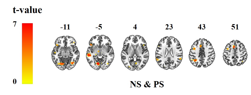
Figure 1. Brain activation for
conjunction analysis of both positive and negative TAF statements (P<0.05,
FDR-corrected for multiple comparisons).
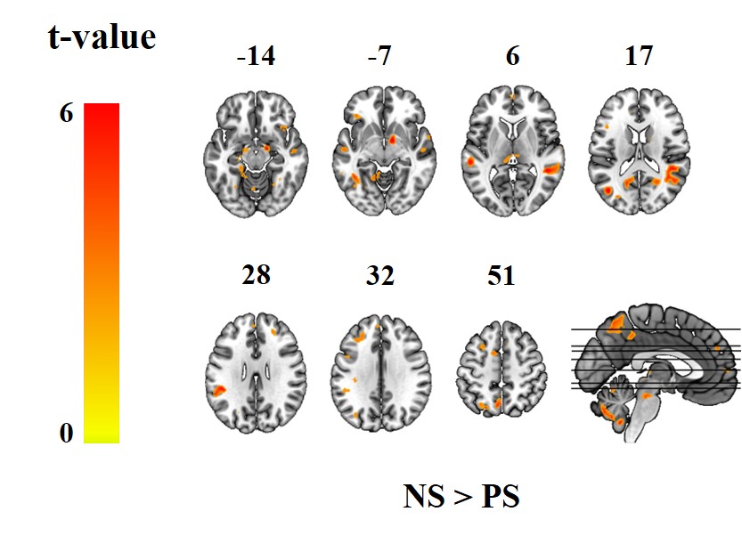
Figure 2. Brain
activation of paired t-test between positive and negative TAF statements (P<0.05,
FDR-corrected for multiple comparisons).
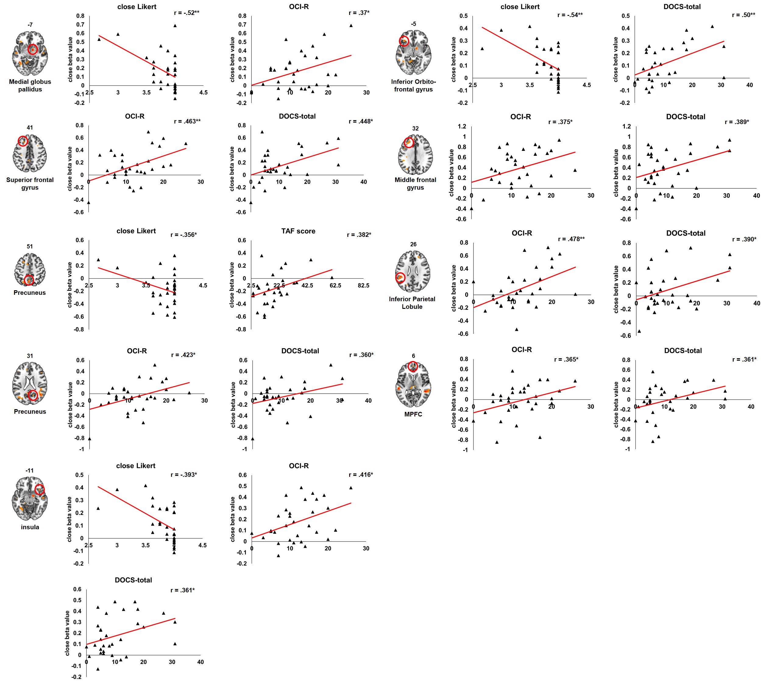
Figure 3. Relationship between
regional activity and psychological measures.