1518
Community-informed connectomics of cortical intrinsic organization in participants with subjective cognitive decline
Qian Chen1, Jilei Zhang2, and Bing Zhang1
1Department of Radiology, Drum Tower Hospital, Clinical College of Nanjing Medical University, Nanjing, China, 2Philips Healthcare, Shanghai, China
1Department of Radiology, Drum Tower Hospital, Clinical College of Nanjing Medical University, Nanjing, China, 2Philips Healthcare, Shanghai, China
Synopsis
Individuals with subjective cognitive decline (SCD) were at higher risk of Alzheimer’s disease. Based on community-informed connectome analysis, we observed decreased intra-module connectivity in superior occipital gyrus of the visual network and decreased inter-module connectivity in supramarginal gyrus of the dorsal attention network in SCD subjects. The SCD group also showed increased intra-module connectivity in the dorsolateral prefrontal cortex and middle frontal gyrus of the frontoparietal network. The altered network measures showed significant correlations with cognitive performance on memory and language. Our findings may benefit a better understanding of the neural basis underlying early cognitive decline in the SCD stage.
Introduction
Cognitive impairment in Alzheimer’s disease (AD) is a disconnection syndrome closely associated with functional disconnection of coordinated neural networks1. Subjective cognitive decline (SCD), considered to be the preclinical stage of AD, is essential for the early diagnosis and intervention of symptomatic AD2. Thus, we aimed to investigate the intrinsic alterations of cortical circuitry in SCD individuals via resting-state functional magnetic resonance imaging (rs-fMRI) connectome analysis and to determine its associations with cognitive performance.Methods
Thirty-six elderly SCD subjects and 32 normal controls (NCs) underwent neuropsychological tests and rs-fMRI scanning. Based on the prior template and 10 predefined communities proposed by Gordon3, we established full connection matrices of 333 cortical parcels and calculated the within-module degree (WMD) and participation coefficient (PC) of each node4. The WMD is a measure of connectivity to other parcels within the same community, and the PC is a measure of the diversity of connectivity across different communities5. Then we calculated the between-group differences in WMD and PC values and further analyzed the correlations with cognitive variables in the SCD group.Results
Compared to the NCs, SCD subjects showed increased WMD in the dorsolateral prefrontal cortex (DLPFC, p < 0.05, FDR corrected) and the middle frontal gyrus (p < 0.005, uncorrected) of the right frontoparietal network (FPN). The SCD group was also observed decreased WMD in the superior occipital gyrus (p < 0.05, FDR corrected) of the left visual network (VN), and decreased PC in the supramarginal gyrus (p < 0.005, uncorrected) of the left dorsal attention network (DAN). The WMD value in the DLPFC was positively correlated with the Auditory Verbal Learning Test (AVLT) short-delayed memory (r = 0.364, p =0.029), AVLT recognition (r = 0.364, p =0.029), and Boston Naming Test (BNT; r = 0.356, p = 0.033) performance. Moreover, the PC value in the supramarginal gyrus was positively correlated with the BNT score (r = 0.413, p = 0.012).Discussion
Cortical network imbalance characterized by decreased intra-module connectivity of VN and decreased inter-module connectivity of DAN in SCD subjects may be a potential indicator for distinguishing individuals at higher risk of incipient AD dementia from subjects with normal aging. Furthermore, increased intra-module connectivity of FPN, especially in the right DLPFC, may serve in a compensatory way for the early cognitive decline.Conclusion
Our results may benefit future researches on the early diagnosis, preventative intervention, and prognostic evaluation of preclinical AD.Acknowledgements
This work was supported by the National Natural Science Foundation of China (81720108022 B.Z., 81971596, X.Z., 82071904, Z.Q.); the Fundamental Research Funds for the Central Universities, Nanjing University (2020-021414380462); the key project of Jiangsu Commission of Health (K2019025); Key medical talents of the Jiangsu province, the "13th Five-Year" health promotion project of the Jiangsu province (ZDRCA2016064); Jiangsu Provincial Key Medical Discipline (Laboratory) (ZDXKA2016020); the project of the sixth peak of talented people (WSN -138). The funders had no role in the study design, data collection and analysis, decision to publish, or preparation of the manuscript.References
- Delbeuck X, Van der Linden M, Collette F. Alzheimer's disease as a disconnection syndrome? Neuropsychol Rev, 13(2), 79-92.
- Jessen F, Amariglio RE, Buckley RF, et al. The characterisation of subjective cognitive decline. Lancet Neurol, 19(3), 271-278.
- Gordon EM, Laumann TO, Adeyemo B, et al. Generation and Evaluation of a Cortical Area Parcellation from Resting-State Correlations. Cereb Cortex, 26(1), 288-303.
- Hwang K, Bertolero MA, Liu WB. The Human Thalamus Is an Integrative Hub for Functional Brain Networks. 37(23), 5594-5607.
- Wang Z, Qiao K, Chen G, et al. Functional Connectivity Changes Across the Spectrum of Subjective Cognitive Decline, Amnestic Mild Cognitive Impairment and Alzheimer's Disease. Front Neuroinform, 13, 26.
Figures
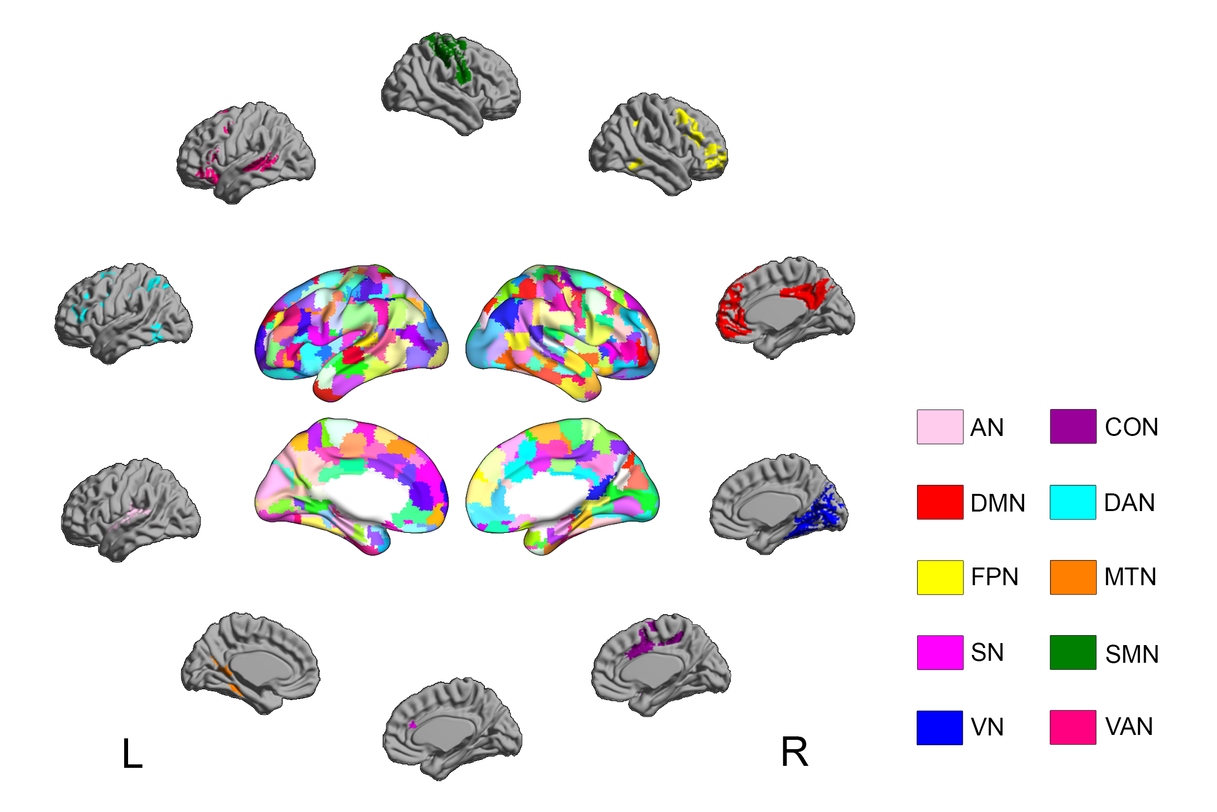
Figure 1. Gordon’s 333
cortical parcels and 10 predefined communities. AN: auditory network; DMN:
default mode network; FPN: frontoparietal network; SN: salience network; VN:
visual network; CON: cingulo-opercular network; DAN: dorsal attention network;
MTN: medial temporal network; SMN: sensorimotor network; VAN: ventral attention
network.
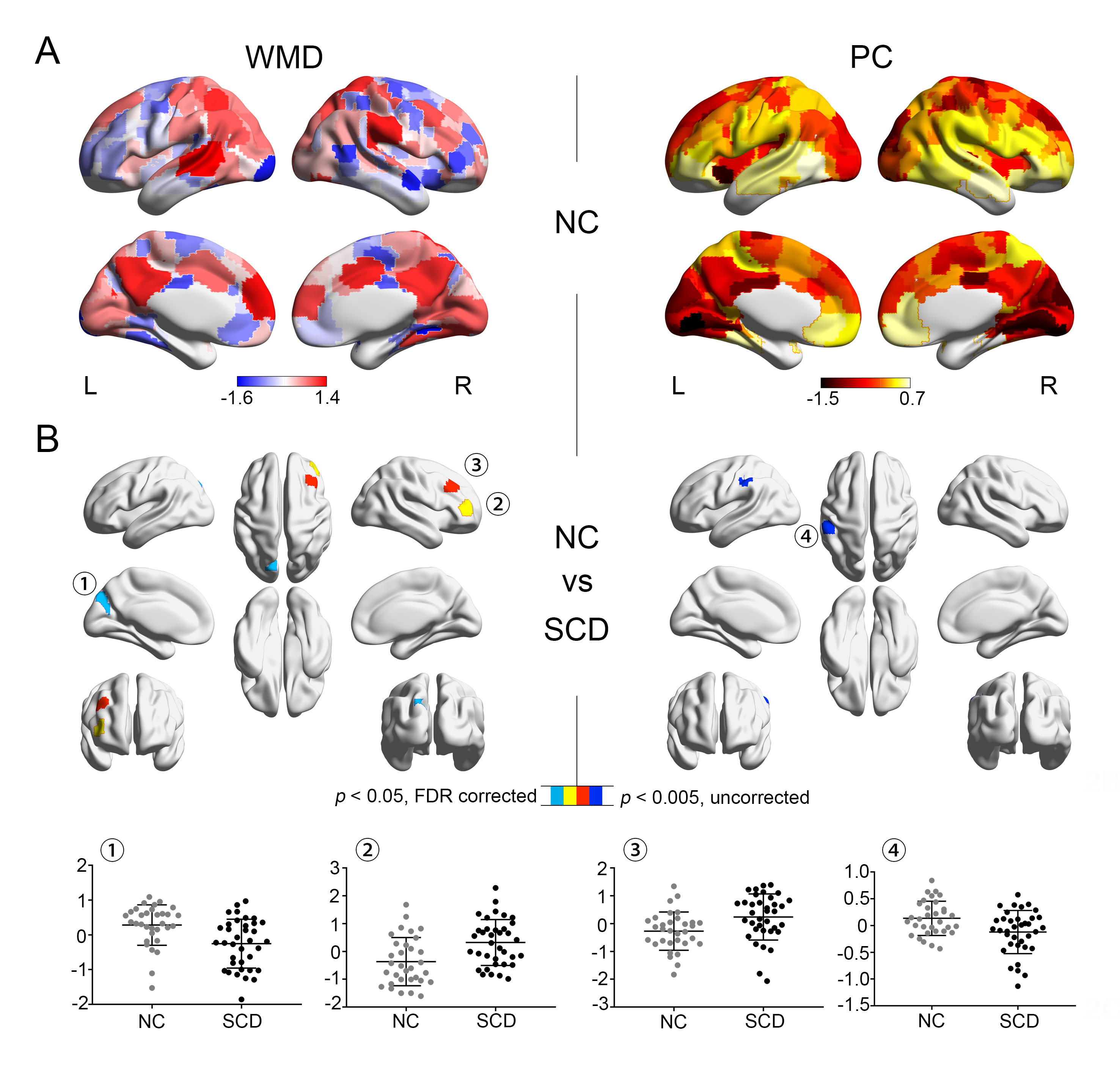
Figure 2. A Distribution of cortical within-module
degree (WMD) and participation coefficient (PC) averaged across 32 normal
controls (NC). B Differences between NC (gray) and 36 participants with
subjective cognitive decline (SCD) (black). Scatterplots represent WMD/PC
values in significant clusters. Findings were adjusted for age, gender, and
years of education.
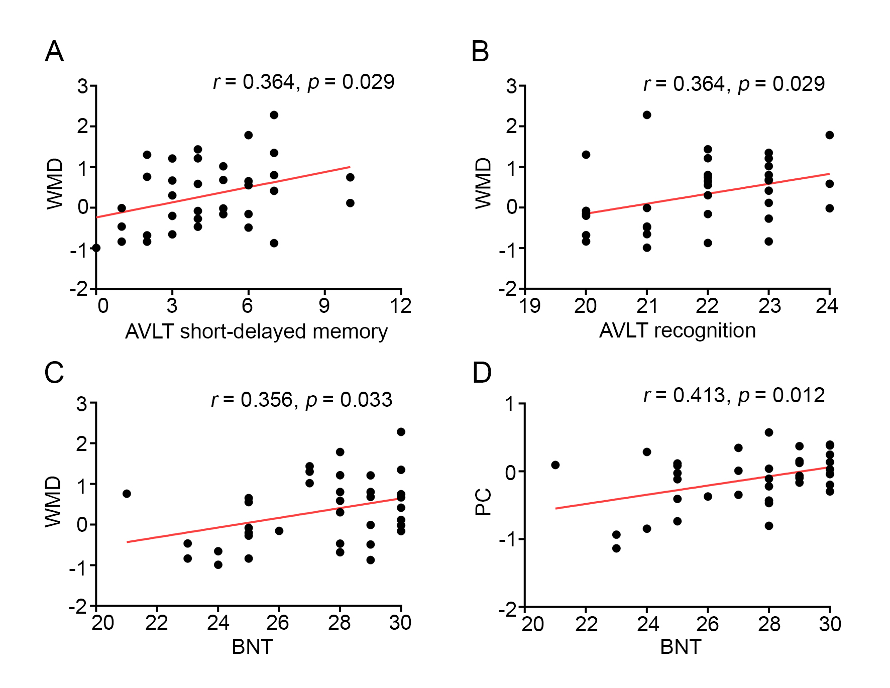
Figure 3. Correlations
between network measures and cognitive variables in the subjective cognitive
decline (SCD) group. WMD: within-module degree; PC: participation coefficient;
AVLT: auditory verbal learning test; BNT: Boston naming test.