1513
Altered voxel-level whole-brain functional connectivity in multiple system atrophy patients with depression symptoms1Radiology, Renmin Hospital of Wuhan University, Wuhan, China, 2MR Research, GE Healthcare, Wuhan, China, 3The First Affiliated Hospital of China Medical University, Shenyang, China
Synopsis
Depression is a common non-motor system symptom in patients with multiple system atrophy (MSA). Its pathogenesis is unknown. Multimodal functional magnetic resonance imaging (fMRI) is the main tool to explore the mechanism of pure depression and other diseases with depressive symptoms. Applications of fMRI on patients with MSA in accompanied with depressive symptoms is expected to reveal the difference to the MSA-involved depression mechanism different to the pure depression-involved brain network. Our results found altered secondary network in MSA patients with depression and provided a new idea for determination on treatments to MSA with depression. This study provided neuroimaging evidence for a clinical understanding of non-pure depression and possible reference for treatment measures.
Introduction
Multiple system atrophy is characterized by prominent non-motor system symptoms and autonomic dysfunction symptoms. With anincidence rate of about 70%1, depressive symptoms are common non-motor symptoms in MSA patients, leading to a serious decline in the life quality of patients. At present, the mechanism of MSA with depression symptoms is unclear, which makes the clinicians mainly prescribe the anti-depression therapy similar to the pure depression diseases to patients with MSA in accompanied with depression. However, different to pure depression, the neurotransmitter disorder in patients with MSA in accompanies with depression dominantly damages dopaminergic neurons supplemented by serotonin (5-HT), norepinephrine and other neurotransmitter2. Therefore, digging out the depression-modulated pathogenesis in the MSA is helpful to determine treatment. fMRI can explore the mechanism changes of depression in vivo3-5. Therefore, this study aims to explore the changes of brain resting-state network in MSA patients in accompanied with and without depression using the degree centrality (DC) along with seed-based functional connectivity.Materials and Methods
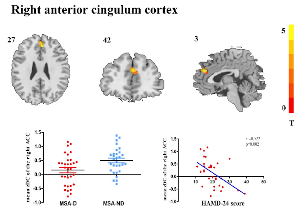
62 patients with MSA with depression (N=32) and without depression (N=30) were included in this study. All patients underwent the resting state function magnetic resonance BOLD sequence and T1 structural image on 3.0T MRI scanner. The changes of degree centrality (DC) and functional connectivity (FC) network results were analyzed by DPABI software embedded in MATLAB. The relationship between these changes and the severity of depression was further analyzed. The severity of depression symptoms was evaluated by the 24 terms Hamilton Depression Scale (HAMD-24). A voxel-wise Pearson correlation analysis was used to identify brain regions significantly associated with clinical depression symptoms in MSA patients.Results
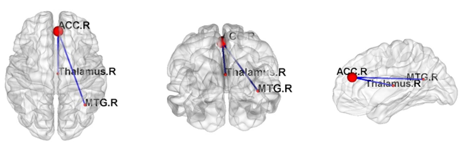
The changes of degree centrality in the right anterior cingulate cortex (rACC) was the main manifestation of MSA patients with depression, which was positively correlated with the severity of depression. A secondary FC analysis based on the rACC as a seed revealed a decreased functional connectivity between the rACC and middle temporal cortex and thalamic network. In addition, only altered DC in the right ACC was found to significantly associate with HAMD-24 scores.Discussion and conclusions:
MSA patients with depression mainly showed the changes of central hub and functional connectivity mainly occurred in temporal lobe and subcortical thalamic nuclei of MSA patients with depression compared to those only with MSA. In addition, the importance of rACC as a hub decreased and rACC-based FC network dysfunction in the right middle temporal lobe and right thalamus. The distinct network impairment between MSA patients with and without depression indicated different transmitter system dysfunctions were involved. The results of present study showed altered secondary network in MSA with depression and provided a new idea for determination on treatments to MSA patients with depression. This study provided neuroimaging evidence for a clinical understanding of non-pure depression and possible reference for treatment measures.Acknowledgements
No acknowledgement found.References
1. Zhang, L.Y., Cao, B., Zou, Y.T., Wei, Q.Q., Ou, R.W., Zhao, B., Wu, Y., Shang, H.F., 2018. Depression and anxiety in multiple system atrophy. Acta Neurol Scand 137, 33-37.
2. Berardelli I, Belvisi D, Pasquini M, Fabbrini A, Petrini F, Fabbrini G. 2019. Treatment of psychiatric disturbances in hypokinetic movement disorders. Expert Rev Neurother 19: 965-81
3. Misquitta K, Dadar M, Louis Collins D, Tartaglia MC, Alzheimer's Disease Neuroimaging I. White matter hyperintensities and neuropsychiatric symptoms in mild cognitive impairment and Alzheimer's disease. Neuroimage Clin. 2020;28:102367.
4. Qiu YH, Huang ZH, Gao YY, Feng SJ, Huang B, Wang WY, et al. Alterations in intrinsic functional networks in Parkinson's disease patients with depression: A resting-state functional magnetic resonance imaging study. CNS Neurosci Ther. 2020.
5. Yamashita A, Sakai Y, Yamada T, Yahata N, Kunimatsu A, Okada N, et al. Generalizable brain network markers of major depressive disorder across multiple imaging sites. PLoS Biol. 2020;18(12):e3000966.
Figures