1511
A survey of brainstem reticular activation system connectivity in myalgic encephalomyelitis / chronic fatigue syndrome (ME/CFS)1Griffith University, Southport, QLD, Australia, 2University of Sunshine Coast, Sunshine Coast, QLD, Australia
Synopsis
For 44 ME/CFS and 26 HC, resting state and Stroop task fMRI were spatially normalized with a method optimized for the brainstem. After physiological signal (RETROICOR) denoising in CONN, BOLD correlations were computed between 12 brainstem reticular activation system (RAS) ROI seeds. A survey of connections showed fewer were significant in ME/CFS than HC at Rest, indicating impaired RAS function. During Task, although the same number of connections were active for the two groups, many were between different ROIs, suggesting compensatory mechanisms may be involved. Nerve signal conduction in the brainstem RAS was shown to be impaired in ME/CFS.
introduction
This work was motivated by structural MRI observations in myalgic encephalomyelitis / chronic fatigue syndrome (ME/CFS) of abnormal MRI correlations with symptom severity and autonomic metrics that inferred impaired nerve signal conduction within the brainstem reticular activation system (RAS)1-3. Here we analyse fMRI BOLD correlations to survey RAS connectivity in terms of the number of significant pair-wise correlations between a set of 12 regions for HC, and ME/CFS during resting state and task.methods
Resting and task functional MRI (fMRI) were acquired for 45 ME/CFS (Fukuda criteria) and 27 healthy controls (HC). The fMRI data were acquired sagitally on a 3T MRI scanner (Skyra,Siemens) with a 64 channel head-neck coil using a multiband echo-planar imaging (EPI) pulse sequence with 72 slices, multiband factor = 8, TR = 798 ms, TE = 30 ms, flip angle = 40°, acquisition matrix 106 x 106 and voxel size 2 x 2 x 2 mm. A total of 1100 resting state (rfMRI) volumes were acquired in 15 min while the subject was awake and viewing a fixed stationary cross through goggles to reduce the likelihood of falling asleep. Then 1100 task fMRI ( tfMRI) volumes were acquired in 15 min while the subject was performing a sequence of Stroop colour-word tasks4 chosen to test the attention and concentration difficulties frequently reported by CFS/ME patients5. For both resting state and task, after distortion correction and motion correction6, spatial normalization was optimised for the brainstem in two steps using FSL’s ‘FLIRT’6: the single band reference image was coregistered (9DF affine) to the T2 MNI template and then coregistration was repeated constrained within a mask created to isolate the brainstem and diencephalon but excluding the ventral half of the pons and midbrain where signal dropout was seen in many subjects. The two deformations were merged and applied to the 1100 EPI volumes. Covariates were generated (RETROICOR, CompuCor) to isolate physiological noise from the ROI BOLD time series within the CONN package7. We selected limited RAS regions-of-interest (ROIs) based on our previous structural MRI findings in a different ME/CFS cohort (bilateral rostral medulla and midbrain cuneiform nucleus and left cerebellum culmen)2, and from brain atlases the dorsal Raphe, bilateral pontine nuclei, and two subcortical ROIs reported to have rich brainstem connections (bilateral hippocampus subiculum and thalamus intralaminar nucleus). Figure 1 shows 4 of the RAS ROIs. Connectivity analysis was performed with CONN7. 1st-level analysis performed correlations between denoised BOLD time series from all pairs of ROIs. 2nd-level analysis ran 1-sample statistics for HC and ME/CFS groups for both resting state and Task data sets. For each of the 12 ROIs, correlation coefficients with another ROI for all subjects in the group were tested for statistical inference of the null hypothesis. This was repeated for all of the other 11 ROIs. The number of ROI pairs (connections) with p-FDR < 0.05 were counted.results
The number of significant connections counted for each ROI for each group are presented in Table 1 and are displayed as connectograms in Fig 2. The maximum significant connections for each ROI was 11. At Rest the number of significant RAS connections for ME/CFS was reduced relative to HC, whereas during the task HC and ME/CFS showed the same number of connections, although many were between different RAS regions.discussion
In the resting state, the connections surveyed for significance within and from the brainstem RAS were fewer in ME/CFS than HC, while during the task the numbers were the same. This indicates RAS function in the ambient state is impaired. In response to the Stroop Task, although the number of significant connections was the same, they were between different regions. Indeed, we have reported reduced connectivity during Task between several RAS nuclei8. This suggests compensatory mechanisms may be activated during task, in line with observations in the cortex9.conclusion
This preliminary survey of reticular activation system connections showed fewer were active in ME/CFS than HC at Rest. During Task, although the same number of connections were active for the two groups, they were different suggesting compensatory mechanisms may be involved. Nerve signal conduction in the brainstem RAS appears to be impaired in ME/CFS.Acknowledgements
We thank the patients and healthy controls who donated their time and effort to participate in this study. This study was supported by the Stafford Fox Medical Research Foundation, the Judith Jane Mason Foundation (MAS2015F024), Mr. Douglas Stutt, and the Blake-Beckett Foundation, Ian and Talei Stewart, Buxton Foundation and McCusker Charitable Foundation. Their financial support did not affect any aspect of the study.
Kevin Finegan, Tim Ireland and Sandeep Bhuta effected MRI acquisition.
References
1 Barnden, L., Crouch, B., Kwiatek, R., Burnet, R. & Del Fante, P. Evidence in Chronic Fatigue Syndrome for severity-dependent upregulation of prefrontal myelination that is independent of anxiety and depression. NMR Biomed 28, 404-413, doi:10.1002/nbm3261 (2015).
2 Barnden, L., Kwiatek, R., Crouch, B., Burnet, R. & Del Fante, P. Autonomic correlations with MRI are abnormal in the brainstem vasomotor centre in Chronic Fatigue Syndrome. NeuroImage: Clinical 11, 530-537, doi:10.1016/j.nicl.2016.03.017 (2016).
3 Barnden, L. R. et al. Hyperintense sensorimotor T1 spin echo MRI is associated with brainstem abnormality in chronic fatigue syndrome. NeuroImage. Clinical 20, 102-109, doi:10.1016/j.nicl.2018.07.011 (2018).
4 Lamers, M. J., Roelofs, A. & Rabeling-Keus, I. M. Selective attention and response set in the Stroop task. Mem Cognit 38, 893-904, doi:10.3758/mc.38.7.893 (2010).
5 Carruthers, B. et al. Myalgic encephalomyelitis: International Consensus Criteria. J Intern Med 270, 327 - 338 (2011).
6 Jenkinson, M., Bannister, P., Brady, M. & Smith, S. Improved optimization for the robust and accurate linear registration and motion correction of brain images. Neuroimage 17, 825-841 (2002).
7 Whitfield-Gabrieli, S. & Nieto-Castanon, A. Conn: a functional connectivity toolbox for correlated and anticorrelated brain networks. Brain connectivity 2, 125-141, doi:10.1089/brain.2012.0073 (2012).
8 Barnden, L. et al. Intra-brainstem connectivity is impaired in chronic fatigue syndrome. Neuroimage Clinical 24, 102045, doi:https://doi.org/10.1016/j.nicl.2019.102045 (2019).
9 Shan, Z. Y. et al. Brain function characteristics
of chronic fatigue syndrome: A task fMRI study. NeuroImage. Clinical 19,
279-286, doi:10.1016/j.nicl.2018.04.025 (2018).
Figures
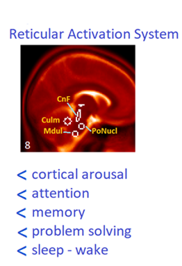
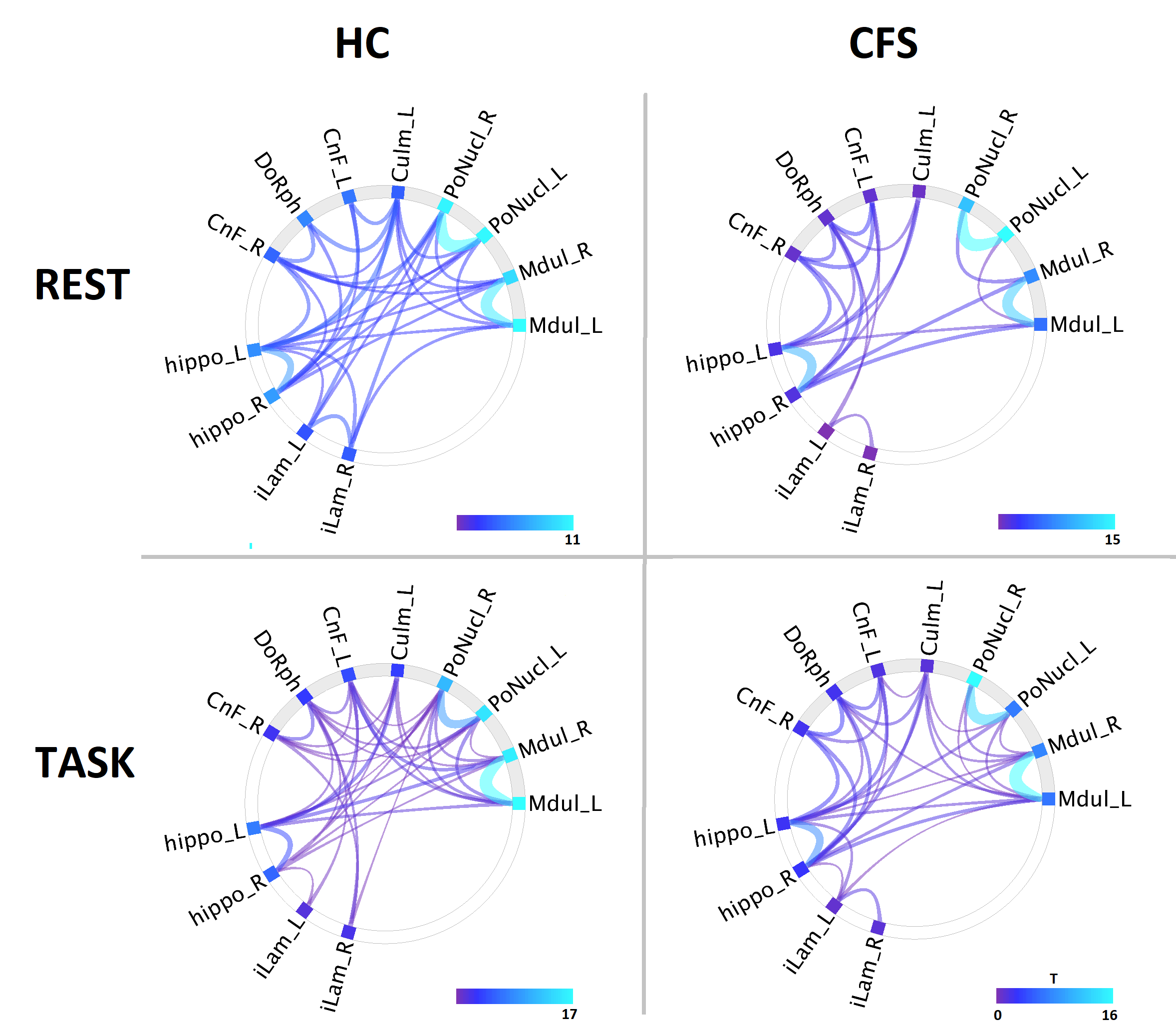
Figure 2 Reticular Activation System connectograms for HC and CFS during Resting State and Task. Reduced connections are seen for CFS at Rest, particularly for PoNucl.
Mdul=medulla; PoNucl=pontine nuclei; Culm=culmen;CnF=cuneiform nucleus; DoRph=dorsal Raphe; hippo=subiculum of hippocampus; iLam=thalamus intralaminar nucleus; L=left; R=right.
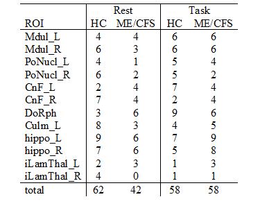
Table 1 Number of significant Connections (p-FDR < 0.05)