1510
Resting-State Functional Connectivity Predicts Cognitive Impairment Related to Type 2 Diabetes Mellitus1Department of Radiology & Functional and Molecular Imaging Key Lab of Shaanxi Province, Department of Radiology & Functional and Molecular Imaging Key Lab of Shaanxi Province, Tangdu Hospital, Fourth Military Medical University (Air Force Medical University), Xi'an, Shaanxi, China, 2Department of thoracic surgery, Tangdu Hospital, Air Force Medical University., Xi'an, China
Synopsis
Resting-state functional connectivity (RSFC) patterns of the human brain show unique inherent or intrinsic characteristics, similar to a fingerprint. There is significant interest in using RSFC to predict human behavior. Inspired by previous RSFC fingerprinting studies, we adopted whole-brain RSFC as discriminative features to predicted the MoCA scores in 102 individuals with T2DM, using a connectome-based predictive modeling (CPM). We find that, the identified CPM, based on whole-brain RSFC patterns, are strong for predicting the MoCA scores in T2DM. The application of CPM to predict neurocognitive abilities can complement conventional neurocognitive assessments and aid the management of people with T2DM.
Introduction
Resting-state functional connectivity (RSFC) has emerged as a powerful network-level approach to significantly advance our understanding of individual differences in human cognitive ability and personality traits. Many studies have revealed robust and reliable patterns of RSFC within many well-known networks spanning the brain, which extensively overlap with coactivation patterns induced by relevant task demands. Therefore, an analysis of the connectivity patterns based on the whole-brain RSFC, in comparison with the techniques based on traditional connectivity, could provide us a much more comprehensive understanding on the neural mechanism of certain cognitive disorders. Rather than constraining to specific areas of interest, whole-brain RSFC records enormous functional interaction information between any pair of brain nodes across the whole brain, which enriches the individual phenotypic prediction, which are used as fingerprints to identify and predict individuals of different behaviors and cognitions1-3. Whole-brain RSFC have previously been used in a number of studies addressing cognitive disorders. Previous studies have reported that patients with Alzheimer's Disease or mild cognitive impairment had abnormal connectivity patterns 4, 5. Whole-brain RSFC have also been used to successfully predict individual behavioral and cognitive phenotypes in recent fMRI studies, such as identification of psychiatric disorders 6、attention ability 2, 7、verbal creativity 8、intelligence ability 1, 9 and age estimation 10. In addition, Zeng et al detected disorder-related connectivity patterns from RSFC and then used them to discriminate major depressed patients from matched healthy subjects by means of machine learning6. Similarly, Li et al utilized a machine learning method based on RSCF to extract and analyze classification features that characterized differential connectivity patterns between the schizophrenia group and the healthy control group11.All in all, these results suggest that an individual’s functional connectome—his or her unique pattern of whole-brain RSFC— contains important behavioral and clinical information. RSFC patterns within each individual are both highly unique and reliable, similarly to a fingerprint, serving to underlie individual differences in personality traits or cognitive functions1, 2. Thus, it can be speculated that some of connectivity patterns could be regarded as potential biomarkers to either evaluate or identify cognitive impairment in patients with Type 2 diabetes mellitus (T2DM). T2DM is typically accompanied by cognitive impairments and is associated with a much higher risk of dementia 12, 13. Previous studies have identified neural correlates of cognitive impairment related to T2DM using resting-state functional imaging14-17. The present study examined whether machine learning techniques could utilize whole-brain RSFC patterns to predicts cognitive impairment related to T2DM with a high degree of accuracy.
Methods
Resting-state fMRI data were acquired from 102 individuals with T2DM and their degree of cognition was assessed by the Montreal cognitive assessment (MoCA). A new technique, connectome-based predictive modeling (CPM) was used to identify RSFC biomarkers to predicting the MoCA scores related to T2DM. CPM is an algorithm for building predictive models based on participants’ RSFC matrices, and for testing these models using cross-validation of novel data1, 18. We computed RSFC patterns using a functional brain atlas19 that comprised 264 nodes covering the whole brain. Specifically, we calculated the Pearson’s correlation coefficient of the average blood oxygenation level-dependent time series between each possible pair of nodes and transformed it to approximate a Gaussian distribution using Fisher’s z transformation. Subsequently, we constructed a 264 × 264 symmetrical connectivity matrix for each subject, with each element in the matrix representing the strength of the RSFC between two nodes. All of these processes were performed using the BRANT toolbox. Finally, three matrices that reflected RSFC patterns in each of the different scanning conditions were generated for each subject. Predictive accuracy was assessed via the pearson's correlation between predicted and actual scores (r predicted- actual).Results
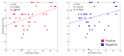
We found that CPM successfully and reliably predicted the MoCA scores from T2DM (positive network: r=0.40, Pr=0.0083, Pp=0.0038; negative network: r=0.36, Pr=0.0176, Pp=0.0066), demonstrating that patterns in RSFC reveal cognition-level measures of T2DM. CPM also revealed predictive networks that exhibit some anatomical patterns consistent with past studies and potential new brain areas of interest in cognition related to T2DM.Discussion
RSFC have received increasing attention as a promising neuromarker for cognitive decline in aging population and individuals with other psychiatric disorders, based on its ability to reveal functional differences associated with cognitive impairment across individuals. This study shows that RSFC predicts the MoCA scores related to T2DM using CPM. Previous studies have identified neural correlates of cognitive impairment related to T2DM using resting-state functional imaging14-17. However, to our knowledge, this is the first study to predict the MoCA scores related to T2DM from an fMRI scan using CPM. Furthermore, it is worth emphasizing that this study predicted the MoCA scores using resting-state instead of task-based fMRI data. Resting-state fMRI may be less taxing for participants than task-based fMRI or neuropsychological tests and it could alleviate burdens associated with performing tasks in the scanner, thus allowing prediction on individuals who might have difficulty doing.Conclusion
Our study provides promising evidence that whole-brain RSFC might provide potential neuroimaging-based information for clinically predicting the MoCA scores from T2DM and can reveal cognitive impairment in middle-aged and elderly people with T2DM, although more in-depth research and more development is needed for clinical application.Acknowledgements
This research was supported by the National Natural Science Foundation of China (81771815). S.A. performed the data analysis and wrote the draft. S.A. conceived and designed the experiments, and rewrote some paragraphs in Introduction and Discussion parts. T.X. revised the draft. All authors read, revised, and approved the final version of the manuscriptReferences
1. Finn, E.S., et al., Functional connectome fingerprinting: identifying individuals using patterns of brain connectivity. Nat Neurosci, 2015. 18(11):1664-71.
2. Rosenberg, M.D., et al., A neuromarker of sustained attention from whole-brain functional connectivity. Nat Neurosci, 2016. 19(1):165-71.
3. Drysdale, A.T., et al., Resting-state connectivity biomarkers define neurophysiological subtypes of depression. Nat Med, 2017. 23(1):28-38.
4. Liu, Z., et al., Investigation of the effective connectivity of resting state networks in Alzheimer's disease: a functional MRI study combining independent components analysis and multivariate Granger causality analysis. NMR Biomed, 2012. 25(12):1311-20.
5. Liu, Z., et al., Altered topological patterns of brain networks in mild cognitive impairment and Alzheimer's disease: a resting-state fMRI study. Psychiatry Res, 2012. 202(2):118-25.
6. Zeng, L.L., et al., Identifying major depression using whole-brain functional connectivity: a multivariate pattern analysis. Brain, 2012. 135(Pt 5):1498-507.
7. Yoo, K., et al., Connectome-based predictive modeling of attention: Comparing different functional connectivity features and prediction methods across datasets. Neuroimage, 2018. 167:11-22.
8. Sun, J., et al., Verbal Creativity Correlates with the Temporal Variability of Brain Networks During the Resting State. Cereb Cortex, 2019. 29(3):1047-1058.
9. Jiang, R., et al., Gender Differences in Connectome-based Predictions of Individualized Intelligence Quotient and Sub-domain Scores. Cereb Cortex, 2020. 30(3):888-900.
10. Tian, L., L. Ma, and L. Wang, Alterations of functional connectivities from early to middle adulthood: Clues from multivariate pattern analysis of resting-state fMRI data. Neuroimage, 2016. 129:389-400.
11. Li, J., et al., Machine learning technique reveals intrinsic characteristics of schizophrenia: an alternative method. Brain Imaging Behav, 2019. 13(5):1386-1396.
12. Kodl, C.T. and E.R. Seaquist, Cognitive dysfunction and diabetes mellitus. Endocr Rev, 2008. 29(4): 494-511.
13. McCrimmon, R.J., C.M. Ryan, and B.M. Frier, Diabetes and cognitive dysfunction. The Lancet, 2012. 379(9833):2291-2299.
14. Macpherson, H., et al., Brain functional alterations in Type 2 Diabetes - A systematic review of fMRI studies. Front Neuroendocrinol, 2017. 47:34-46.
15.Tan, X., et al., Altered functional connectivity of the posterior cingulate cortex in type 2 diabetes with cognitive impairment. Brain Imaging Behav, 2019. 13(6):1699-1707.
16. Liu, Z., et al., Identification of Cognitive Dysfunction in Patients with T2DM Using Whole Brain Functional Connectivity. Genomics Proteomics Bioinformatics, 2019.17(4):441-452.
17. Liu, H., et al., Changes in default mode network connectivity in different glucose metabolism status and diabetes duration. Neuroimage Clin, 2019. 21:101629.
18. Shen, X., et al., Using connectome-based predictive modeling to predict individual behavior from brain connectivity. Nat Protoc, 2017. 12(3):506-518.
19. Power, J.D., et al., Functional network organization of the human brain. Neuron, 2011. 72(4):665-78.
Figures
