1503
Brain amplitude of low frequency fluctuation alterations in optic neuritis patients: a 3-year follow-up study
Jing Huang1, Juan Wei2, and Jie Lu1
1Xuanwu Hospital, Capital Medical University, Beijng, China, 2GE Healthcare, MR Research, Beijng, China
1Xuanwu Hospital, Capital Medical University, Beijng, China, 2GE Healthcare, MR Research, Beijng, China
Synopsis
The amplitude of low frequency fluctuation (ALFF) in middle temporal gyrus and middle frontal gyrus might play important role in the prognosis of visual acuity in optic neuritis, which might be used as a potential imaging marker to predict the outcome of visual acuity.
Introduction
The prognosis of visual acuity in optic neuritis (ON) patients varies greatly from individual to individual. Visual acuity (VA) was significantly restored by remodeling neurons of visual pathway in some patients after acute phase, but in the others, VA gradually declined until blindness.1 The relationship between visual prognosis and brain function alterations in ON patients remain unclear. The objective of this study was to investigate the potential links between brain activity amplitude alteration and VA, during a 3-year follow-up period.Methods
A total of 43 ON patients and 44 healthy controls (HCs) were examined twice, with a 3-year interval. Based on the VA at the third year, ON patients were divided into two groups: 26 ON patients with good visual outcome (the 4m logarithm of the minimum angle of resolution (logMAR) VA<0.3), 17 ON patients with poor visual outcome (LogMAR VA>0.3). Resting-state fMRI collected to investigate the amplitude of low frequency fluctuation (ALFF) difference among the three groups. The relationship between ALFF in regions with significant group differences and VA was further explored.Results
Significantly decreased mALFF were observed in visual cortex (left cuneus and left middle occipital gyrus), cognition-related areas (right paracentral lobule), and right anterior cingulate cortex, and bilateral middle temporal gyrus, left middle frontal gyrus in ON patients than HCs. While increased mALFF were occurred in several regions in the frontal and temporal lobes (P < 0.001, FDR corrected). We found positive correlations between VA and ALFF in bilateral middle temporal gyrus and significantly negatively related to ALFF in left middle frontal gyrus.Discussion
In this study, we explored the brain activity amplitude alteration in patients with ON by using resting state fMRI data. Furthermore, we examined the relationships between brain activity amplitude alteration and VA. Significantly decreased mALFF were observed in bilateral middle temporal gyrus, left middle frontal gyrus in ON patients than HCs, which provided imaging evidence of cortical neuroplasticity. The positive correlations were found in bilateral middle temporal gyrus and significantly negatively related to ALFF in left middle frontal gyrus, which was suggested that these regions might play important role in the prognosis of visual acuity in ON.Conclusions
From this follow-up study, we found the ALFF in middle temporal gyrus and middle frontal gyrus might play important role in the prognosis of visual acuity in ON. These findings provide important insights into the underlying neural mechanisms of ON.Acknowledgements
Thanks for the MR Research of GE Healthcare for the support of MR technology in this paper.References
1. Pau D, Al Zubidi N, Yalamanchili S, et al. Optic neuritis. Eye (Lond) 2011; 25:833-42.
2. Rombouts SA, Lazeron RH, Scheltens P, et al. Visual activation patterns in patients with optic neuritis: an fMRI pilot study. Neurology 1998;50:1896–1899.
Figures
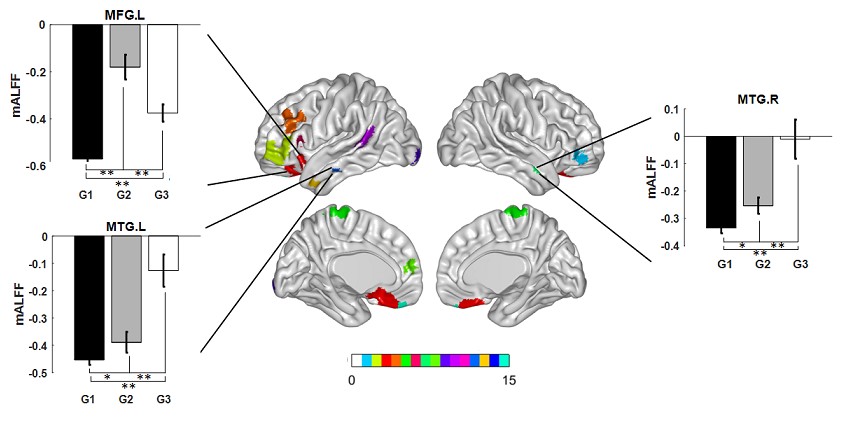
The differences in
ALFF among three groups. G1: healthy control group, G2: ON patients with good visual outcome,
G3: ON patients with poor visual outcome. Significantly
decreased mALFF were observed in left cuneus and left middle occipital gyrus,
right paracentral lobule,
right anterior cingulate cortex, and
bilateral middle temporal gyrus, left middle frontal gyrus in ON
patients than HCs. While increased mALFF
were occurred in several
regions in the frontal and temporal lobes (P< 0.001, FDR corrected).
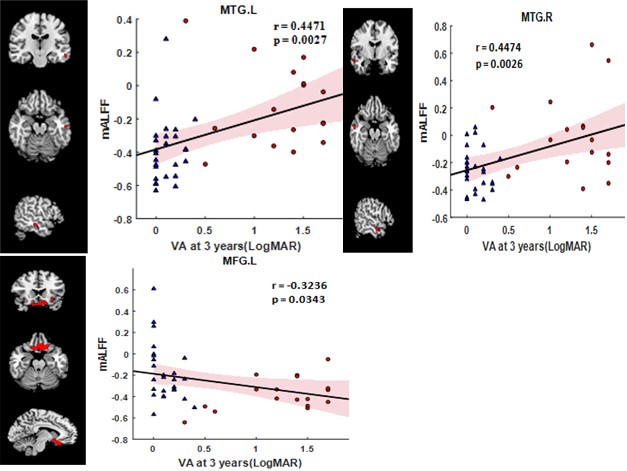
The correlations between the VA and ALFF in ON. The positive correlations were found
in bilateral
middle temporal gyrus and significantly negatively
related to ALFF in left middle frontal gyrus.