1499
Locus coeruleus degeneration is associated with disorganized functional topology in Parkinson’s disease1Zhejiang University, Hangzhou, China
Synopsis
A major emphasis has been placed on the symptom related brain alterations caused by dopamine deficiency, the brain organization underlying the norepinephrine deficiency is largely unknown. We used the neuromelanin sensitive-magnetic resonance imaging for evaluating the degeneration of LC, and using graph theory-based network analysis for characterizing the brain functional topology in 94 PD patients and 68 healthy controls.Relationships between LC degeneration, network disruption and cognitive/motor manifestations in PD patients were assessed.An independent PD subgroup with MRI scanning before and after levodopa administration was enrolled to clarify whether LC degeneration related network disruption were independent of dopamine deficiency.
Objective
Locus coeruleus (LC) degeneration is recognized as a critical hallmark of Parkinson's disease (PD). However, whether LC degeneration contribute to cognitive/motor manifestations through modulating brain functional organization remains unknown, though recent studies reported that LC-norepinephrine have crucial effects on brain functional organization.Methods
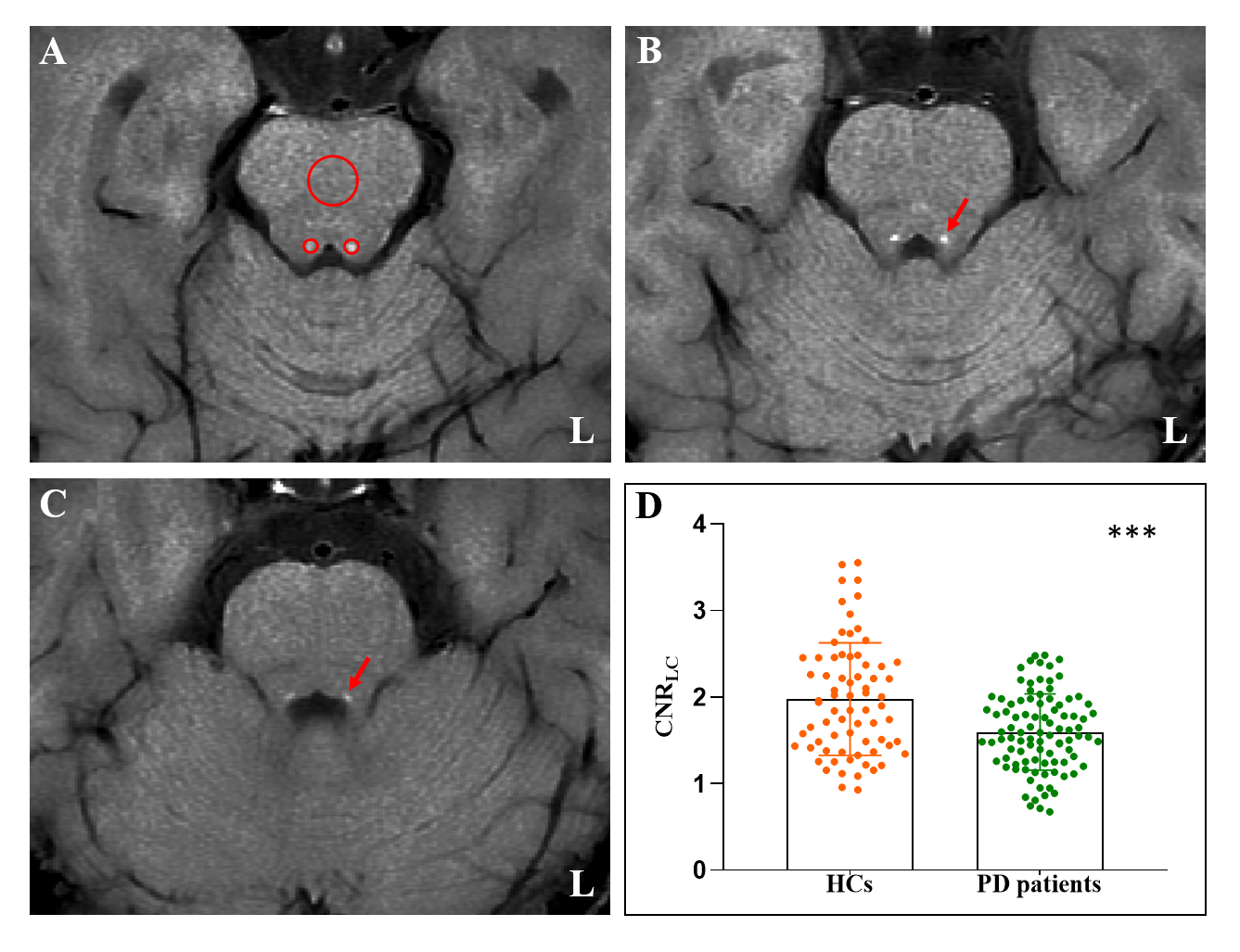
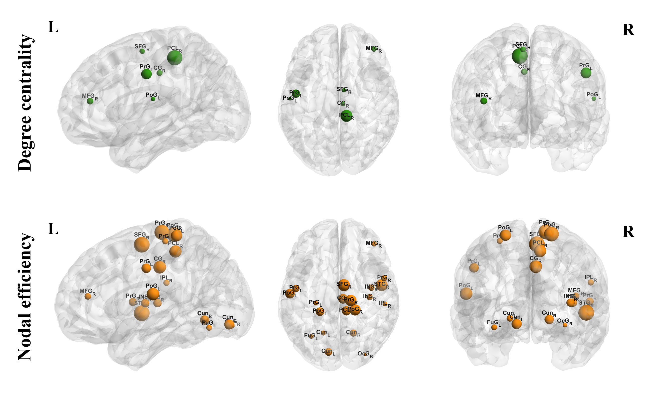
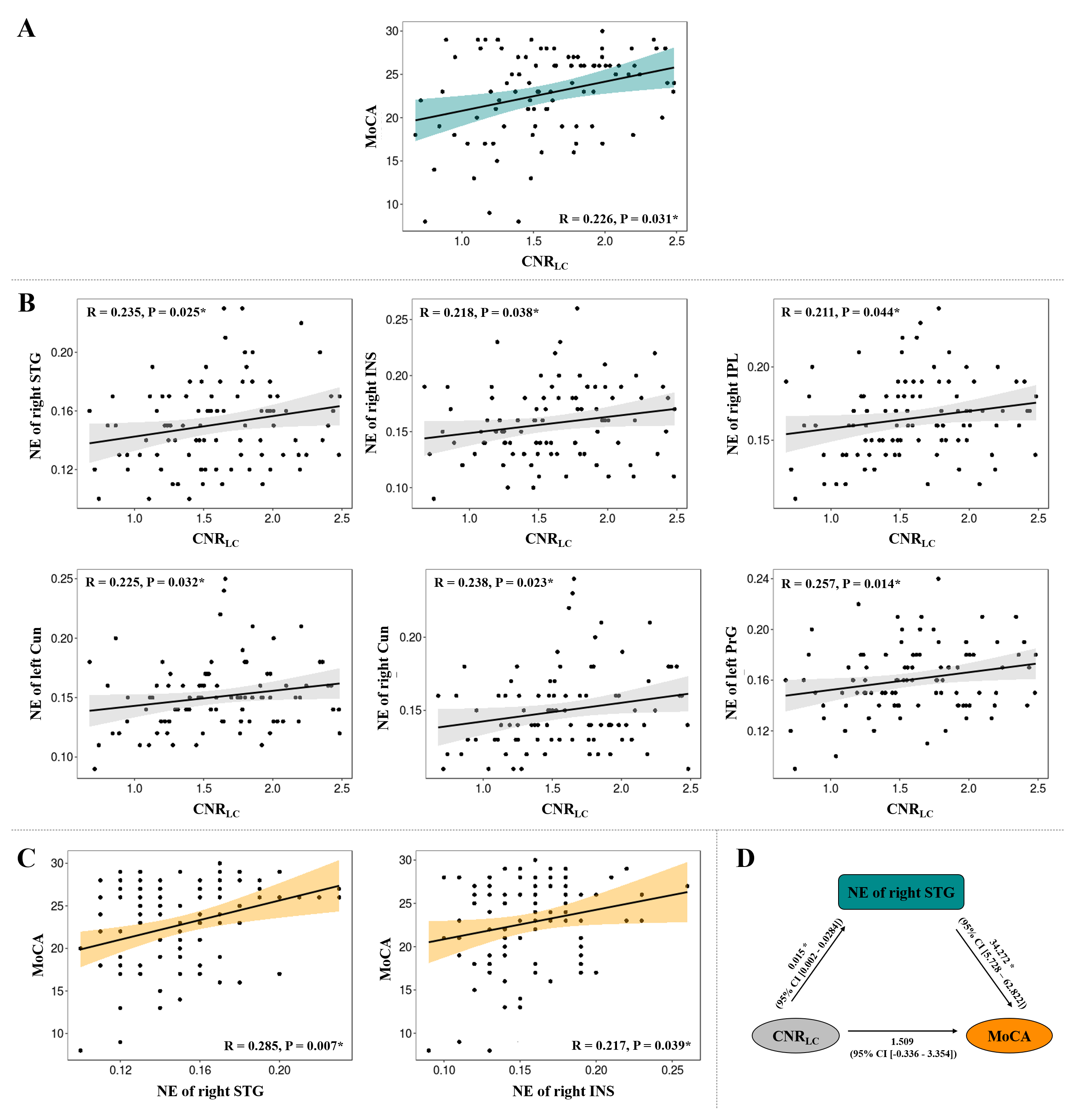
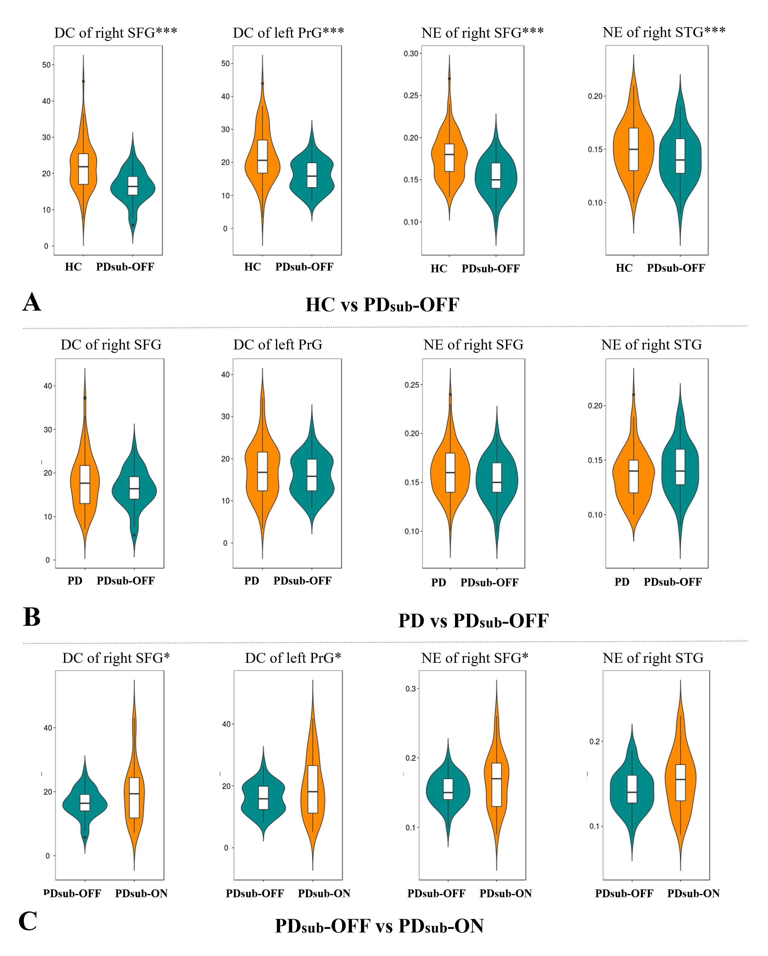
Ninety-four PD patients and 68 healthy controls (HC) were enrolled. LC integrity was measured using the contrast-to-noise ratio of LC (CNRLC) calculated from neuromelanin sensitive magnetic resonance imaging. Graph theory-based network analysis was used to characterize the functional organization (degree centrally, nodal efficiency). Relationships between LC degeneration, network disruption and cognitive/motor manifestations in PD patients were assessed. Mediation analysis was conducted to test whether network disruption was a mediator between LC degeneration and cognitive/motor impairments. An independent PD subgroup (n = 35) with MRI scanning before and after levodopa administration was enrolled to clarify whether those LC degeneration related network attributes were independent of dopamine deficiency.Results
We found significant LC degeneration in PD group (Figure 1). Extensive network disruption in cognitive and motor related cortex were found in PD patients, which is consist with previous knowledge of widespread functional disorganization and have an incremental value (Figure 2). CNRLC was positively correlated with Montreal Cognitive Assessment (MoCA) score (Figure 3A) and the nodal efficiency of several cognitive related regions (Figure 3B). Decreased nodal efficiency in superior temporal gyrus was a mediator between LC degeneration and cognitive impairment in PD patients (Figure 3C, D). And we demonstrated that network attributes of frontal and motor cortex were normalized by levodopa administration while decreased nodal efficiency of superior temporal gyrus (mediator) could not be modulated (Figure 4).Conclusion
Our findings suggested that LC degeneration was a key pathway for PD cognitive decline through associating with the disorganized functional topology.Acknowledgements
We thank all patients with Parkinson’s disease patients and healthy controls who participated in this study.References
1. Betts MJ, Kirilina E, Otaduy MCG, et al. Locus coeruleus imaging as a biomarker for noradrenergic dysfunction in neurodegenerative diseases. Brain 2019;142:2558-2571.
2. Espay AJ, LeWitt PA, Kaufmann H. Norepinephrine deficiency in Parkinson's disease: the case for noradrenergic enhancement. Movement disorders : official journal of the Movement Disorder Society 2014;29:1710-1719.
3. Mather M, Harley CW. The Locus Coeruleus: Essential for Maintaining Cognitive Function and the Aging Brain. Trends in cognitive sciences 2016;20:214-226.
4. Zerbi V, Floriou-Servou A, Markicevic M, et al. Rapid Reconfiguration of the Functional Connectome after Chemogenetic Locus Coeruleus Activation. Neuron 2019;103:702-718 e705.
5. Borchert RJ, Rittman T, Rae CL, et al. Atomoxetine and citalopram alter brain network organization in Parkinson's disease. Brain communications 2019;1:fcz013.
6. Rae CL, Nombela C, Rodríguez PV, et al. Atomoxetine restores the response inhibition network in Parkinson's disease. Brain 2016;139:2235-2248.
7. Joshi S, Li Y, Kalwani RM, Gold JI. Relationships between Pupil Diameter and Neuronal Activity in the Locus Coeruleus, Colliculi, and Cingulate Cortex. Neuron 2016;89:221-234.
8. Oertel WH, Henrich MT, Janzen A, Geibl FF. The locus coeruleus: Another vulnerability target in Parkinson's disease. Movement disorders : official journal of the Movement Disorder Society 2019;34:1423-1429.
9. Sara SJ. The locus coeruleus and noradrenergic modulation of cognition. Nature reviews Neuroscience 2009;10:211-223.
Figures