1494
Differential task-induced brain activation and functional connectivity patterns between OCD and GAD: Preliminary study on verbal memory11Advanced Institute of Aging Science, Chonnam National University, Gwangju, Korea, Republic of, 2Department of Psychiatry, Massachusetts General Hospital and Harvard Medical School, Boston, MA, United States, 3Department of Radiology, Chonnam National University Medical School, Gwangju, Korea, Republic of
Synopsis
This current study supports that abnormalities in task-induced functional connectivity and localized brain activation patterns in explicit, verbal short-term memory processing are potentially linked with cognitive and memory impairment. These findings will be helpful for understanding the differential neural mechanisms associated with the severity of clinical symptoms in OCD and GAD.
Introduction
Although obsessive-compulsive disorder(OCD) has been separated into the category of obsessive-compulsive and related disorder(OCRD) in DSM-5 since 2013, it has an intersectional characteristic of anxiety disorder with generalized anxiety disorder(GAD). To date, therefore, most of neuroimaging studies on the cognitive or memory tasks in patients with OCD has not ruled out the nature of anxiety. There are primary differences between OCD and GAD, such as nature of thought involved and the subjective behavioral response to negative concerns. Thus, this study aimed to compare neural activation patterns and the functional connectivity(FC) underlying verbal memory tasks with explicit recall between patients with OCD and patients with GAD. In a subsequent aim, we statistically evaluated the correlation between task-induced FC and clinical symptom severity scores.Methods
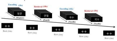
Twelve healthy control participants (mean age, 35.25±7.20 years), 12 patients with OCD (mean age, 29.50±8.42 years), and 12 patients with GAD (mean age, 35.00±9.65 years) participated in this study. All subjects underwent imaging with a 3-T Magnetom Verio MR Scanner (Siemens Medical Solutions, Erlangen, Germany) with an 8-channel birdcage-type of head coil. The high-resolution T1-weighted images were acquired using a three-dimensional magnetization prepared rapid gradient-echo (3D MP-RAGE) sequence with repetition time (TR)/echo time (TE) = 1900/2.35 ms, field-of-view (FOV) = 22×22 cm2, matrix size = 256×256, number of excitations (NEX) = 1, and slice thickness = 1 mm. Functional images were acquired using a gradient-echo echo planner image (GRE-EPI) with the following parameters: TR/TE = 2000/30 ms, flip angle = 90°, FOV = 22×22 cm2, matrix size = 64×64, NEX = 1, number of slices = 25, and slice thickness = 5 mm without a slice gap. A series of explicit memory tasks with two-syllable words was performed as follows: rest (14 s), first encoding (18 s), rest (14 s), first retrieval (18 s), rest (14 s), second encoding (18 s), rest (14 s), second retrieval (18 s), and rest (14 s) (Fig 1). Two fixation crosses were displayed in the rest periods. In each encoding period, six different two-syllable words were displayed for 3 s per slice. In the retrieval periods, either new words or the repeated words were given in the encoding period. Functional imaging data were analyzed by using SPM12 software (Wellcome Department of Cognitive Neurology, London, UK) and CONN-fMRI functional connectivity toolbox (ver.17e).Results
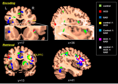
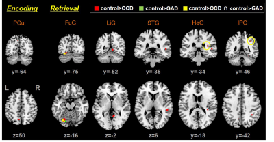
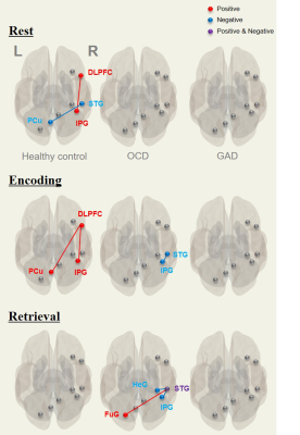
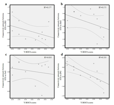
The commonly activated brain areas in all groups during the memory tasks included the insula (Ins) during the encoding period and the dorsolateral prefrontal gyrus (DLPFC) during the retrieval period (p<0.005, t=3.12) (Fig. 2). Compared with healthy controls, the OCD group showed significantly lower activations in the precuneus (PCu) during the encoding period and in the fusiform gyrus (FuG), lingual gyrus (LiG), superior temporal gyrus (STG), Heschl gyrus (HeG), and inferior parietal gyrus (IPG) during the retrieval period (p<0.001, t=3.37), whereas the GAD group showed lower activation in the FuG (p<0.001, t=3.37) during the retrieval period (Fig.3; ANCOVA and post-hoc t-test). We also observed task-induced functional connectivity in response to the verbal short-term memory tasks with explicit recall (Fig. 4). During the rest period, healthy controls showed a positive connection of the DLPFC and IPG, but a negative connection of the STG and PCu. During the encoding period, healthy controls showed positive connections of the DLPFC, IPG, and PCu, and the OCD group showed a negative connection of the STG and IPG. However, no functional connectivity patterns were observed in the GAD group. During the retrieval period, functional connectivity was observed in the OCD group only: a positive connection of the STG and FuG and negative connections of the STG with the HeG and IPG. These observed functional connectivities were negatively correlated with clinical symptom severity in the OCD group (fig 5). It is noteworthy, however, that no such correlations were observed during either the memory retrieval or encoding in the GAD group.Conclusion
This current study supports that abnormalities in task-induced functional connectivity and localized brain activation patterns in explicit, verbal short-term memory processing are potentially linked with cognitive and memory impairment. These findings will be helpful for understanding the differential neural mechanisms associated with the severity of clinical symptoms in OCD and GAD.Acknowledgements
This work was supported by a National Research Foundation (NRF) grant funded by the Korea government (MSIT) (2018R1A2B2006260) and the Chonnam National University Research Fund for CNU Research Distinguished professor (2017-2022).References
1. Koçak, O. M., Özpolat, A. Y., Atbaşoğlu, C., & Çiçek, M. Cognitive Control of a Simple Mental Image in Patients With Obsessive--Compulsive Disorder. Brain Cogn 76, 390-399, https://doi.org/10.1016/j.bandc.2011.03.020 (2011).
2. Coles, M. E., Turk, C. L., & Heimberg, R. G. Memory Bias for Threat in Generalized Anxiety Disorder: The Potential Importance of Stimulus Relevance. Cogn Behav Ther 36, 65-73, https://doi.org/10.1080/16506070601070459 (2007)
Figures
