1445
Radiomics and Machine Learning on multiparametric MRI for breast cancer diagnosis1Radiology, Memorial Sloan Kettering Cancer Center, NEW YORK, NY, United States, 2Memorial Sloan Kettering Cancer Center, New york, NY, United States
Synopsis
Radiomics coupled with machine learning is based on the extraction of signatures from medical images that are invisible to the human eye to create models which would improve breast cancer diagnosis. Radiomics features extracted from dynamic contrast-enhanced MRI and diffusion-weighted imaging can be combined in multiparametric MRI. We hypothesize that radiomics features extracted from multiparametric MRI would allow for an improved model affording a more accurate breast cancer diagnosis. We developed a multiparametric model that achieved the best accuracy for breast cancer diagnosis compared to models based on dynamic contrast-enhanced MRI or diffusion-weighted imaging.
INTRODUCTION
Dynamic contrast-enhanced MRI (DCE-MRI) is an image modality with very high sensitivity but low specificity for breast cancer diagnosis (1). The reason is the overlap in the image characteristics of benign and malignant enhancing lesions which entails a high number of false positive diagnoses and unnecessary biopsies. Multiparametric MRI with diffusion-weighted imaging (DWI) has demonstrated to increase the specificity for breast cancer diagnosis compared to DCE-MRI alone (2). Currently, radiomics coupled with machine learning (ML) has mainly focused on extracting features from DCE-MR images with promising results (3). However, a multiparametric approach could further improve breast cancer diagnosis. With only a few published studies investigating the performance of radiomics in multiparametric MRI (4) (5) (6), the aim of our work was to evaluate the diagnostic value of radiomics analysis coupled with ML on multiparametric MRI for the evaluation of suspicious enhancing breast tumors.METHOD AND MATERIALS
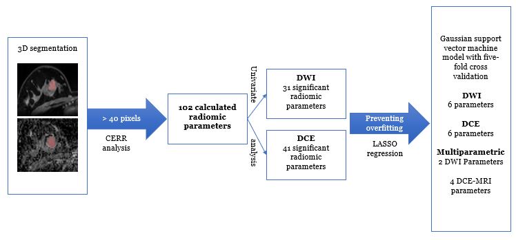
In this IRB- approved retrospective study 93 women with 104 biopsy-proven suspicious breast lesions (58 benign, 46 malignant) were included. Three-dimensional tumor segmentation was conducted on DCE-MR images from first post-contrast T1-weighted and ADC maps derived from DWI by two dedicated breast radiologists with five years of experience in breast imaging. Radiomics features derived from DCE and DW images were calculated using the publicly available computational environment for radiological research software based on first-order statistics, gray level co-occurrence matrix (GLCM), run length matrix (RLM), size zone matrix (SZM), neighborhood gray level dependence matrix, and neighborhood gray tone difference matrix. Univariate and multivariate analysis were performed to identify significant radiomic features to be included in a ML model to discriminate between malignant and benign breast lesions. Least absolute shrinkage and selection operator (LASSO) regression was performed to prevent overfitting and a medium Gaussian support vector machine model with five-fold cross validation was employed to develop the predictive models. A flowchart of the radiomics analysis is shown in Figure 1. Measures of sensitivity, specificity, negative predictive value (NPV), positive predictive value (PPV) and accuracy were estimated for DCE-MRI, DWI and multiparametric MRI models. Classification performance was evaluated using the receiver operating characteristic curve.RESULTS AND DISCUSSION
102 radiomics parameters were calculated. Subsequently, univariate modelling rendered 31 and 41 significant radiomic parameters for ADC and DCE-MRI respectively. After LASSO regression and multivariate modelling, 6 parameters from DCE-MR images (entropy, total energy, zln, Joint Variance, inverse variance and coarseness) and 6 parameters from the DW images (kurtosis, min, entropy, gln Normalized sze and Irhgle) were used into a medium Gaussian support vector machine model with five-fold cross validation to develop a final robust ML model. We achieved sensitivities of 79.6%, 76.3% and 83.4% and specificities of 69.7%, 78.2% and 81.5% for ADC, DCE-MRI and multiparametric MRI models respectively. PPV and NPV were 68.4%, 74.3%, 78.8 % and 80.6 %, 80.1%, 85.6% for ADC, DCE-MRI and multiparametric MRI models respectively. The diagnostic accuracy was 74.2 %, 77.4 % and 82.4% for ADC, DCE-MRI and multiparametric MRI models respectively. The area under the curve (AUC) was 0.80, 0.84 and 0.86 for ADC, DCE-MRI and multiparametric MRI models respectively. Radiomics features extracted from multiparametric MRI allowed the development of a model which maximized diagnostic accuracy and AUC. These results are in accordance with other group who developed a multiparametric scheme that achieved an AUC of 0.87 outperforming DCE (4). Other groups have incorporated pharmacokinetic analysis to the multiparametric model (5) and DWI kurtosis (6) achieving even better results with AUCs over 0.9. The multiparametric model achieved a better specificity and PPV compared to the other two models which indicates that multiparametric models not only have the potential to aid in breast cancer diagnosis, but also in clinical decision making to prevent unnecessary biopsies.CONCLUSIONS
We developed a radiomic model derived from multiparametric MRI that yielded the best diagnostic results for breast cancer diagnosis maximizing diagnostic performance and achieving the best accuracy, highest specificity and PPV compared to radiomic models based solely on DCE and DWI extracted features. Radiomics coupled with ML combining features extracted from multiparametric MRI may help improve breast cancer diagnosis and prevent unnecessary breast biopsies.Acknowledgements
This work was financially supported by the NIH/NCI Cancer Center Support Grant (P30 CA008748), the Breast Cancer Research Foundation, and the Spanish Foundation Alfonso Martin Escudero.References
1. Mann RM, Kuhl CK, Moy L. Contrast-enhanced MRI for breast cancer screening. J Magn Reson Imaging. 2019;50(2):377–90.
2. Marino MA, Helbich T, Baltzer P, Pinker-Domenig K. Multiparametric MRI of the breast: A review. J Magn Reson Imaging. 2018;47(2):301–15.
3. Lee S-H, Park H, Ko ES. Radiomics in Breast Imaging from Techniques to Clinical Applications: A Review. Korean J Radiol. 2020 Jul;21(7):779–92.
4. Hu Q, Whitney HM, Giger ML. A deep learning methodology for improved breast cancer diagnosis using multiparametric MRI. Sci Rep. 2020 29;10(1):10536.
5. Parekh VS, Jacobs MA. Integrated radiomic framework for breast cancer and tumor biology using advanced machine learning and multiparametric MRI. NPJ Breast Cancer. 2017;3:43.
6. Zhang Q, Peng Y, Liu W, Bai J, Zheng J, Yang X, et al. Radiomics Based on Multimodal MRI for the Differential Diagnosis of Benign and Malignant Breast Lesions. J Magn Reson Imaging. 2020;52(2):596–607.
Figures