1428
Reduction of coupling and noise by ultrahigh dielectric constant (uHDC) materials for phase array coil at 3T1Neurosurgery, Pennsylvania State University, Hershey, PA, United States, 2Radiology, Pennsylvania State University, Hershey, PA, United States, 3Pennsylvania State University, Hershey, PA, United States, 4Center for Magnetic Resonance Research, Department of Radiology, University of Minnesota, Minneapolis, MN, United States, 5Materials Science and Engineering, Pennsylvania State University, State College, PA, United States
Synopsis
Prior work has examined the effects of uHDC material upon phased array coils. Several beneficial effects have been observed: reduction of per-channel noise and inter-channel noise correlations, and enhancement of SNR. Due to the high dielectric constant the electric field will be confined to the dielectric material, which leads to a reduction of the primary noise source, the conservative electric field. As a result the non-conservative field becomes more dominant within the sample, with a resulting increase of B1 field intensity and more substantial SNR enhancement.
Introduction
It has been shown that the usage of ultra-high dielectric materials (uHDC) with RF coils can enhance the B1 field and signal-to-noise ratio (SNR) across a range of human magnets (3T - 10.5T) due to the strong displacement current induced in the uHDC material(1,2). In addition, a significant reduction of noise by the uHDC material has also been observed as a contributing factor to the SNR enhancement(3,4). This de-noising effect is thought to be induced by the reduction of the conservative electric field within the sample by uHDC materials(3). The goal of this investigation is to determine this effect upon receive array coils commonly used for clinical MRI at 3T.Methods
Theory: The electric field created by the RF coil consists of two components, a conservative and a non-conservative field:$$ \overrightarrow{E}_{total} = \overrightarrow{E}_{Conservative} + \overrightarrow{E}_{NonConservative} = -\overrightarrow{\triangledown\phi} - \frac{\partial \overrightarrow{A}}{\partial t} (1)$$
where $$$\phi$$$ is the electric scalar potential, $$$\overrightarrow{A}$$$ is the vector potential. Both E-field components produce currents that induce B field and losses in the tissue samples during transmission and reception. However, the conservative E-field is undesirable because it does not interact with the magnetization of the nuclear spin system and only generates noise. We have shown as a proof of concept that using uHDC material, the conservative E-field in the sample can be reduced, leading to a noise reduction and ultimate SNR improvement with a surface coil. The goal of this study is to demonstrate and apply this effect for a commonly used 3T 4 channel surface phased array coil.
Experiment: A 4 channel Flex array with each coil dimension 20 cm x 11 cm was used on a 3T system (PrismaFit, Siemens Healthineers, Erlangen, Germany). Data were acquired both without and with uHDC ceramic blocks with εr~880 (Figure 1). The dimension of the uHDC blocks was 8.7 x 10.8 x 2.7 (W x L x H cm3). Four blocks were placed in the middle of each loop. A 2D interleaved spoiled GRE sequence (2 x 2 x 4 mm3, FA = 2 degrees) with integrated noise pre-scans was acquired to calculate SNR maps. Flip angle maps were acquired with a 2D pre-saturated TurboFlash sequence(5) (Saturation Angle = 60 degrees), with scan geometry matched to that of the GRE scan. The noise standard deviation and noise correlation matrices were calculated from the noise pre-scans for both baseline and uHDC blocks. The SNR was calculated with the SNR Units Method divided by the B1+ map to remove the flip angle effects(6).
Results and Discussion
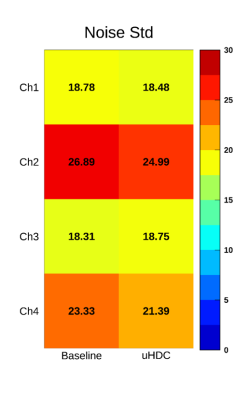
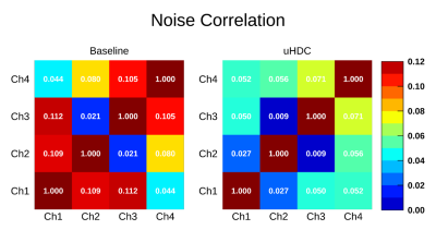
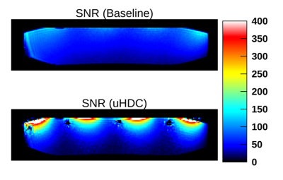
As can be seen in Figure 2, in the presence of the uHDC the Noise Standard Deviation (Std) for each individual coil element was reduced. The noise correlation between adjacent coil elements showed an even more significant reduction with uHDC material (Figure 3). This effect of the uHDC material is due to the shielding of the conservative E component by the uHDC material (3). The induced E field not only decreases the conservative component and associated noise but also create a strong displacement current that increases the B1 field within the sample. Therefore, the SNR increases synergistically. As shown in Figure 4 the SNR was increased up to 4-fold next to the surface of the phantom. However, an SNR drop around the edges of the dielectric material was also observed.Conclusion
We demonstrated experimentally that employing the uHDC materials in a standard clinical phase array coil could reduce the total noise as well as enhance the B1 field and thereby increase the SNR substantially. We showed that proper placement of uHDC materials with respect to the phase array coil could effectively reduce the noise correlation by decreasing the conservative E component.Acknowledgements
This work is supported by NIH grant U01EB026978.References
1. Sica CT, Rupprecht S, Hou RJ, Lanagan MT, Gandji NP, Lanagan MT, Yang QX. Toward whole-cortex enhancement with a ultrahigh dielectric constant helmet at 3T. Magnetic Resonance in Medicine 2019;0(0).
2. Navid P. Gandji, Myung-Kyun Woo, Sebastian Rupprecht, Michael Lanagan, Bei Zhang, Riccardo Lattanzi, Russell L. Lagore, Jerahmie Radder, Gregor Adriany, Kamil Ugurbil, Brian Rutt a, Yang QX. The displacement current wave on a high dielectric constant (HDC) helmet and its effect on B1 Field at 10.5 T (447 MHz). Virtual 2020.
3. Chen W, Lee BY, Zhu XH, Wiesner HM, Sarkarat M, Gandji NP, Rupprecht S, Yang QX, Lanagan MT. Tunable Ultrahigh Dielectric Constant (tuHDC) Ceramic Technique to Largely Improve RF Coil Efficiency and MR Imaging Performance. IEEE Transactions on Medical Imaging 2020;39(10):3187-3197.
4. Navid Pourramzangandji , Christopher Sica , Byeong-Yeul Lee , Hannes Wiesner , Maryam Sarkarat , Michael Lanagan , Xiao-Hong Zhu , Wei Chen , Yang Q. Large denoising effect of ultrahigh dielectric constant (uHDC) materials. ISMRM. Virtual2020.
5. Chung S, Kim D, Breton E, Axel L. Rapid B1+ mapping using a preconditioning RF pulse with TurboFLASH readout. Magn Reson Med 2010;64(2):439-446.
6. Kellman P, McVeigh ER. Image reconstruction in SNR units: A general method for SNR measurement†. Magnetic Resonance in Medicine 2005;54(6):1439-1447.
Figures


