1402
UVC Based Wireless Patient Bore Disinfection Utilizing Scanner RF Transmission
Devavrat Likhite1, Rob Amerling1, Leon Lee1, and Saban Kurucay1
1GE Healthcare, Waukesha, WI, United States
1GE Healthcare, Waukesha, WI, United States
Synopsis
In the recent times, cleaning protocols after every patient, regular disinfection of medical equipment and use of single-use devices have become a common practice. MRI equipment has been one of the most difficult imaging modalities to clean. Here, we present a simple technique that uses UV light to disinfect the MRI system, without the use of any external power or connecting cables. This technique uses the power from the MRI system and thereby provides a simple and wire-free UV disinfection solution that does not need external electrical source.
Background
Magnetic Resonance (MR) imaging equipment has been one of the most difficult imaging modalities to clean. This can be a time-consuming task for the MR technician, trying to single-handedly scan patients in limited time. Moreover, the long patient bore coupled with the presence of a strong magnetic field severely restricts the options available for cleaning and disinfection of the system after every patient.Ultraviolet (UV) light has been used for disinfection over a few decades now. The entire UV spectrum can be divided into three bands, namely, UVA (315-400nm), UVB (280-315nm) and UVC (200-280nm)1. Of these three bands, UVC has been known to possess germicidal disinfection properties, due to its ability to hinder replication by affecting the DNA/RNA of the microbes.
In this article, we present a technique that uses UV light to disinfect the MRI system, without the use of any external power or connecting cables. This technique harnesses the electric fields generated by the MR system and thus provides a simple and wire-free UV disinfection solution. Since the presented method uses no additional hardware besides the UVC generating tubes, it can be replicated easily at a low cost.
Methods
In previous studies, researchers have determined the UVC dosage required to inactivate various pathogens 2. It is reported that a UVC dose >40mJ/cm2 is sufficient to achieve reasonable inactivation of the most common pathogens that may be present on the MR equipment.Moreover, UVC has been previously shown to be effective at inactivation of different members of the coronavirus family i.e. SARS-CoV-1 and MERS-CoV 3,4. Although the scientific community is still learning about SARS-CoV-2, it is believed that a similar result can be expected while using UVC against the COVID-19 virus 5 .
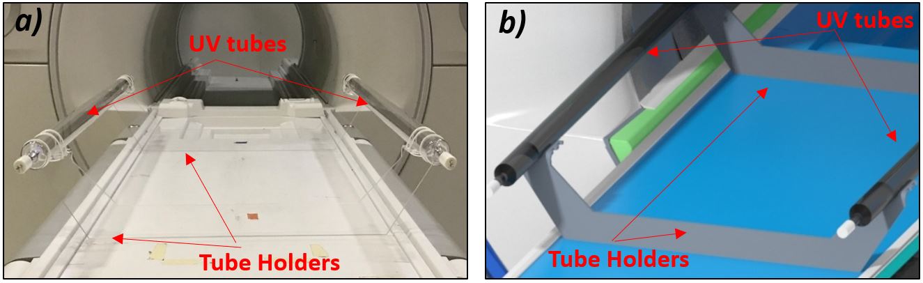
The technique presented, uses a simple UVC fixture constructed with materials available for purchase online.
- GX48L UVC lamps – Qty. 2. Source : 6
- UVC 100 dots (disposable dosimeter stickers) – Qty. As per need. Source : 7 [Figure 2]
- Tube holder -- Made in-house by cutting and bending readily available transparent polycarbonate. Design drawing shared at 8
Six UVC dosimeter stickers were positioned along the inner circumference of the bore-wall and the patient table. These dosimeter stickers were placed ~50cm along the Z-axis from the start of the scanner bore. Figure 3 shows an axial schematic of this placement. A landmark was placed at the center of the fixture. A 2D Gradient Echo sequence was prescribed with TR=12 ms, TE=7ms, FOV=40cm, 256x256 resolution, Receive Bandwidth =16kHz, Slice thickness= 10mm. In our experiments we used the same protocol for both 1.5T and 3.0T scanners. However, changed the flip angle of the excitation pulse to 180 degrees for the 1.5T system. Note that the goal of the protocol is to maximize the rms E field delivered to the UVC probes. Further details of the imaging parameters can be found at 8 .
The protocol was prescribed, and the Transmit Gain was manually set to maximum using the manual pre-scan window. The scan was run for 2-minutes on a 3T system and for 6-minutes on a 1.5T system. Figure 4 shows the operational fixture disinfecting the bore and patient table on a 3T Signa Premier system.
The amount of UVC dose delivered was measured using the disposable dot shaped UVC dosimeter stickers 7. The UVC dosimeter stickers used in this testing were able to indicate a UVC dose of 25mJ/cm2, 50mJ/cm2, 75mJ/cm2 and 100mJ/cm2. Figure 2 shows the dose reference chart received with the dosimeter stickers.
Results
After the scan duration, the disposable dosimeter stickers were carefully removed. The changed color of the dosimeter stickers was compared to the UVC dose reference chart (Figure 2) for a quantitative estimation of the delivered dose. Table 1 shows the pictures and corresponding UVC dose measured on various GE MR systemsConclusion
The findings show that the technique presented in this article can deliver a UVC dose greater than the required 40 mJ/cm2 in 2-minutes on a 3T GE MR system and in 6-minutes on a 1.5T GE MR system. Although, our study was limited to GE 70cm bore configurations, utilizing the methodology presented here, it is straightforward to experimentally optimize for other commercially available 1.5T and 3.0T systems. The fixture is lightweight and easy to use.Acknowledgements
No acknowledgement found.References
- "https://iuva.org/What-is-UV," [Online].
- A. &. M. M. &. C. B. &. B. J. Malayeri, "Fluence (UV Dose) Required to Achieve Incremental Log Inactivation of Bacteria, Protozoa, Viruses and Algae.," IUVA News, pp. 4-6, 18.
- M. E. D. et.al., "Inactivation of the coronavirus that induces severe acute respiratory syndrome, SARS-CoV," Journal of Virological Methods, vol. 121, no. 1, pp. 85-91, 2004.
- M. H. et.al., "Ultraviolet irradiation doses for coronavirus inactivation – review and analysis of coronavirus photoinactivation studies," GMS Hygiene and Infection Control, vol. 15, p. Doc08, 2020.
- C. S. H. et.al., "Susceptibility of SARS-CoV-2 to UV irradiation," American Journal of Infection Control, vol. 48, no. 10, pp. 1273-1275, 2020.
- "https://www.buyultraviolet.com/ultraviolet-lamps-uv-bulbs-gx48l-05-1311-r?gclid=CjwKCAjwt-L2BRA_EiwAacX32UMgXNqEJeb5--l2RWM81h3Q3UP22qqceXGa-x6JaElYMpqpsCEvGhoCYb8QAvD_BwE," [Online].
- "https://uvcdosimeters.com/uvc-100-dosimeter/," [Online].
- "https://www.dropbox.com/s/tkmyu6b0014ufz3/ISMRM%20Online%20Content_Drawing-Prototcol.pdf?dl=0," [Online].
Figures
Figure
1 a) Assembled fixture on a Signa
Premier system. b) 3D
rendering of the fixture
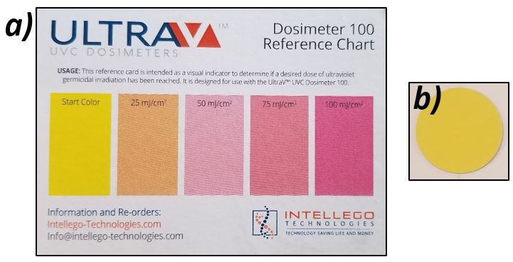
Figure 2 a) UVC dose
reference chart for the UVC dots 100 7.
b) Initial appearance of the UVC dot (dosimeter sticker)
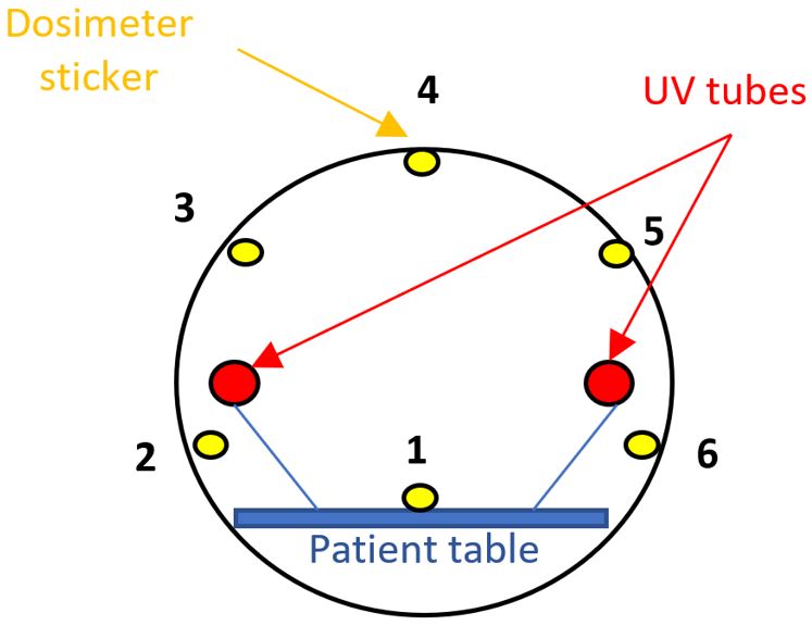
Figure
3 An axial cross-section schematic of the bore for the test
setup
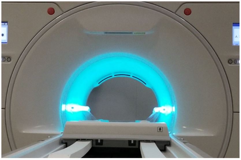
Figure
4 UVC fixture operating and disinfecting the inner surfaces of the
bore and patient table on a 3T Signa Premier system
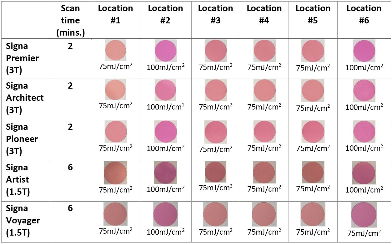
Table
1 shows the used UVC dots and corresponding UVC dose measured on various GE MR
systems(Color representation
in the table may not be accurate due to image capture challenges. Refer to the corresponding
numerical dose value presented)