1386
High Resolution PET/MR Imaging Using Anatomical Priors & Motion Correction1Radiology, Stanford University, Stanford, CA, United States, 2Engineering Dept., GE Healthcare, Waukesha, WI, United States, 3Bioengineering, Stanford University, Stanford, CA, United States, 4Neurology, Stanford University, Stanford, CA, United States
Synopsis
Using the anatomical priors in PET image reconstruction by exploiting the correlation between similar voxels in addition to the correlation between neighboring voxels to control noise, will improve the image quality and resolution. However, motion can degrade the outcome if they anatomical priors are not perfectly registered with PET images. Here we have combined PET image reconstruction with anatomical priors and rigid motion correction for PET/MR brain images to address this. The results show improved image resolution in addition to higher signal-to-noise ratio.
Objectives
Recently a new PET image reconstruction method has been introduced to use anatomical priors by updating the penalty function of penalized maximum likelihood PET reconstruction methods such as Block Sequential Regularized Expectation Maximization (BSREM).1 The penalty function is updated to exploit the correlation between similar voxels (determined from anatomical priors) in addition to the correlation between neighboring voxels. However, just like all other PET reconstruction methods using anatomical priors, this method relies on the co-registration of anatomical priors and PET images. Here we have combined this method with another recently published method for rigid motion correction of PET/MR brain images2 to address the co-registration problem. We will show how combining these two methods can improve the quality and image resolution of PET/MR brain images.Methods
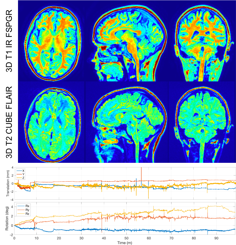
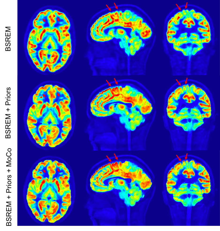
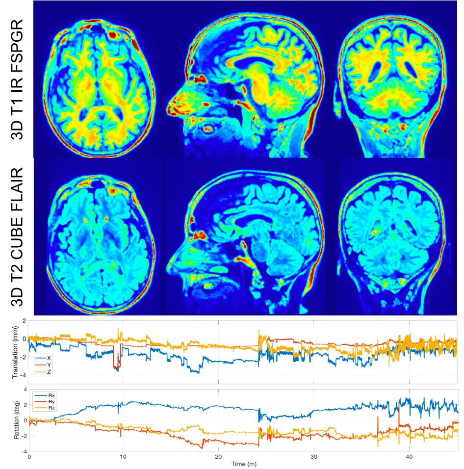
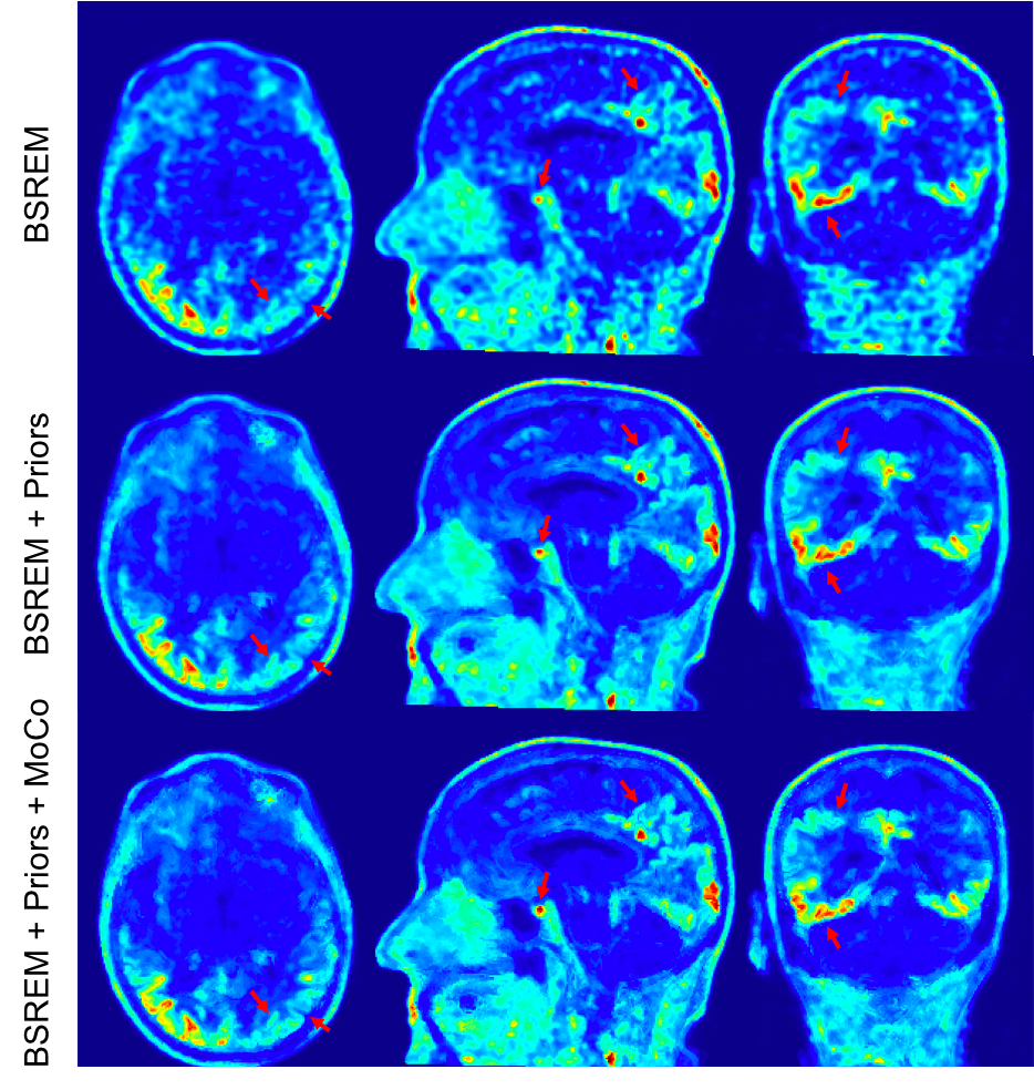
One subject was injected with 15 mCi of 11C-UCB-J and underwent a 90-minute brain scan on a SIGNA PET/MR (GE Healthcare, Waukesha, WI). Another subject was injected with 8.5 mCi of 18F-PI2620 (tau tracer) and underwent a 30-minute brain scan after 45 min uptake time. Both studies were approved by Stanford's Institutional Review Board and all subjects provided written consent. 3D T1 IR FSPGR and 3D T2 FLAIR CUBE images were acquired simultaneously with PET. The head motion was recorded for both subjects using the HobbitView camera.2 The PET images were reconstructed with TOF-BSREM (β = 25) with 1 mm isotropic resolution. The anatomical images were co-registered with the PET volume and corrected for background bias and normalized to their mean intensity over all tissues. Using the anatomical priors, the PET images were reconstructed with MRgTOF-BSREM method1 with β=15 and βm=150. Then using the recorded motion from the HobbitView camera and the anatomical priors, the PET images were reconstructed with MRgTOF-BSREM method1 (β=15, βm=150) and motion correction.2 Finally all PET reconstructed images were co-registered with the motion corrected PET images for comparison.Results
Figures 1 and 3 show the anatomical images and recorded motion plots for the first (injected with 11C-UCB-J) and the second (injected with 18F-PI2620) subjects. The anatomical images are corrected for bias and co-registered to PET images. Figures 2 and 4 show a comparison of PET images reconstructed with TOF-BSREM (β=25), MRgTOF-BSREM (β=15, βm=150) and MRgTOF-BSREM (β=15, βm=150) with motion correction. MRgTOF-BSREM images show improved signal-to-noise ratio (SNR) and resolution compared to TOF-BSREM as expected;1 however, motion correction has improved the image resolution and sharpness in both subjects. The first subject’s PET images show more improvement with motion correction because there are more counts (and therefore more SNR) due to the higher dose, increased uptake, and longer scan-time.Discussions
Anatomical priors can be used in PET image reconstruction to improve the image resolution and control image noise1; however, having perfectly matched anatomical priors with PET events will improve the outcome image quality. Motion can be recorded by an external camera or can be estimated directly from PET data. We have demonstrated the use of anatomical priors and motion correction on PET/MR brain imaging and future work would address other anatomies.Acknowledgements
GE Healthcare.References
1. Khalighi MM, Deller T., et Al. High Quality Isotropic Whole-body PET Imaging Using MR Priors. J Nucl Med May 1, 2020 vol. 61 no. supplement 1 1477
2. Spangler-Bickell M. et al., "Rigid Motion Correction for Brain PET/MR Imaging using Optical Tracking", IEEE Transactions on Radiation and Plasma Medical Sciences, Oct 2018.
Figures