1379
Measuring extracranial magnetic field changes due to head motion during multi-slice EPI acquisition1Sir Peter Mansfield Imaging Centre, University of Nottingham, Nottingham, United Kingdom
Synopsis
A novel approach to motion tracking has been successfully tested during a EPI scan in a 7T scanner. A set of NMR field probes, placed in between the standard head transmit and receiver coils, was used to measure the extra-cranial field changes due to head movement every 150 ms during quiet periods of the EPI sequence. These measurements were used in conjunction with a regression method to predict head motion parameters. This represents a step forward in integrating a marker-less motion correction technique with standard imaging.
Introduction
In previous work we used the 16 probes of an NMR field camera1 to measure the extra cranial magnetic field changes produced by head movement2. By comparing the field changes to simultaneous measurements of head movement made using an optical camera, the association between field change and head position could be learned, allowing subsequent prediction of head movement based on the field measurement alone. However, in the previous experiments the field probes were mounted in a holder that replaced the 32-channel receiver coil, so that imaging performance was compromised, and measurements were made in the absence of imaging. Here we therefore extend the previous work by making measurements of head movement and extra-cranial field changes in quiet periods of a standard multi-slice EPI acquisition, with the field probes sited between the transmit and receiver RF head coils. This represents a step forward in integrating a marker-less motion correction technique with standard imaging.Methods
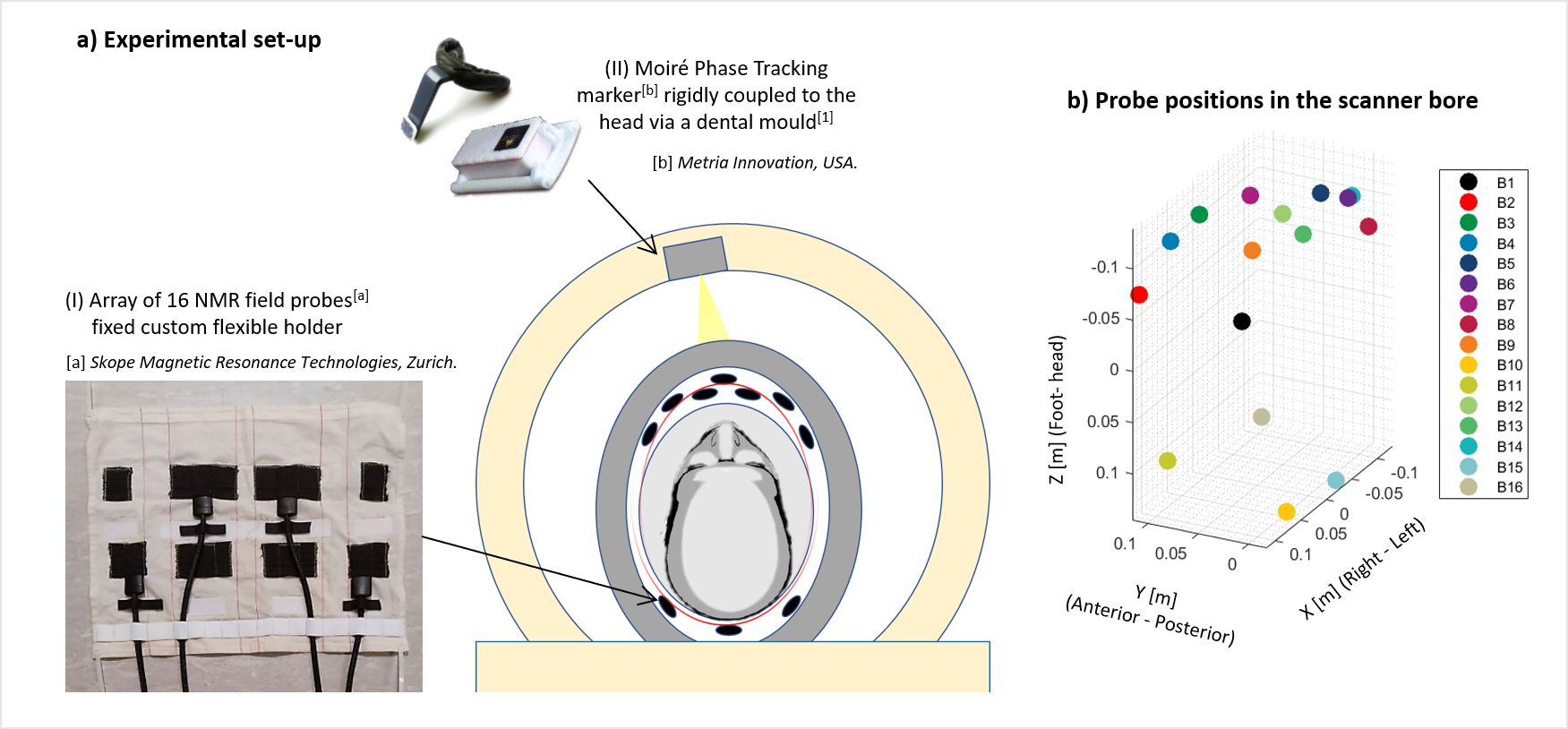
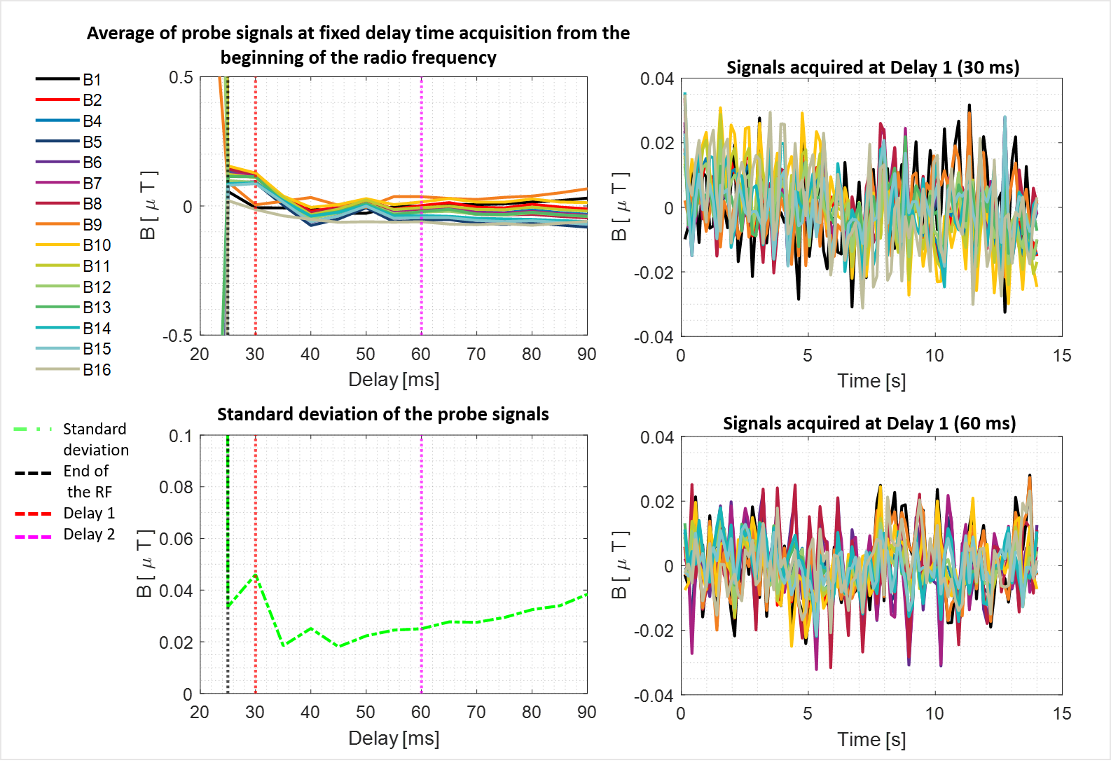
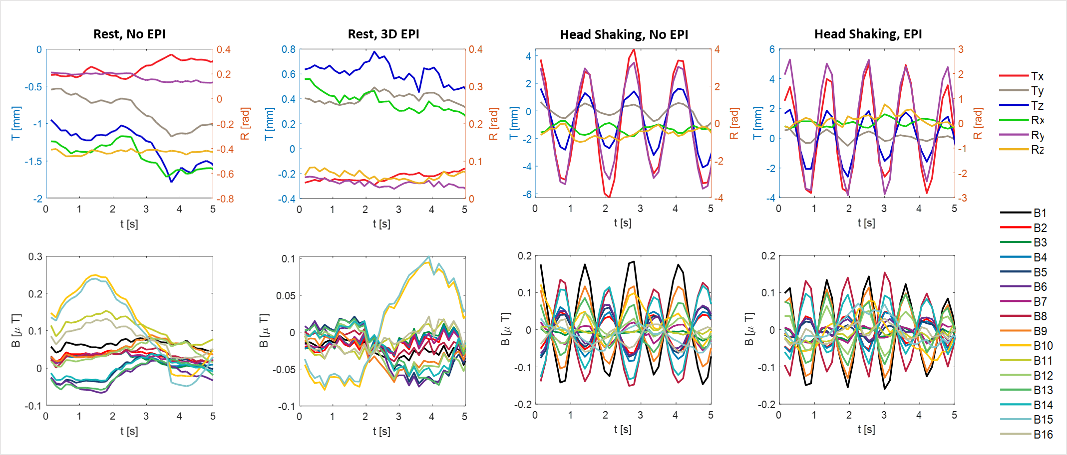
The field probes were held in a customized probe holder which was positioned in the small space were between the cylindrical volume transmit and 32 channel helmet-shaped receiver coil of the 7T Philips Achieva scanner (Figure 1.a). Images were acquired using a multi-slice EPI sequence (48 slices, 3 mm isotropic resolution, TR = 3.6s, TE = 20 ms) with a slice TR of 75 ms. The field camera was triggered to make field measurements 30 ms after the RF excitation every 150 ms (i.e. in a quiet period of the sequence after every second slice acquisition). The usable timing of the acquisition was found by sampling the magnetic field at different delays (Figure 2) to find a minimum delay at which the probe signals are not significantly affected by eddy currents from the image acquisitions. In order to test the motion detection technique, an optical camera was fixed into the magnet bore and used to track a marker fixed onto a customized mouthpiece fixed to the subject’s teeth using a dental mould. Simultaneous measurements of head motion parameters and magnetic field changes were acquired with and without simultaneous scanning while the subject performed several types of head movement (rest, head shaking and head nodding). Head motion parameters were then predicted from the extra-cranial field measurements using a non-linear method2.Results
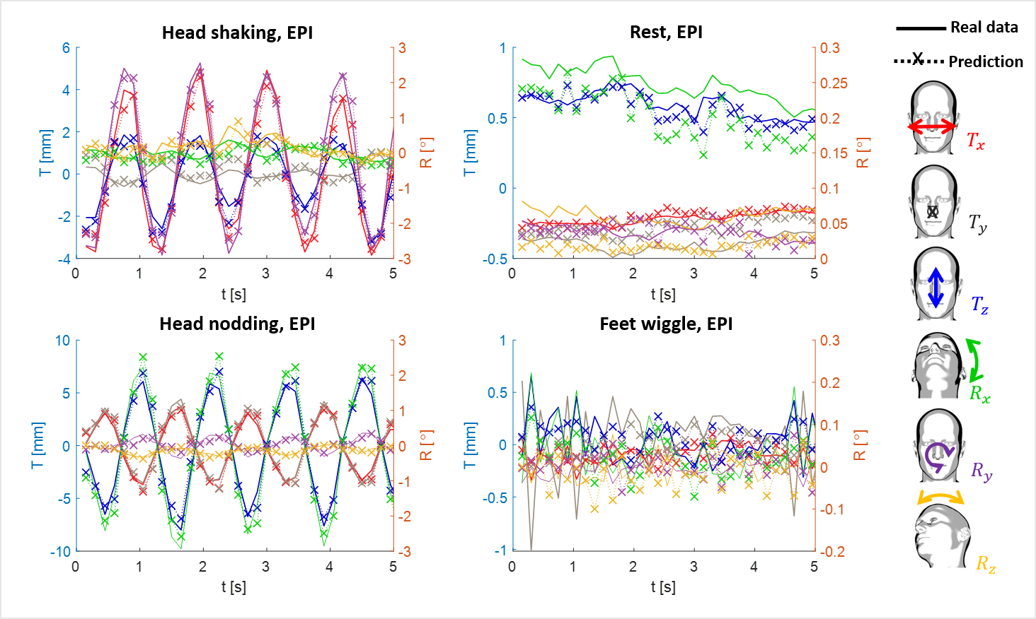
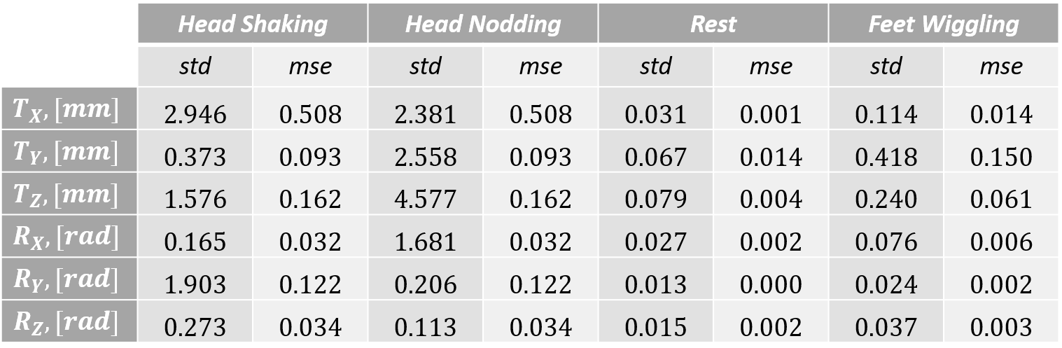
The delay was chosen equal to 30 ms as the probe signal characteristics are similar to those found in the absence of scanning at this delay (Figure 2). The magnetic field sampled during the EPI sequence is similar to that measured for similar motion in the absence of scanning (Figure 3). The time series acquired was then used to perform a prediction on the head motion parameters. (Figure 4). Results (Table 1) shows that the prediction was successful and with a good accuracy.Discussion
The results reported here confirm that measurements of extra-cranial field changes made with a field camera can be used to monitor head position in a 7T MRI scanner during EPI scan acquisition. A non-linear (NARX) regression method provides good prediction over a range of different movements of head motion parameters. This approaches require the acquisition of less than 20 minutes of training data where head movement parameters are measured with a different approach (here, an optical camera and an MPT marker attached to a dental mould have been used) in concomitance as field variation is measured.Conclusions
Head motion during EPI scanning can be predicted by using a field camera to make interleaved measurements of the extra-cranial field changes due to head movement. The method currently needs to be appropriately trained before application using a secondary instrument to measure head motion parameters, but in future this training could be based on analysis of recorded image data. This work represents a step towards the full development of a marker-less technique for head motion tracking that doesn’t not require modification to the image acquisition sequence.Acknowledgements
No acknowledgement found.References
1. Dietrich BE, Brunner DO, Wilm BJ, et al. A field camera for MR sequence monitoring and system analysis. Magnetic Resonance in Medicine (MRM). 2015.
2. Bortolotti L, Bowtell R. Measurement of head motion using a field camera in a 7 T scanner, ISMRM 2020, Abstract number: 0464.
3. Wezel J, Boer VO, van der Velden TA, et al. A comparison of navigators, snap-shot field monitoring, and probe-based field model training for correcting B_0 induced artifacts in T_2 weighted images at 7T. Magnetic Resonance in Medicine. November 2017;78:1373-1382.
4. Babayeva M, Kober T, Knowles B, et al. Accuracy and Precision of Head Motion Informationin Multi-Channel Free Induction Decay Navigatorsfor Magnetic Resonance Imaging. IEEE TRANSACTIONS ON MEDICAL IMAGING. September 2015;34:1879.
5. Eschelbach M, Aghaeifar A, Bause J, et al. Comparison of prospective head motion correction with NMR field probes and an optical tracking system. Magnetic Resonance in Medicine (MRM). July 2018.
6. Aranovitch A, Haeberlin M, Gross S, et al. Motion detection with NMR markers using real‐time field tracking in the laboratory frame. Magnetic Resonance in Medicine. December 2019;84:89-102.
7. Zanche ND, Barmet C, Nordmeyer‐Massner JA, Pruessmann KP. NMR probes for measuring magnetic fields and field dynamics in MR systems. Magnetic Resonance in Medicine (MRM). June 2008.
8. Maclaren J, Herbst M, Speck O, Zaitsev M. Prospective motion correction in brain imaging: a review. Magneti Resonance in Medicine. 2013;69:621-636.
9. Aranovitch A, Haeberlin M, Gross S, et al. Prospective motion correction with NMR markers using only native sequence elements. Magnetic Resonance in Medicine (MRM). August 2017.
Figures