1369
SMS-EPI real-time motion correction by receiver phase compensation and coil sensitivity interpolation1Department of Diagnostic Radiology and Nuclear Medicine, University of Maryland, Baltimore, Baltimore, MD, United States, 2U.S. Food Drug Administration, Silver Spring, MD, United States
Synopsis
We investigated SMS-EPI real-time motion correction using phase compensation and SMS reconstruction with coil sensitivity interpolation for severe movements. A prospective motion correction system was used to provide motion information and update sequence parameters in real time. The receiver phase needs to match the cycled phase variation, and, when motion updates alter slice positions, an additional phase modulation is required. Coil sensitivity was initially calculated using single-slice reference images and then recalculated by spline interpolation for new slice position. Split slice-GRAPPA and SENSE with updated coil sensitivity maps show significantly improved results.
Introduction
Simultaneous multislice EPI (SMS-EPI) can reduce volume acquisition times in brain MRI and is widely used in fMRI and diffusion MRI.1 However, the gain in scanning efficiency is often used to acquire more complex scanning protocols (such as multiple b-values, more diffusion directions), which may lead to artifacts due to patient movement. Of particular concern are larger motions, which may change coil sensitivity maps. Therefore, we investigated real-time motion correction (MoCo) enabled SMS-EPI with split slice-GRAPPA (SSG) and SENSE reconstruction 2-4 with coil sensitivity interpolation.Materials and Methods
The study was performed on a 3 T whole-body MR imaging system (Siemens Prisma, Erlangen, Germany) using a 20-channel head coil. A blipped-CAIPIRINHA SMS sequence was used.4 Unlike prior implementations, 4,5 the blip scheme of the sequence has no prewinding lobe; see Fig. 1 for two simultaneous slices (bottom slice at isocenter). The phase variation at top slice edges is $$$\theta(z',N_{y})=\sum_{n_{y}=1}^{N_{y}}rA_{blip}(n_{y})z'$$$, with slice offset z' and blip area along phase direction $$$A_{blip}(n_{y})$$$. The interslice image shift is $$$\frac{FOV}{4}$$$, and the phase variation at slice edges is $$$\frac{\pi}{2}\pm\theta$$$ (Fig. 1a). The $$$G_{z}$$$ blip cycle is 1:1:(-3):1 (Fig. 1b). When MoCo alters slice positions, an additional phase is imposed by the $$$G_{z}$$$ blips (Fig. 1c). Accordingly, the receiver phases need to be altered; this was implemented in real-time by updating the phases of ADC events (lines along $$$k_{x}$$$).To test the accuracy of the real-time phase compensation scheme, we injected 10mm slice offsets while acquiring a two-slice single-shot SMS-EPI time series in a stationary phantom. Single-band reference images were acquired with EPI parameters matched to the SMS acquisition. SSG was used to separate the simultaneously acquired slices.3
Next, motion experiments were performed in a phantom with an internal structure using tracking markers. The phantom was scanned inside the head coil with the SMS-EPI sequence (thickness 5 mm, SMS-factor 2, 10 mm slice-to-slice distance, TR/TE 3000/37 ms, FOV 280 × 280 mm2, matrix 64 × 64, bandwidth 2170 Hz/pixel, 20 repetitions). Phantom motion started at the 10th repetition, primarily x-rotations (up to 20°) and z-translations (up to 50mm). Prospective motion updates (slice offsets and rotation matrix) were applied, and the receiver phase was additionally compensated in real-time as noted.
For SENSE and SSG, raw data were reconstructed twice, using:
1. original coil sensitivity maps (oCSM) or GRAPPA weights from single-band reference images, and
2. updated coil sensitivity maps (uCSM) calculated by interpolation from oCSM for altered slice positions, or updated GRAPPA weights, calculated for altered slice positions from single-band reference images with uCSM.
Additionally, raw data from the first repetition (stationary phantom) was used to simulate and quantify motion in SSG reconstructions, from 1-50mm translations and 1-50° rotations. With no-motion images (Istandard) as the gold standard, motion-induced errors were quantified for each reconstructed image (Iestimated) using the cumulative normalized mean squared error (cNMSE), 6 $$cNMSE=100\times\sum_{s=1}^S\frac{\sum_r{\mid{\mid{I_{estimated}^s}(r)}\mid-\mid{I_{standard}^s}(r)\mid\mid}^{2}}{\sum_r\mid{I_{standard}^s}(r)\mid^{2}},$$ where r denotes the voxel position, and S is the slice number. Next, the movement was reversed (i.e., motion correction was performed) and cNMSE recalculated.
Results
Fig. 2 shows the SMS-EPI images with and without real-time receiver phase compensation. The left column is the aliased image of two slices with $$$\frac{FOV}{4}$$$ blipped-CAIPI interslice shift, and the right two columns show the SSG reconstructions of the two slices acquired simultaneously. Shifting the slices by 10mm without compensating the receiver phase caused substantial aliasing artifact in the individual images (Fig. 2a). Conversely, ghost artifacts were eliminated when the receiver phase was updated in real-time (Fig.2b).Fig. 3a shows the SMS reconstruction of SSG and SENSE methods with oCSM and uCSM, while the motion was at its maximum (motion parameters within TR period in Fig.3b). The reconstructed images using oCSM show residual aliasing artifacts that obscure the inner structures and outer boundaries. Conversely, images reconstructed with uCSM show improved delineations of inner structure and boundaries as well as much less aliasing artifacts.
Fig. 4a shows the results of the two reconstruction methods towards the end of the scan when the phantom was stationary (x/y/z-translation -2.4/6.4/21.5mm and x/y/z-rotation 10.5°/5.2°/-0.5°). Again, residual slice-aliasing artifacts appear in most slices of the SSG with oCSM (solid arrows). Conversely, the images reconstructed by SSG with uCSM show much less artifact and clearer phantom inner structure and boundaries. SENSE with oCSM shows aliasing artifact (red boxes), but SENSE with uCSM shows clear slice structures, and there is no slice-aliasing artifact.
Fig. 5 shows the cNMSEs comparisons between images with oCSM and uCSM for SSG reconstructions (for translations 1-50mm and rotations 1-50°). The cNMSE (black curves) with original kernels rise supra-linearly with translation and rotation increases (black curves). When coil sensitivity maps are corrected, the cNMSE (red curves) show minimal dependence on motion parameters.
Discussion
For larger movements, SSG and SENSE with dynamically updated sensitivity maps (uCSM) show marked improvements in aliasing artifacts and image quality compared to reconstructions using the original sensitivity maps (oCSM). By interpolating spatial sensitivity profiles, the two techniques have the ability to separate slices from collapsed multislice images even in the presence of severe motion. Of note, a larger number of slices, thinner slices and smaller slice gaps will most likely result in smoother uCSMs and ultimately better reconstructions.Acknowledgements
This work was supported by NIH grant 1R01 DA021146 (BRP). We also would like to acknowledge Drs. Pan Su and Tobias Kober from Siemens Healthineers for providing technical assistance and expertise.
References
[1]. Feinberg DA, Moeller S, Smith SM, Auerbach E, Ramanna S, Gunther M, Glasser MF, Miller KL, Ugurbil K, Yacoub E. Multiplexed echo planar imaging for sub-second whole brain FMRI and fast diffusion imaging. PLoS One 2010;5(12):e15710.
[2]. Pruessmann KP, Weiger M, Scheidegger MB, Boesiger P. SENSE: sensitivity encoding for fast MRI. Magn Reson Med 1999;42(5):952-962.
[3]. Cauley SF, Polimeni JR, Bhat H, Wald LL, Setsompop K. Interslice leakage artifact reduction technique for simultaneous multislice acquisitions. Magn Reson Med 2014;72(1):93-102.
[4]. Setsompop K, Gagoski BA, Polimeni JR, Witzel T, Wedeen VJ, Wald LL. Blipped-controlled aliasing in parallel imaging for simultaneous multislice echo planar imaging with reduced g-factor penalty. Magn Reson Med 2012;67(5):1210-1224.
[5.] R. G. Nunes JVH, X. Golay, D. J. Larkman. Simultaneous slice excitation and reconstruction for single shot EPI. Simultaneous slice excitation and reconstruction for single shot EPI. In: Proceedings of the 14th annual meetng of ISMRM, Seattle, Washington, USA, 2006. p 293.
[6]. Ying L, Sheng J. Joint image reconstruction and sensitivity estimation in SENSE (JSENSE). Magn Reson Med 2007;57(6):1196-1202.
Figures
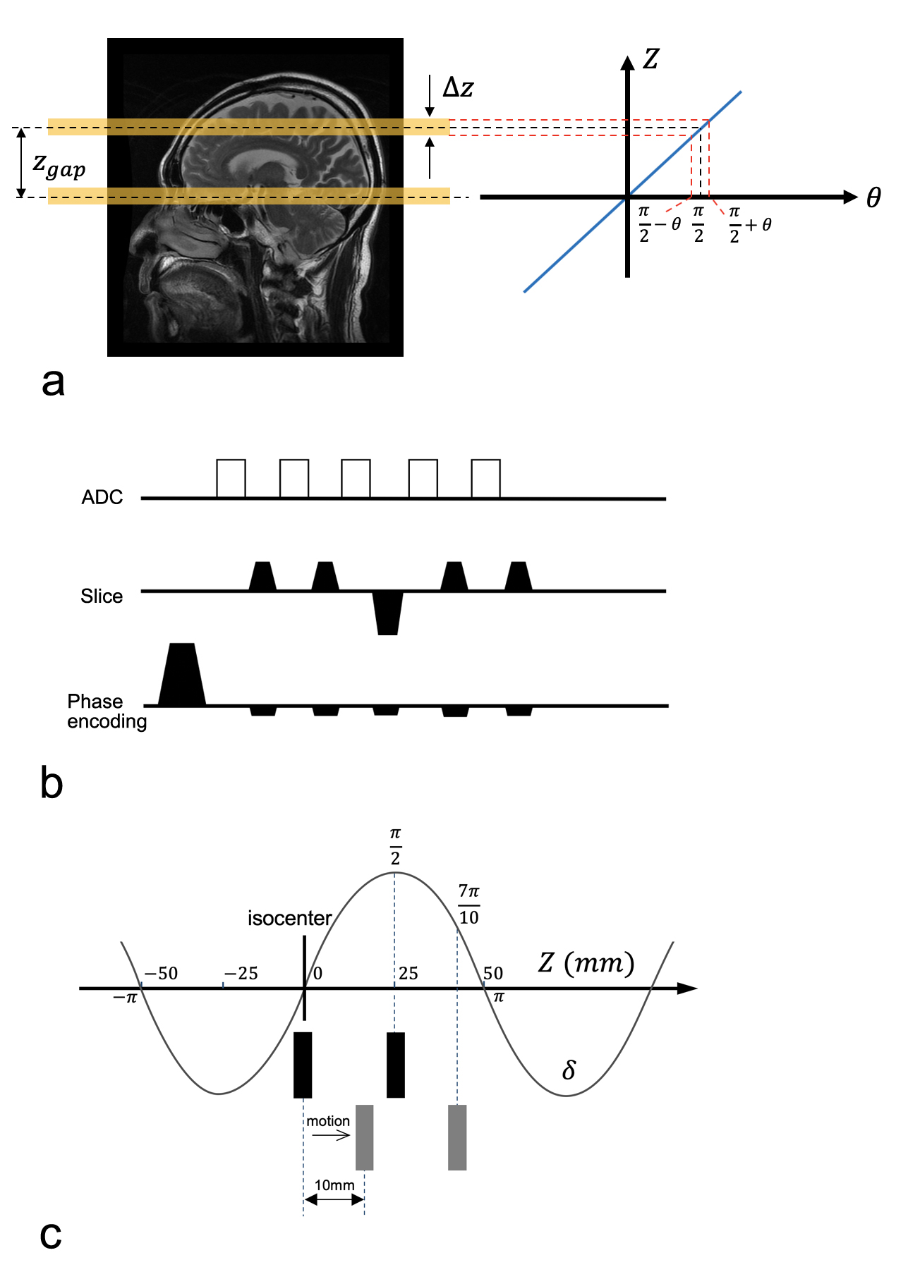
Fig.1. Diagram of blipped-CAIPI sequence (a,b), and (c) phase modulation due to Gz blips along the FOVsms in z (Δz=5 mm slice thickness; Zgap=25 mm slice-to-slice distance). The black boxes represent 2 slices before motion, and gray ones after 10mm z-translation. The phase increment is 2π/FOVsms/mm. The SMS phase variation of the off-center slice is π/2, and, once the 10mm motion is detected, the receiver phase needs to be compensated immediately by δ=2π*10/FOVsms.