1361
Motion Correction and Registration Networks for Multi-Contrast Brain MRI
Jongyeon Lee1, Byungjai Kim1, Wonil Lee1, and HyunWook Park1
1Korean Advanced Institute of Science and Technology, Daejeon, Korea, Republic of
1Korean Advanced Institute of Science and Technology, Daejeon, Korea, Republic of
Synopsis
Deep learning techniques have been applied to motion artifact correction without motion estimation or tracking. We previously studied the motion correction method for the multi-contrast brain MRI using NMI maximization and the multi-input neural network. However, as the previous work suffered from a prolonged alignment time and a training inconvenience, we adopt the registration network to reduce alignment time and the multi-output neural network to be trained only once. Our proposed method successfully reduces motion artifacts in the multi contrast images.
Introduction
Motion during MRI scan typically hinders image quality by producing motion artifacts1. To reduce the motion artifacts, various motion correction techniques have been developed. Prospective motion correction methods tracked motions during acquisition using a navigator2 or external sensors3 and updated a sequence in real time4 to keep a patient’s relative position stationary. In contrast, retrospective motion correction methods estimated motion information after scan using self-navigated trajectories5 to correct inconsistencies among k-space lines. However, these previous techniques are typically lacking generality due to their dependency on MRI system or specialized pulse sequences, and require additional data acquisition to correct motion artifacts6.To overcome these issues in the multi-contrast brain MRI environment, we previously introduced a deep learning technique to correct motion-corrupted images using motion-free images of other contrasts7. This work utilized the multi-contrast MRI with three contrasts including T1-weighted (T1w), T2-weighted (T2w), and T2-weighted FLuid-Attenuated Inversion Recovery (FLAIR)8, applied the multi-modal registration by the normalized mutual information (NMI) maximization, and used a motion correction network. However, the previous work had problems with its prolonged runtime for the registration and its inconvenience of training each network for correction of each contrast image. To resolve these problems, we extend this work to a new framework that utilizes the registration network to reduce time consumed in NMI maximization and employs a multi-output motion correction network, which can be trained only once regardless of the number of contrasts.
Methods
As illustrated in Figure 1-a, the proposed framework consists of two parts: image alignment and motion correction. To correct misalignment between multi-contrast images due to motions, the image alignment is performed with the unsupervised registration network, which uses the normalized cross-correlation9 (NCC) loss to yield the best motion parameters that minimize the loss among the multi-contrast input images, and the NMI maximization to fine-tune the alignment. Their combination facilitates faster and more accurate image alignment. After the aligning process, the motion correction network, which consists of three encoders, one transformer, and three decoders, corrects motion artifacts in the input images using SSIM10 loss and VGG11 loss with the ratio of 1:0.001. This multi-input and multi-output network structure enables to effectively correct motion artifacts for every contrast. The detailed network architecture is described in Figure 1-b. For training, Adam optimizer was used with a learning rate of 0.001. An actual implementation was performed on TensorFlow 2.0 with the Keras library.2D motion-free axial brain MR images with three contrasts were obtained from 41 healthy subjects (age 22.7 ± 2.8 years) with a slice thickness of 5mm using a 3.0T MRI system (MAGNETOM Verio, Siemens) and the 32-channel head coil. The field of view (FOV) was 220mm × 220mm, and the image matrix size was 256 × 256. From 262 motion-free slices, 193 slices were used for training, 34 slices for validation, and 35 slices for test. In order to train, validate, and test the network, we simulated motion artifacts depending on the sequences used for the study and synthesized the datasets with the simulated motion artifacts. Figure 2 shows example cases of the random motion simulation for a) a T1w spin-echo image and b) a T2w turbo spin-echo image. The training process was done only with the synthesized training dataset.
Tests are done using both the synthesized test dataset and the in-vivo test dataset, which contains random real motion artifacts from 5 subjects (age 25.2 ± 1.8 years). To investigate the effectiveness of each process in the proposed method, we perform the experiments that correct motion artifacts without the image alignment, with the network registration only, and with the whole image alignment process including the NMI maximization for the synthesized test dataset.
Results
Figure 3 shows the results of the example test images for a) the synthesized dataset and b) the real motion dataset, where an image of the target contrast is motion-corrupted and the other contrast images are motion-free. For the real motion dataset, it is visually addressed that the motion artifacts are greatly reduced as shown in the areas within the colored boxes.The importances of the registration network and the NMI maximization are presented in Figure 4 with the average SSIM and NMI scores for the motion-corrected images of the synthesized test dataset. The test scores without any image alignment are the lowest, whereas the test scores with the whole image alignment process are the highest. Example images are also presented for these experiments in Figure 5, where the experiment with the whole image alignment shows the best performance among the experiments for all contrasts as the highest SSIM scores and the lowest NRMSE scores of the motion-corrected images indicate.
Discussion and Conclusion
Although our method is currently limited to the correction of intra-slice motion artifacts in the given contrast images, the work is likely to be extended to dealing with through-plane motion artifacts and other contrast images.We proposed the framework with the image alignment and the motion correction using deep learning, and the proposed method successfully reduced motion artifacts for all contrasts as the quantitative scores and the tests with the real motion datasets present.
Acknowledgements
This research was supported by a grant of the Korea Health Technology R&D Project through the Korea Health Industry Development Institute (KHIDI), funded by the Ministry of Health & Welfare, Republic of Korea (grant number: HI14C1135) and by Institute for Information & communications Technology Promotion (IITP) grant funded by the Korea government (MSIT) (No.2017-0-01778).References
- Zaitsev M, Maclaren J, Herbst M. Motion artifacts in MRI: A complex problem with many partial solutions. Journal of Magnetic Resonance Imaging. 2015;42(4):887-901.
- Firmin D, Keegan J. Navigator echoes in cardiac magnetic resonance. Journal of Cardiovascular Magnetic Resonance. 2001;3(3):183-193.
- Zaitsev M, Dold C, Sakas G, Hennig J, Speck O. Magnetic resonance imaging of freely moving objects: prospective real-time motion correction using an external optical motion tracking system. Neuroimage. 2006;31(3):1038-1050.
- Herbst M, Maclaren J, Weigel M, Korvink J, Hennig J, Zaitsev M. Prospective motion correction with continuous gradient updates in diffusion weighted imaging. Magnetic Resonance in Medicine. 2012;67(2):326-338.
- Pipe JG. Motion correction with PROPELLER MRI: application to head motion and free‐breathing cardiac imaging. Magnetic Resonance in Medicine. 1999;42(5):963-969.
- Godenschweger F, Kägebein U, Stucht D, et al. Motion correction in MRI of the brain. Physics in Medicine & Biology. 2016;61(5):R32.
- Lee J, Kim B, Jeong N, Park H. Motion correction for a multi-contrast brain MRI using a multi-input neural network. In Proceedings of the 28th Annual Meeting of ISMRM, 2020.
- Lu H, Nagae‐Poetscher LM, Golay X, Lin D, Pomper M, Van Zijl, PC. (2005). Routine clinical brain MRI sequences for use at 3.0 Tesla. Journal of Magnetic Resonance Imaging, 2005;22(1):13-22.
- Zhao F, Huang Q, Gao W. Image matching by normalized cross-correlation. IEEE International Conference on Acoustics Speech and Signal Processing Proceedings. 2006.
- Wang Z, Bovik AC, Sheikh HR, Simoncelli, EP. Image quality assessment: from error visibility to structural similarity. IEEE Transactions on Image Processing. 2004;13(4):600-612.
- Simonyan K, Zisserman A. Very deep convolutional networks for large-scale image recognition. In International Conference on Learning Representations. 2015.
Figures
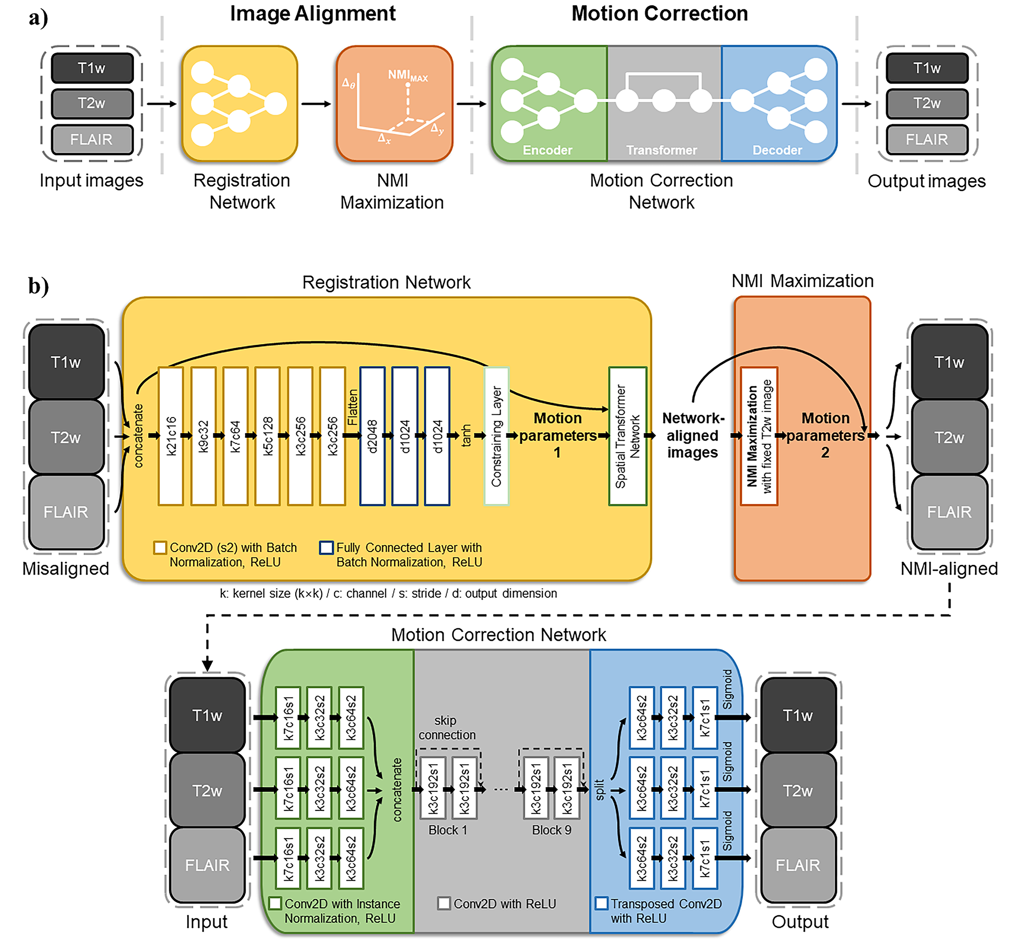
Figure 1: a) An overview of the proposed method. The input images
can contain motion-free images and motion-corrupted images. The output images
are the motion-corrected images if the corresponding input images are
motion-corrupted, whereas the outputs are almost the same as the input images
if the corresponding input images are motion-free. b) The detailed framework of the proposed network with the network
hyperparameters.
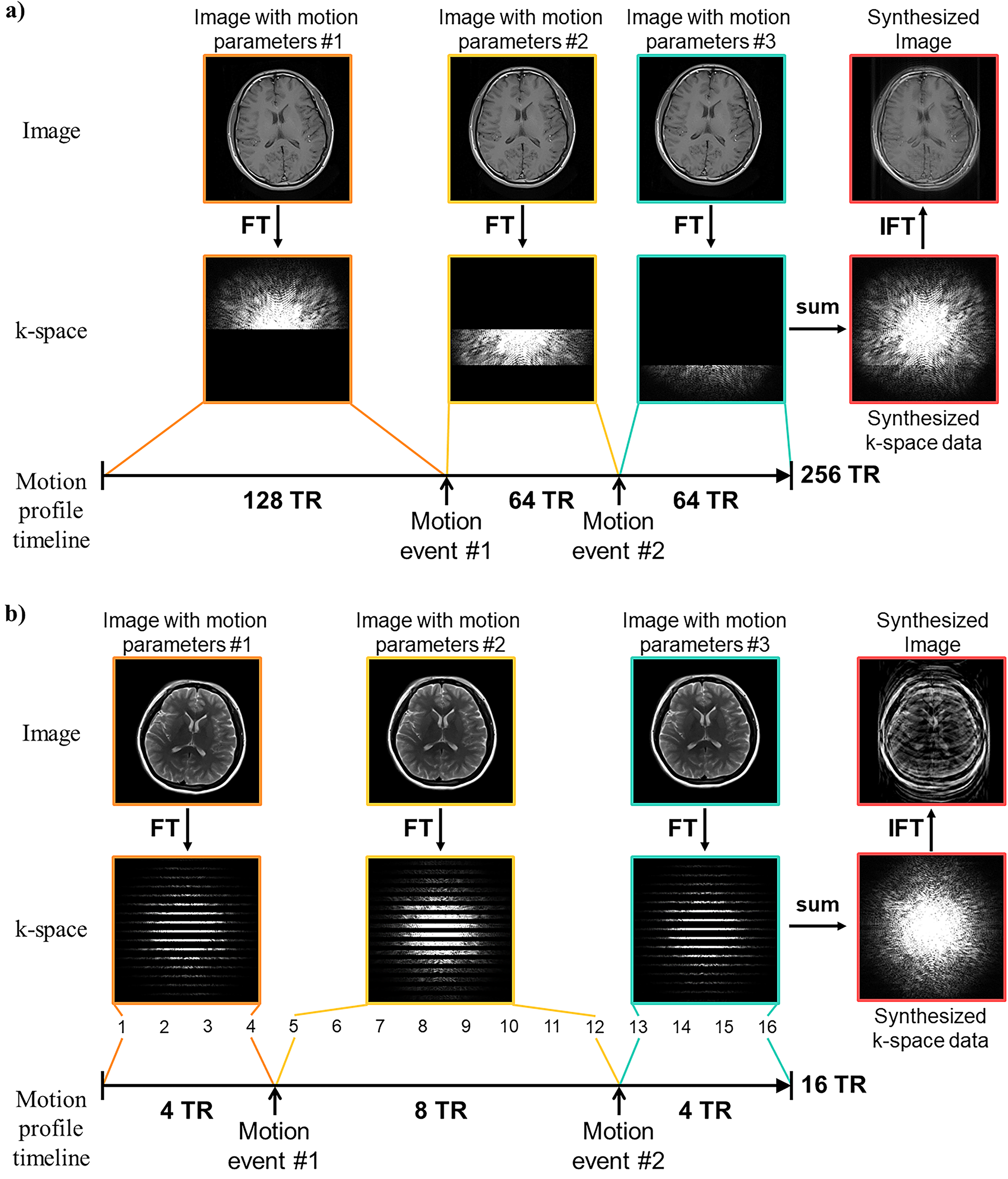
Figure 2: An
illustration of synthesis of motion-corrupted images for a) T1w image (spin-echo) and b)
T2w image (turbo spin-echo). The number of motion events, their timing, and
their motion parameters are randomly selected. With randomly generated motion
profiles, motion-corrupted k-space data are synthesized and are inversely
Fourier transformed to generate motion-corrupted images.
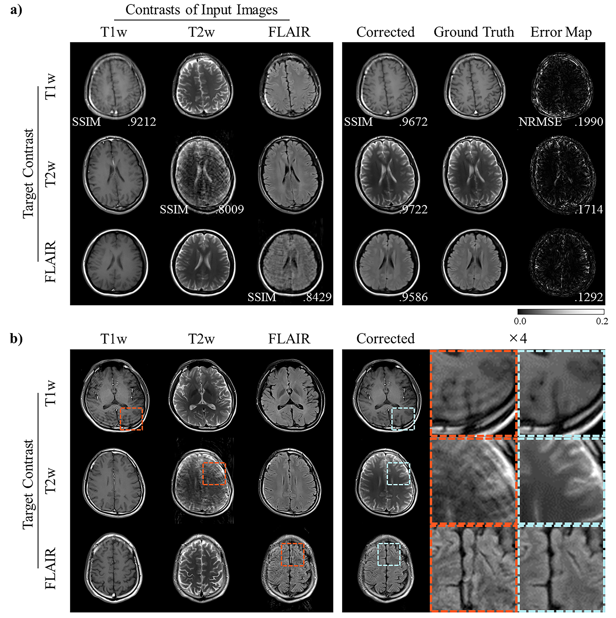
Figure 3: Example images of the baseline experiment for a) the synthesized test data and b) the real motion data in T1w, T2w, and FLAIR correction cases.
SSIM and NRMSE scores are written on the motion-corrected images. For the real
motion test, the red and blue boxes highlight the performance of the proposed method.
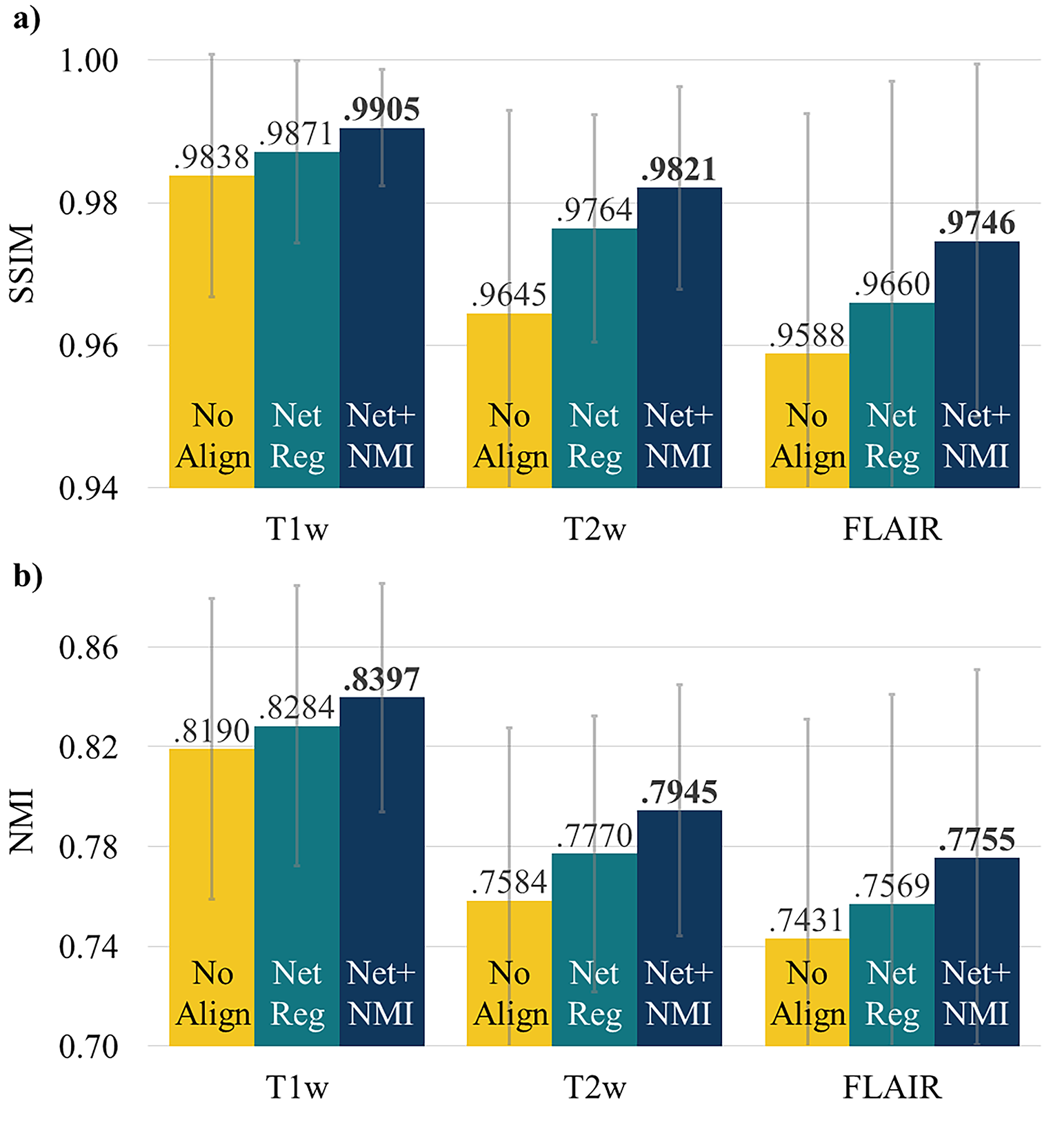
Figure 4: Quantitative evaluation results of the proposed method with various alignment
processes. a) The average SSIM and b) NMI scores are provided with their
standard deviations. “No Reg” indicates the experiment without any
registration, “Net Reg” indicates the experiment with the network registration
only, and “Net+NMI” indicates the experiment with both the registration network
and the NMI maximization process.
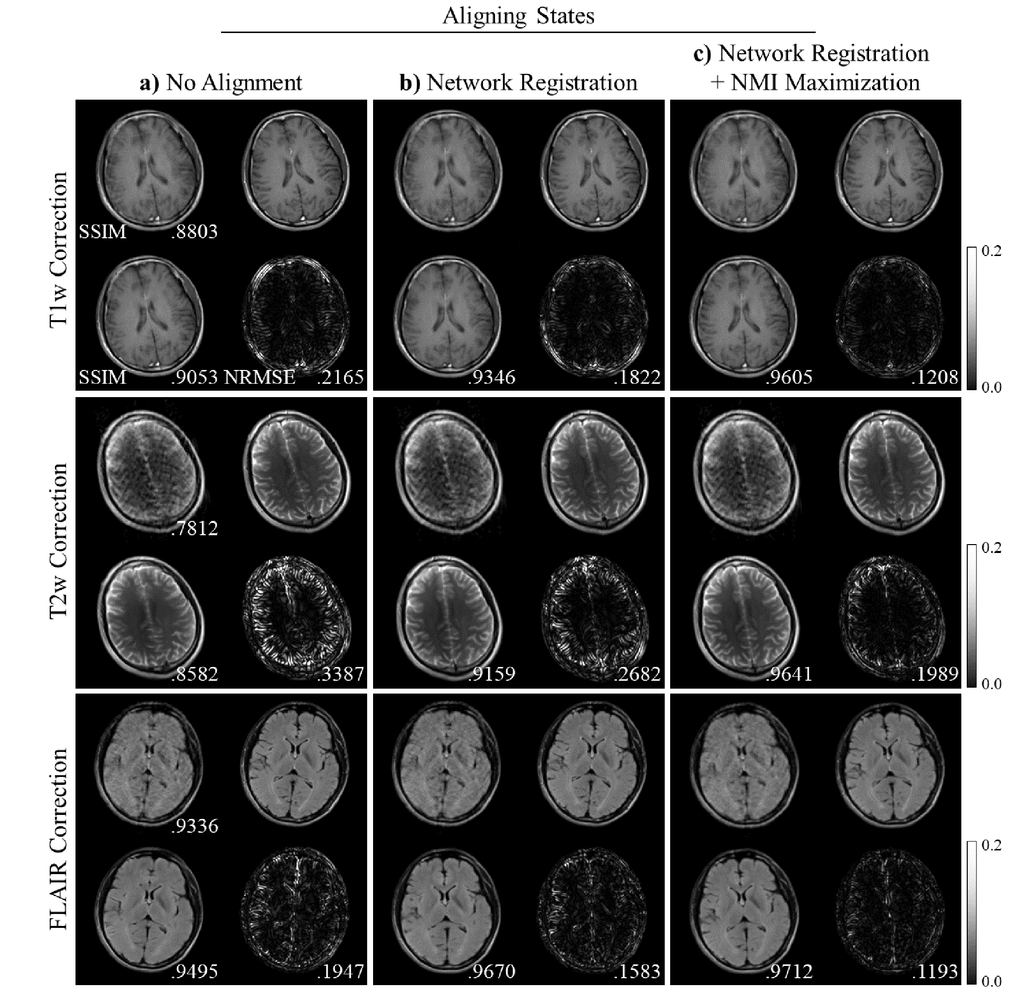
Figure 5: Example
images from the proposed method a)
without any alignment, b) with the
registration network only, and c)
with both the registration network and the NMI maximization process for T1w,
T2w, and FLAIR images. For each block, the motion-corrupted input image (left-top),
the motion-corrected image (left-bottom), the ground truth motion-free image
(right-top), and the error map (right-bottom) between the motion-corrected
image and the corresponding motion-free images are presented.