1343
Is Perfect Filtering Enough Leading to Perfect Phase Correction?1School of Information Science and Technology, Northwest University, Xi'an, China, 2School of Biomedical Engineering, ShanghaiTech University, Shanghai, China, 3Department of Computer Science and Technology, University of Cambridge, Cambridge, United Kingdom, 4Department of Computer Science, The University of Hong Kong, Hong Kong, China, 5State-Province Joint Engineering and Research Center of Advanced Networking and Intelligent Information Services, Xi'an, China, 6Shanghai United Imaging Intelligence Co., Ltd., Shanghai, China, 7Department of Artificial Intelligence, Korea University, Seoul, Korea, Republic of
Synopsis
We cautiously analyze phase correction procedures, unveiling why they falsely introduce dark holes to diffusion-weighted images, and calibrate them with the goal of unbiased white matter microstructure estimation. The calibrated procedures properly rotate image contents to be negative, hence, dark hole issue is effectively avoided.
Purpose
The Diffusion-weighted (DW) magnitude is commonly Rician or non-central $\chi$ distributed, with a signal elevation falsely caused by the noise-floor [1,2]. To mitigate the noise-floor issue, phase correction extracts real-valued Gaussian-distributed diffusion MRI data, eliminating biases of white-matter microstructure estimation (WM-MSE), e.g., fractional anisotropy (FA). Conventional phase correction methods resort to employing spatially-adaptive filters for improving perfromance [3,4]. However, even MPPCA [5], renowned as spatial adaptivity, surprisingly introduces severe artifacts [6]. This unexpected observation unveils underlying issues of phase correction, which inspire us to cautiously analyze and reformulate phase correction procedures.In this abstract, we propose a calibrated procedure for phase correction.Methods
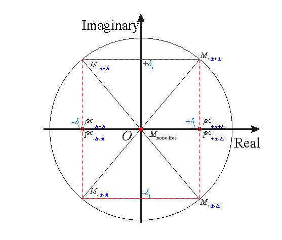
Phase correction complex-rotates actual image contents, $$ I^\text{PC}({x}) = M(x){\rm e}^{j(\varphi({x}) - \varphi_{\text{BG}}({x}))}, $$ Given the magnitude $$$M(x)$$$ and phase $$$\varphi({x})$$$, the phase-corrected result $$$I^\text{PC}({x})$$$ is determined by the background phase $$$\varphi_{\text{BG}}({x})$$$, $$$ \varphi_{\text{BG}}({x}) = \arctan{\frac{f(I_\text{i}({x}))}{f(I_\text{r}({x}))}}, $$$ where $$$x$$$ and $$$f(\cdot)$$$ denote a voxel location and the filtering operator on real $$$I_\text{r}({x})$$$ and imaginary $$$I_\text{i}({x})$$$ parts of DW images, respectively; and $$$I^\text{PC}({x}) = I_\text{r}^{\text{PC}}({x})+j{I}_\text{i}^{\text{PC}}({x})$$$, $$ I_\text{r}^{\text{PC}}({x}) = M({x})\cos({\varphi({x})-\varphi_{\text{BG}}({x})}),\\ I_\text{i}^{\text{PC}}({x}) = M({x})\sin({\varphi({x})-\varphi_{\text{BG}}({x})}). $$ Fig.1 pictorially depicts a noise-free voxel $$$I({x}) = 0 + j0$$$ with $$$M_\text{noise\_free} = 0$$$. When reconstructing $$$M_{-\delta\text{r}-\delta\text{i}}$$$, the noises are typically flipped from negative to positive, and thus result in the noise-floor. By contrast, phase correction results in $$$I^\text{PC}_{-\delta\text{r}-\delta\text{i}} + I^\text{PC}_{+\delta\text{r}+\delta\text{i}} = 0$$$ instead of $$$M_{-\delta\text{r}-\delta\text{i}} = M_{+\delta\text{r}+\delta\text{i}}$$$.In Fig.1, phase correction procedures misleadingly give $$$I^\text{PC}_{-\delta\text{r} + \delta\text{i}}$$$ and $$$I^\text{PC}_{-\delta\text{r}-\delta\text{i}}$$$ the same result. More specifically,$$$I^\text{PC}_{-\delta\text{r} + \delta\text{i}}$$$ is derived only from positive noise in the imaginary axis and negative noise in the real axis, while $$$I^\text{PC}_{-\delta\text{r}-\delta\text{i}}$$$ is derived from negative noises in both axes. $$$I^\text{PC}_{-\delta\text{r} + \delta\text{i}}$$$ is falsely as smaller as $$$I^\text{PC}_{-\delta\text{r} - \delta\text{i}}$$$, and that is the reason why conventional phase correction falsely introduces dark holes.
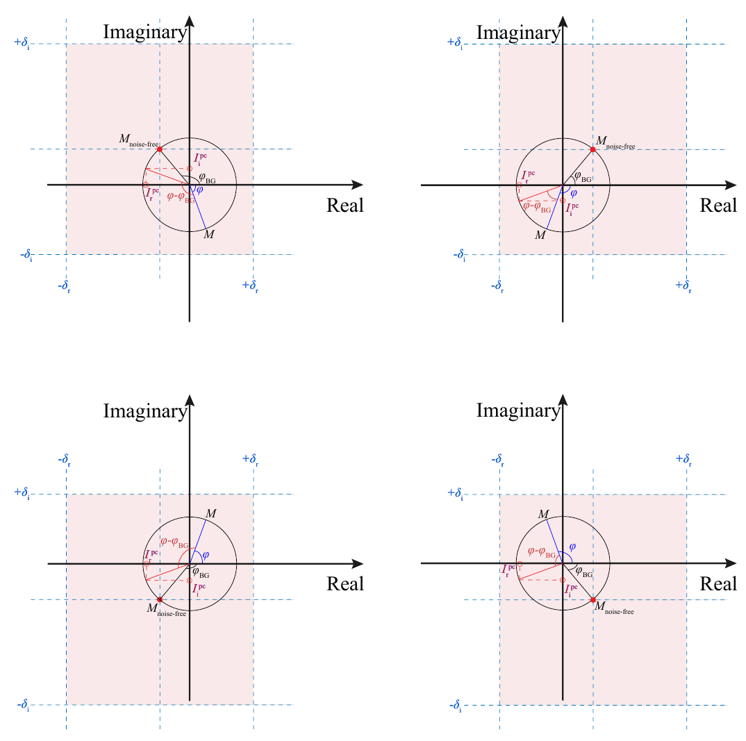
To eliminate the dark holes, we calibrate phase correction procedures, by horizontally flipping $$$I^\text{PC}({x})$$$ with $$$\pi/2 < {\varphi({x})-\varphi_{\text{BG}}({x})} < -3\pi/2$$$ towards the first or the fourth quadrant, in a way analogous to the magnitude reconstruction. Meanwhile, the calibrated phase correction procedures assign negative values to voxels that are most likely to cause the noise-floor; and Fig.2 demonstrates the voxels that correspond to regions of high confidence in inducing noise-floor.
Results
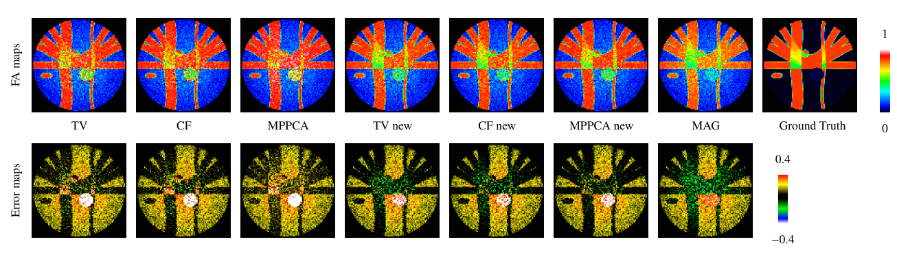
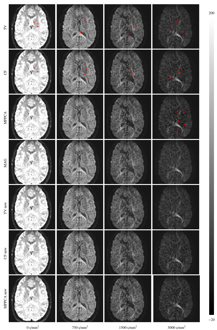
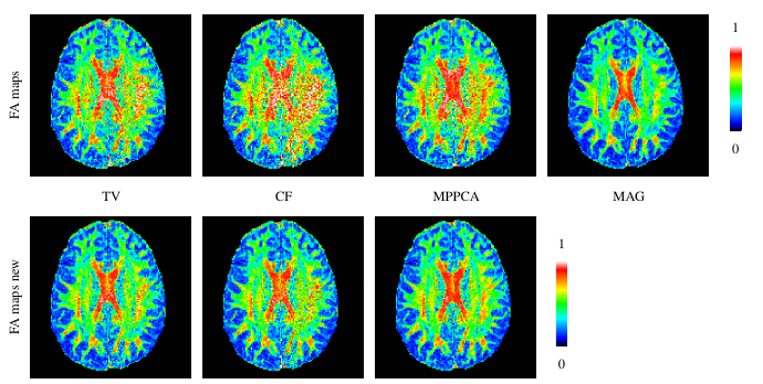
We generated synthetic DW data using Phantom$$$\alpha$$$s [7] with the same gradient vectors of the real data described as follows. We synthesized the background phase according to [2]. With the noise-free DW images, we added spatially varying Gaussian noises to the real and imaginary parts, respectively. The real DW images were acquired using a SIEMENS 3T Magnetom Prisma MR scanner with following protocol, TR =$$$2500 \text{ms}$$$, TE=$$$89 \text{ms}$$$, FoV=$$$210\times 210\,\text{mm}^2$$$, matrix size=$$$140\times140$$$, and $$$b=750,1500,3000\,\text{s/mm}^2$$$ with a total of $$$64$$$ diffusion directions. To demonstrate the improvement, we compared conventional phase correction procedures with the calibrated ones, both of which employed filters with the same parameters, including curvature filtering (CF) [8], total variation (TV) [1], and Marchenko-Pastur PCA (MPPCA) [5].Fig.3 shows synthetic FA maps derived from TV, CF, and MPPCA, with increased FA values of white-matter compared to magnitude (MAG). Furthermore, it can be seen from error maps that the calibrated phase correction procedures yield the results that are closer to the ground-truth, especially for MPPCA. Fig.4 demonstrates the calibrated phase correction procedures introduce significantly fewer dark holes to the phase-corrected real images, as shown in Fig.5 where the artifacts introduced by conventional phase correction are effectively eliminated.
Conclusion
We calibrated the phase correction procedures to avoid arbitrarily specifying negative values, which should only be derived from the noise-floor. The calibrated procedures fully exploit the use of the advanced filter, e.g, MPPCA, which can be effectively employed to mitigate the noise-floor in DW images without introducing additional artifacts.Acknowledgements
This work was supported in part by National Natural Science Foundation of China (No. 62073260, 61971350).References
[1] Eichner, C., Cauley, S.F., Cohen-Adad, J., M¨oller, H.E., Turner, R., Setsompop, K., Wald, L.L., 2015. Real diffusion-weighted MRI enabling true signal averagingand increased diffusion contrast. NeuroImage 122, 373–384.
[2] Pizzolato, M., Fick, R., Boutelier, T., Deriche, R., 2016. Noise floor removal via phase correction of complex diffusion-weighted images: Influence on DTI andq-space metrics, in: MICCAI Workshop on Computational Diffusion MRI, Springer. pp. 21–34.
[3] Pizzolato, M., Deriche, R., 2018. Automatic and spatially varying phase correction for diffusion weighted images, in: 26th annual meeting of the InternationalSociety for Magnetic Resonance in Medicine (ISMRM).
[4] Liu, F., Chen, G., Feng, J., Yap, P.T., Shen, D., 2019. Gaussianization of diffusion mri data using spatially adaptive phase correction, in: 27th annual meeting of theInternational Society for Magnetic Resonance in Medicine (ISMRM).
[5] Veraart, J., Novikov, D.S., Christiaens, D., Ades-Aron, B., Sijbers, J., Fieremans, E., 2016. Denoising of diffusion MRI using random matrix theory. NeuroImage142, 394–406.
[6] Liu, F., Feng, J., Chen, G., Shen, D., Yap, P.T., 2020. Gaussianization of diffusion MRI data using spatially adaptive filtering. Medical Image Analysis .
[7] Caruyer, E., Daducci, A., Descoteaux, M., Houde, J.C., Thiran, J.P., Verma, R., 2014. Phantomas: a flexible software library to simulate diffusion mr phantoms, in:ISMRM.
[8] Gong, Y., Sbalzarini, I.F., 2017. Curvature filters efficiently reduce certain variational energies. IEEE Transactions on Image Processing 26, 1786–1798.
Figures