1342
Robust method for Whole Body DWIBS applied both Image based B0 Shimming and Blip-up Blip-down Distortion Correction1Philips Japan, Tokyo, Japan, 2Philips Healthcare, Tokyo, Japan, 3Department of Radiology, Kumamoto Chuo Hospital, Kumamoto, Japan
Synopsis
DWIBS based on single shot echo-planar imaging (EPI) is a first-choice sequence in routine clinical examinations. However, it sometimes suffers from sever image distortion due to the presence of air within and/or at edge of the FOV, especially at the border of chest and abdomen. On the other hand, image based B0 shimming and blip-up blip-down distortion correction improved for image qualities of DWI. We demonstrated that whole body DWIBS applied both image based B0 shimming and blip-up blip-down distortion correction to provide higher robustness.
Introduction
Diffusion weighted whole body imaging with background suppression (DWIBS)1 can visualize malignant tumor and abnormal lymph nodes and is useful for determining the response of bone metastasis to treatment2-6. DWIBS based on single shot echo-planar imaging (EPI) is a first-choice sequence in routine clinical examinations. However, it sometimes suffers from sever image distortion due to the presence of air within and/or at edge of the FOV, especially at the border of chest and abdomen. On the other hand, a fully automate image based B0 shimming had proposed to optimize the B0 field during acquisition7. In addition, blip-up blip-down distortion correction had been used for purposes EPI distortion correction in diffusion tensor imaging and functional MRI8-10. We expect that image based B0 shimming and blip-up blip-down distortion correction techniques improve the image qualities of whole body DWIBS. The aim of this study is to investigate the feasibility of whole body DWIBS combine with them techniques for enabling more robust.Methods
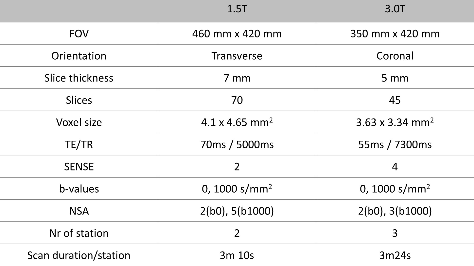
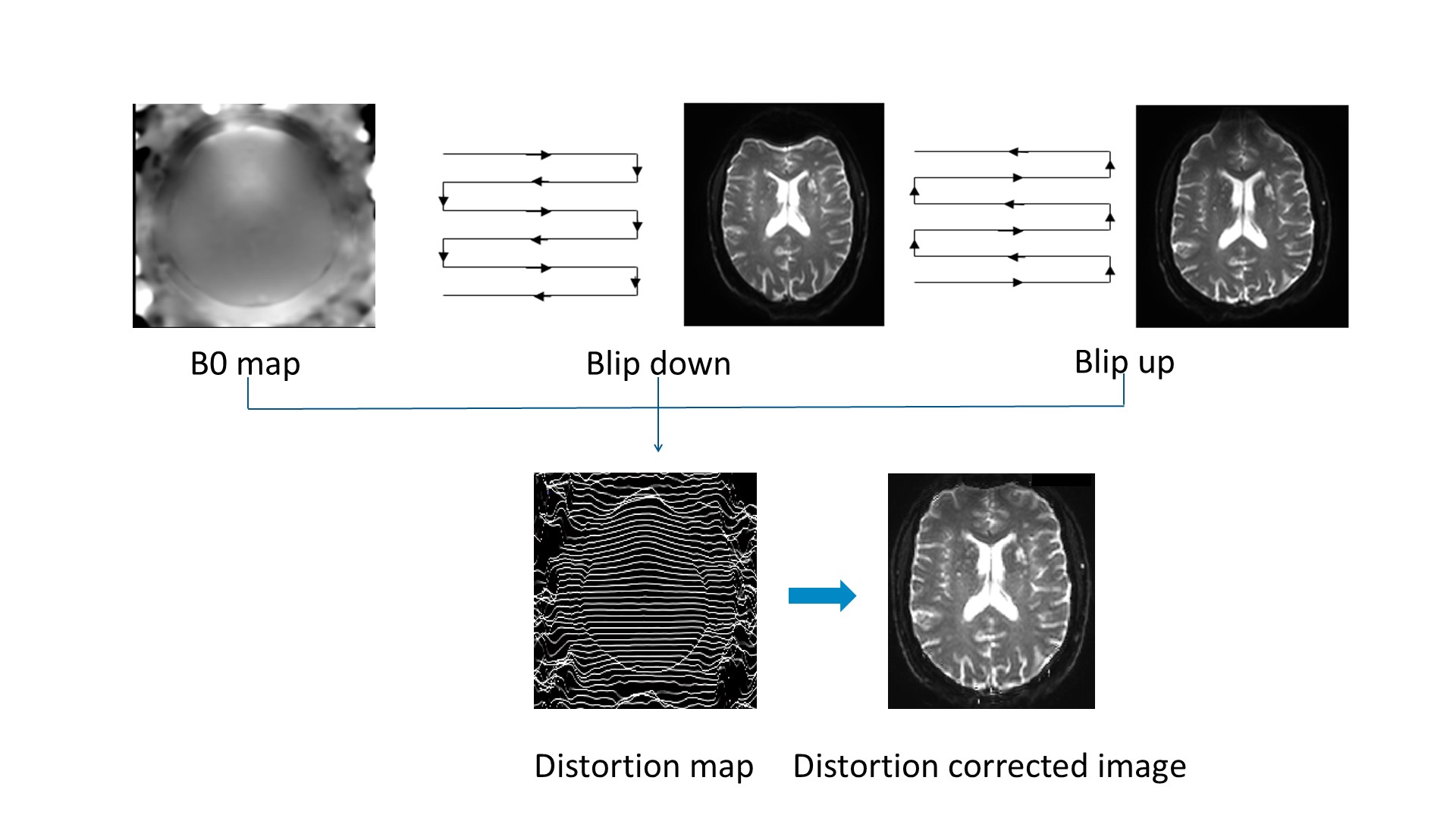
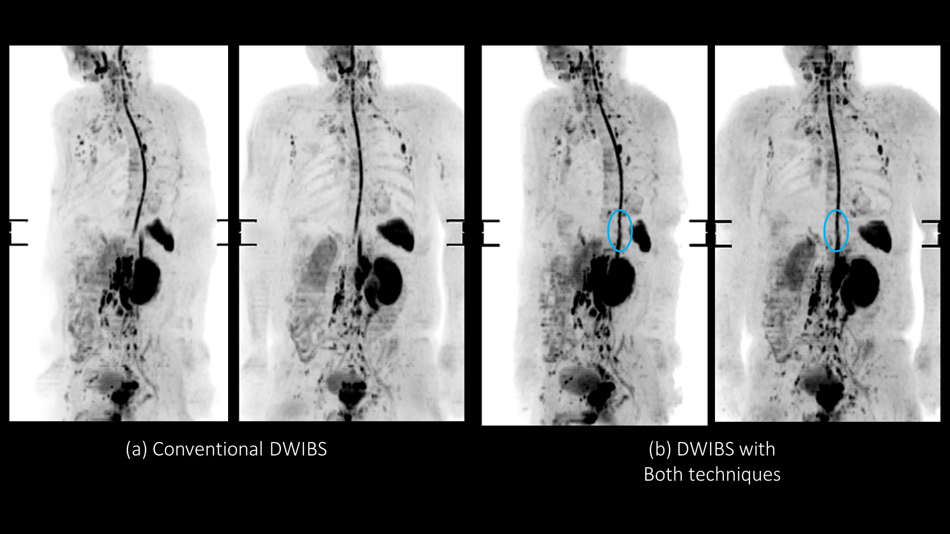
Conventional DWIBS, DWIBS with image based shimming, DWIBS with blip-up blip-down distortion correction and DWIBS with both techniques in one healthy volunteer was obtained on a 3.0T scanner (Ingenia Elition, Philips Healthcare) and clinical case with a patient of bone metastasis was obtained on a 1.5T scanner (IngeniaCX, Philips Healthcare). The volunteer and patient obtained informed consent and approved by institutional review board. Imaging parameter for DWIBS was shown in Table 1. Blip-up blip-down distortion correction algorithm was shown in Figure 1. Next to the standard DWI encoding, data with reversed phase encode blips (with distortions going in opposite direction) and a B0 map were acquired. The susceptibility-induced off-resonance field was estimated using a method similar to that described in [Andersson 2003]8, based on this field map, a DWI image corrected for geometric distortions is generated inline by the MRI scanner software. DWIBS images were obtained in direct coronal with 3 stations at 3.0T, and its were obtained in transverse with 2 stations at 1.5T. To compare the image quality of them, coronal maximum intensity projection (MIP) images were evaluated.Results and Discussion
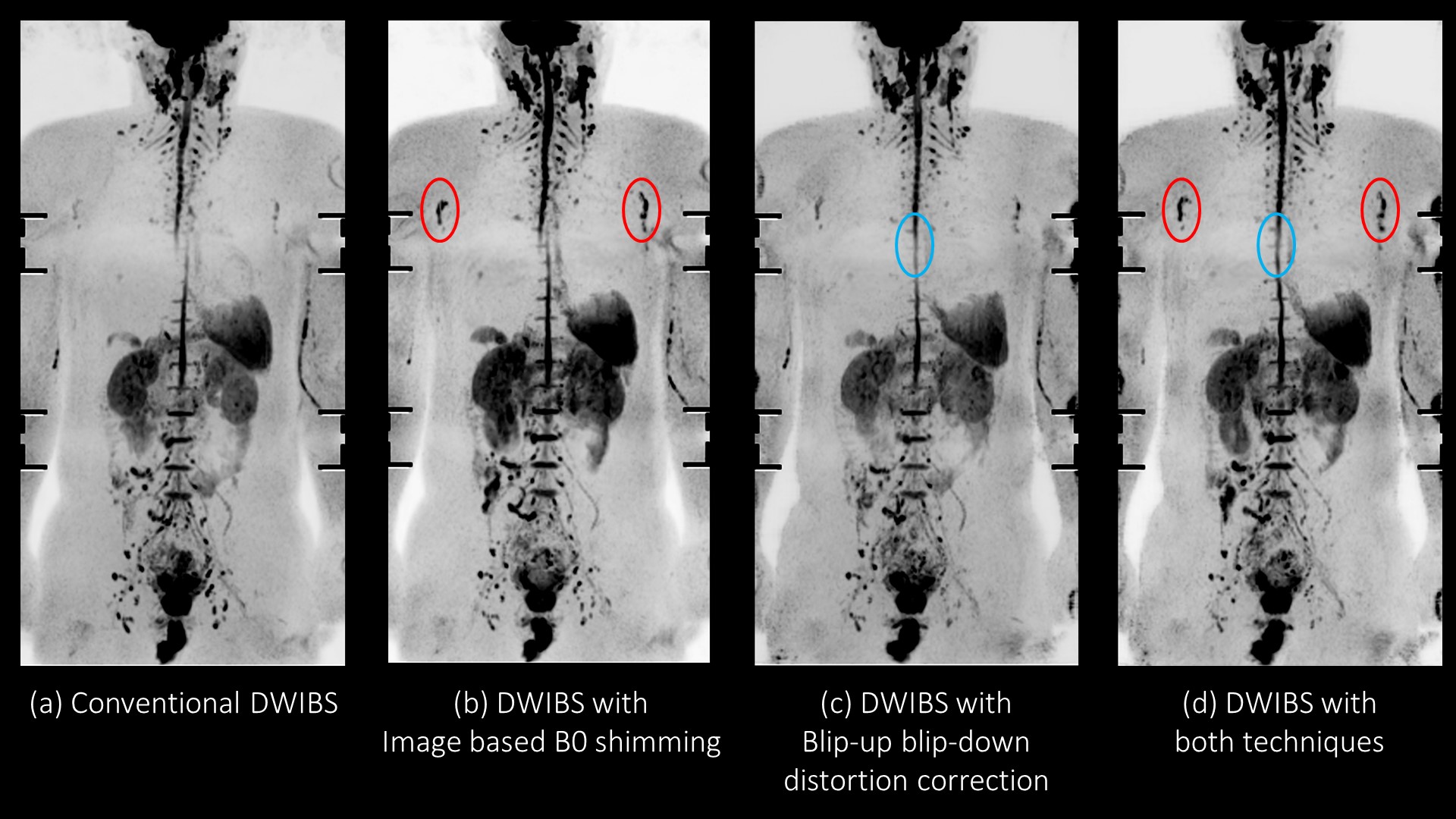
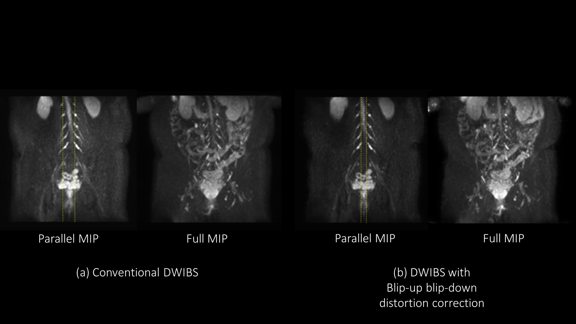
The MIP of direct coronal whole body DWIBS at 3.0T were shown in Figure 2. Image based B0 shimming provided improving depiction of axillary lymph nodes, and DWIBS applied both techniques showed less distortion and no unwanted signal suppression. In Figure 3 shows that DWIBS with bild-up bild-down distortion correction decrease distortion effect due to the presence of air at 3.0T, especially focusing on spinal cord. The MIP of transverse DWIBS at 1.5T were shown in Figure 4. Similar findings to 3.0T were obtained at 1.5T. Blip-up blip-down distortion correction dramatically improved between the each station at both field strength.Conclusion
We demonstrated that image based B0 shimming and blip-up blip-down distortion correction techniques improve the image qualities of whole body DWIBS. Whole body DWIBS applied both image based B0 shimming and blip-up blip-down distortion correction provided higher robustness than that of without them.Acknowledgements
No acknowledgement found.References
1 Taro Takahara et al, Diffusion weighted whole body imaging with background body signal suppression (DWIBS): technical improvement using free breathing, STIR and high resolution 3D display. Radiat Med. Jul-Aug 2004;22(4):275-82
2 Fruehwald-Pallamar J et al, Functional imaging in head and neck squamous cell carcinoma: correlation of PET/CT and diffusion-weighted imaging at 3 Tesla. Eur J Nucl Med Mol Imaging. 2011 Jun;38(6):1009-19
3 Michielsen KL et al, Br J Radiol. Whole-body diffusion-weighted magnetic resonance imaging in the diagnosis of recurrent ovarian cancer: a clinical feasibility study. 2016 Nov;89(1067):20160468
4 Pearce T et al, Bone metastases from prostate, breast and multiple myeloma: differences in lesion conspicuity at short-tau inversion recovery and diffusion-weighted MRI. Br J Radiol. 2012 Aug;85(1016):1102-6
5 Perez-Lopez R et al, Diffusion-weighted Imaging as a Treatment Response Biomarker for Evaluating Bone Metastases in Prostate Cancer: A Pilot Study. Radiology. 2017 Apr;283(1):168-177.
6 Sun M et al, Application value of diffusion weighted whole body imaging with background body signal suppression in monitoring the response to treatment of bone marrow involvement in lymphoma. J Magn Reson Imaging. 2016 Dec;44(6):1522-1529
7 A. W. Simonetti et al, 3D breast segmentation for image based shimming. In:Proc. 17th Annual Meeting of ISMRM, 2009 ; 2114
8 Andersson JL, et al., How to correct susceptibility distortions in spin-echo echo-planar images: application to diffusion tensor imaging . Neuroimage. 2003 Oct;20(2):870-88
9 Smith, S.M et al, Advances in functional and structural MR image analysis and implementation as FSL. Neuroimage. 2004;23 Suppl 1:S208-19
10 Jezzard P, et al. Correction for geometric distortion in echo planar images from B0 field variations. Magn Reson Med. 1995 Jul;34(1):65-73.
Figures