1331
Distortion-free high-resolution diffusion weighted imaging of mouse brain using diffusion-prepared 3D VF-RARE
Qiang Liu1,2, Yuanbo Yang1,2, Xinyuan Zhang1,2, Yingjie Mei1,2,3, Qiqi Lu1,2, Guoxi Xie4, and Yanqiu Feng1,2
1School of Biomedical Engineering, Southern Medical University, Guangzhou, China, 2Guangdong Provincial Key Laboratory of Medical Image Processing, Southern Medical University, Guangzhou, China, 3Philips Healthcare, Guangzhou, China, 4Department of Biomedical Engineering, School of Basic Sciences, Guangzhou Medical University, Guangzhou, China
1School of Biomedical Engineering, Southern Medical University, Guangzhou, China, 2Guangdong Provincial Key Laboratory of Medical Image Processing, Southern Medical University, Guangzhou, China, 3Philips Healthcare, Guangzhou, China, 4Department of Biomedical Engineering, School of Basic Sciences, Guangzhou Medical University, Guangzhou, China
Synopsis
The purpose of the present work was to achieve three-dimensional (3D) high spatial resolution diffusion weighted imaging (DWI) of mouse brain using diffusion-prepared 3D VF-RARE (DP 3D VF-RARE) sequence at 7 Tesla. To employ long echo trains while prospectively controlling signal variation during acquisition, variable flip angles (VF) were implemented in the refocusing pulse train. The results showed improved image quality with less distortion over 2D single-shot EPI and comparable signal-to-noise ratio (SNR). With the application of the presented sequence, undistorted diffusion-weighted mouse brain images were obtained with high spatial resolution and potential SNR efficiency.
Introduction
As an alternative acquisition strategy to EPI which has been applied to improve image quality in diffusion MRI applications, Rapid Acquisition with Relaxation Enhancement (RARE) based sequence provides the advantages of high signal-to-noise ratio (SNR) efficiency and insensitive to B0 inhomogeneity1. With the use of a separated preparation module, the diffusion-sensitized transverse magnetization is stored into longitudinal magnetization, thus can be combined with multi-shot 3D RARE based readout scheme, without suffering from the intershot phase errors caused by motion2. However, high RF power deposition and blurring limit its use at a higher field, especially at 7 Tesla. To overcome these limitations, a series of variable low-flip-angle (VF) nonselective refocusing pulses are used to maintain signal at a constant “target” level over very long echo train. The purpose of study was to assess the feasibility of diffusion-prepared 3D RARE sequence combined with VF to obtain distortion-free, high SNR, high spatial resolution mouse brain diffusion image at 7 Tesla Bruker system.Methods
Sequence: Figure 1 showed the sequence diagram of the DP 3D VF-RARE sequence. DP module was followed by a 3D VF-RARE readout with centric phase-encoding ordering. To obtain the diffusion-sensitized magnetization in the DP module, nonselective RF pulses were used to tip down, refocus and tip up spins, with a pair of diffusion gradients along each side of the refocusing pulse. A spoiler gradient was applied to crush any remaining transverse magnetization. Animal: All experiments were performed under institutional Review Board approval. Adult C56BL/6 mouse was involved in the study. MRI: In vivo MR imaging was performed on a 7T horizontal bore experimental MR scanner (Bruker, Germany). Table 1 summarized the scan protocol of this study. Mean diffusivity (MD) and fractional anisotropy (FA) of both VF-RARE and single-shot EPI (SS-EPI) were calculated for comparison. In addition, we also calculated the image SNR for both sequences.Results
Figure 2 compared the overall DW image quality of DP 3D VF-RARE and SS-EPI at b values of 0 (Figure 2 b, c) and 500 s/mm2 (Figure 2 d, e). Figure 3 showed FA maps (a, b) and color-encoded FA maps (c, d) of an adult mouse brain acquired using DP 3D VF-RARE and SS-EPI. The areas of cortex and hippocampus labelled in different colors were used in the following MD’s and FA’s calculation. Table 2 displayed MD and FA measurements of the cortex and hippocampus of DP 3D VF-RARE sequence and SS-EPI sequence, respectively. The DP VF-RARE sequence had higher MD and FA values overall than the SS-EPI measurements, both in hippocampus and in cortex. For the SNR performance, the mean SNR were 26.11 and 26.86 for DP VF-RARE and SS-EPI, respectively.Discussion and Conclusion
In this study, we investigated the implement of DP 3D RARE sequence using a variable flip-angle strategy to acquire distortion free diffusion weighted images on a small animal pre-clinical 7 T scanner. Compared with SS-EPI, which suffers from the inferior image quality of both distortion induced and T2 decay related signal loss2, RARE based sequence has shown superior due to its insensitive to B0 inhomogeneity. The application of a variable FA scheme also allows efficient acquisition with long echo train length for high spatial resolution and high SNR efficiency2. However, signal loss artifact arising from eddy current3 was observed in this study, and that may account for the little difference of MD values of between our sequence and SS-EPI, which can be further resolved by phase-cycling method1 or other stimulated echo based navigator methods4 in the future study.Acknowledgements
No acknowledgement found.References
1. Alsop DC. Phase insensitive preparation of single‐shot RARE: Application to diffusion imaging in humans. Magn Reson Med 1997;38(4):527-533. 2. Roccia E, Neji R, Benkert T, et al. Distortion‐free 3D diffusion imaging of the prostate using a multishot diffusion‐prepared phase‐cycled acquisition and dictionary matching. Magn Reson Med 2020. 3. Zhang Q, Coolen BF, Versluis MJ, et al. Diffusion-prepared stimulated-echo turbo spin echo (DPsti-TSE): An eddy current-insensitive sequence for three-dimensional high-resolution and undistorted diffusion-weighted imaging. Nmr Biomed 2017;30(7):e3719. 4. Zhang Q, Coolen BF, Nederveen AJ, et al. Three‐dimensional diffusion imaging with spiral encoded navigators from stimulated echoes (3D‐DISPENSE). Magn Reson Med 2018;81(2):1052-1065.Figures
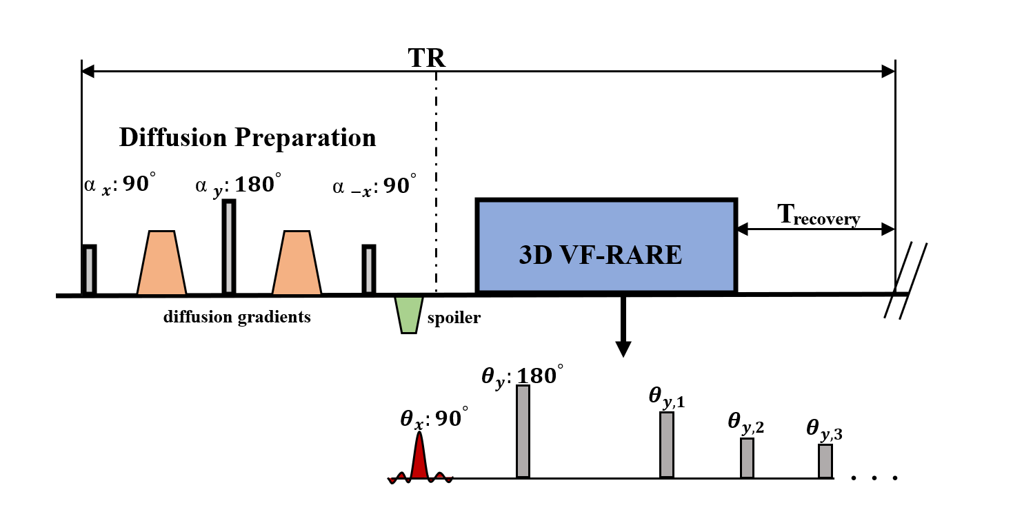
Figure 1. Sequence diagram of Diffusion
Prepared 3D VF-RARE. Diffusion preparation module was followed by a 3D VF-RARE
readout with centric phase-encoding ordering. VF-RARE consists of a
slab-selective (θx)
excitation RF pulse and a nonselective (θy)
refocusing pulse followed by a series of variable low-flip-angle nonselective
refocusing pulses (θy,
1, θy,
2,θy,
3 …). TR = 2000 ms was used in this work.
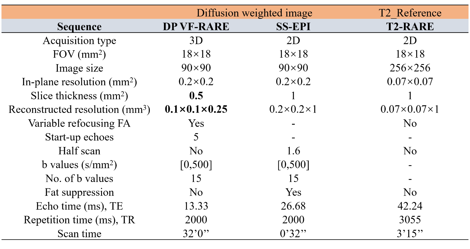
Table 1 Sequence parameters for in vivo
scans
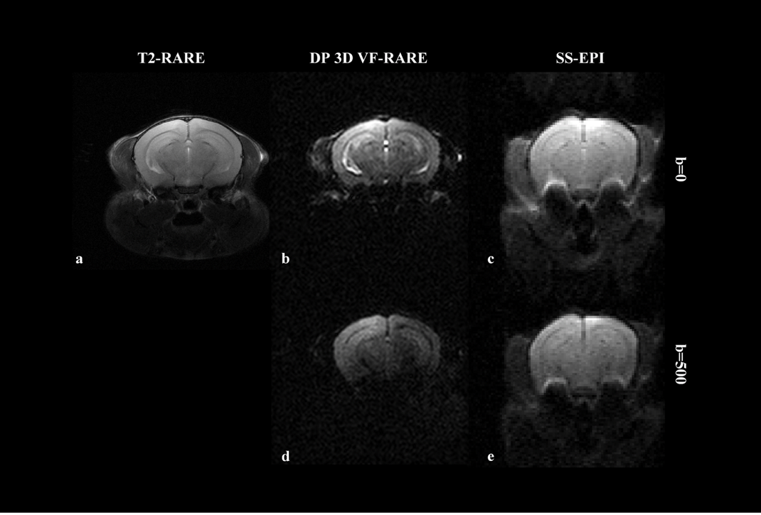
Figure
2 Comparisons of both non-diffusion and
diffusion weighted images of mouse brain acquired using Diffusion-prepared 3D
VF-RARE and single-shot EPI. a: T2-RARE was acquired as anatomy reference. b, c
: b=0 s/mm2 images of sequence DP VF-RARE and SS-EPI. d, e : b=500
s/mm2 images of sequence DP VF-RARE and SS-EPI.
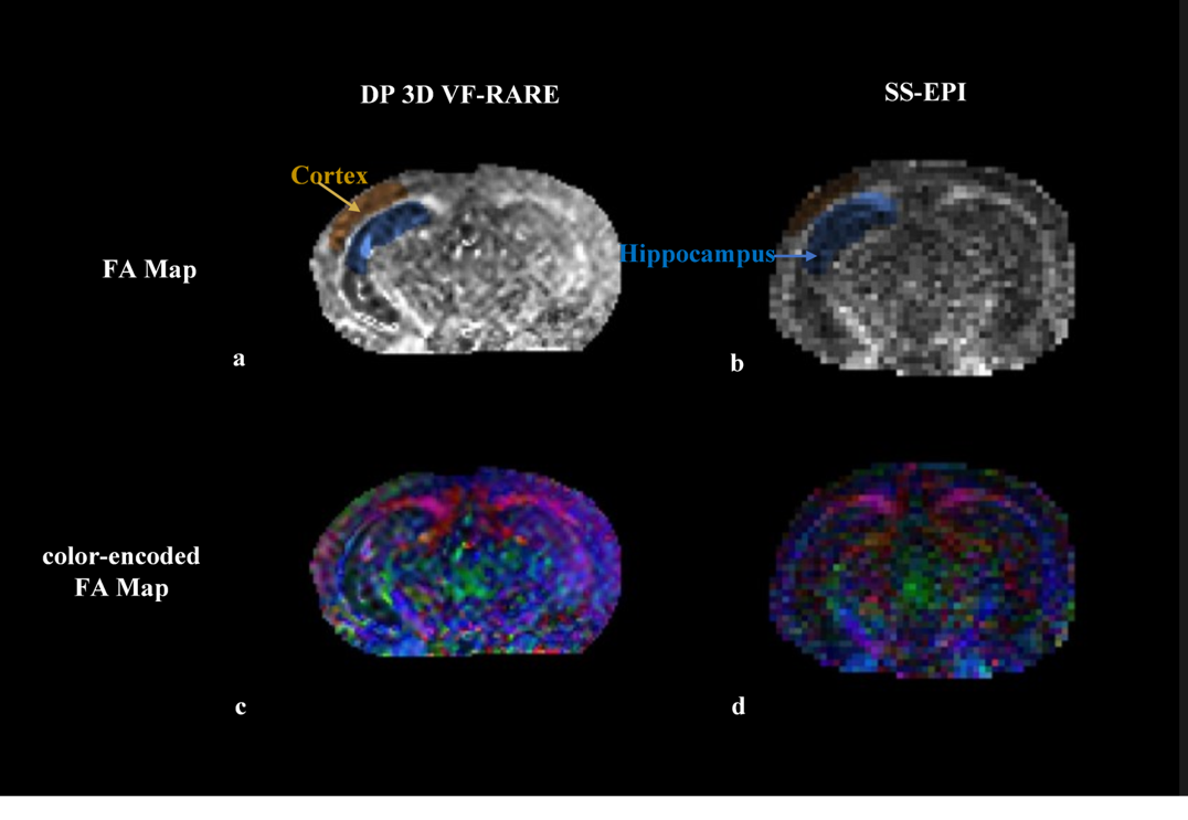
Figure 3 Comparison of in vivo FA maps (a,
b) and color-encoded FA maps (c,d) of an adult mouse brain acquired using DP 3D
VF-RARE and SS-EPI. The areas of cortex and hippocampus were used in the
following MD’s and FA’s calculation.
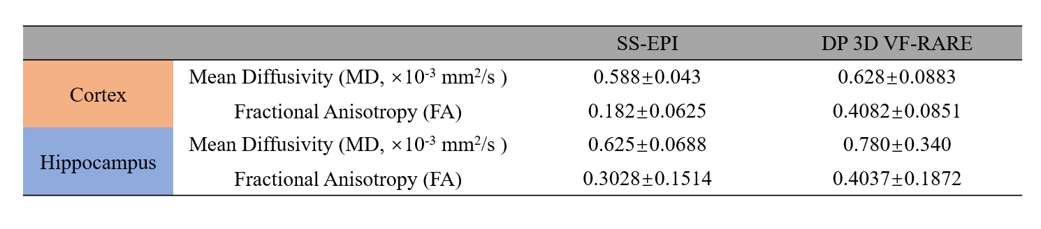
Table 2 Mean Diffusivity (MD) and
Fractional Anisotropy (FA) Measurements of the Adult Mouse Cortex and
Hippocampus. Data are presented as mean ± standard deviation (SD).