1328
Four-shot Navigator-free Spiral Acquisition Strategy for High-resolution Diffusion Imaging1Center for Biomedical Imaging Research, Department of Biomedical Engineering, School of Medicine, Tsinghua University, Beijing, China, 2Center for Magnetic Resonance Research, Radiology, Medical School, University of Minnesota, Minneapolis, MN, United States
Synopsis
Multi-shot acquisition is widely used for high-resolution diffusion weighted imaging (DWI). For multi-shot spiral DWI, one of the challenges is correcting shot-to-shot phase variations. Uniform density spiral (UDS) is a navigator-free acquisition scheme with high efficiency of spatial encoding. Previous studies indicated that POCS-ICE algorithm can iteratively solve the phase errors for navigator-free acquisitions in multi-shot diffusion imaging. Another challenge is off-resonance correction. In this study, we proposed two acquisition strategies to reduce spiral readout duration for alleviating blurring effects. High-resolution diffusion-weighted images can be acquired using a 4-shot navigator-free spiral acquisition.
Introduction
Multi-shot acquisition is a widely utilized technique in high-resolution diffusion weighted imaging (DWI). For multi-shot spiral DWI, one of the challenges is correcting the motion-induced shot-to-shot phase variations. Previous studies indicated that the phase variations can be estimated from self-navigator of variable-density spiral (VDS) acquisition 1-3. Compared to VDS, uniform-density spiral (UDS) is a navigator-free acquisition scheme with higher efficiency of spatial encoding. POCS-ICE algorithm 4 can iteratively solve the phase errors for navigator-free acquisitions. Another challenge is off-resonance correction. Spiral acquisition is sensitive to field inhomogeneity. When higher spatial resolution is required, the blurring and artifacts will become aggravated due to lengthening the readout duration. By using more interleaves, the readout duration can be shortened to reduce blurring effects, but the scan time increases correspondingly. In this study, we proposed two acquisition strategies to reduce spiral readout duration for alleviating blurring effects; then diffusion-weighted images with an in-plane resolution of 1.0 mm can be acquired using a 4-shot navigator-free spiral acquisition.Methods
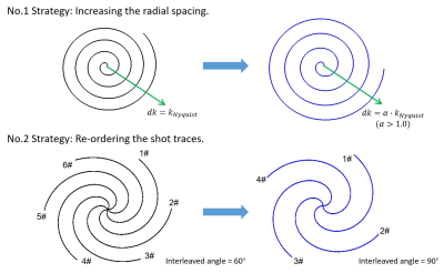
Sampling strategyIn this work, we proposed two strategies (Figure 1) to reduce the readout duration of multi-shot spiral-based diffusion imaging.
1. Increasing the radial spacing: For a base Archimedean spiral, the radial spacing satisfies Nyquist sampling requirement. (i.e. $$$dk=d_{Nyquist}$$$ ). Each interleaf moves a constant distance radially. In order to reduce the readout duration, we can increase the radial spacing appropriately to make the trajectory reach the maximum k-space radius faster. (i.e. $$$dk=$$$$$$\alpha$$$$$$d_{Nyquist}$$$, $$$\alpha$$$ is the radial spacing factor).
2. Re-ordering the shot traces: For conventional N-shot UDS, N interleaves were evenly distributed in the k-space with an interleaved angle of $$$\frac{360}{N} $$$ degrees. Currently, in order to reduce the shot number, m of the N shots were employed to acquire k-space data, then these m interleaves were re-ordered with an interleaved angle of $$$\frac{360}{m} $$$ degrees to cover k-space.
Data acquisition
MR scanning was performed on a Philips Ingenia 3.0T CX scanner using a 32-channel head coil. The maximum gradient amplitude used is 40mT/m and slew rate is 200mT/m/ms.
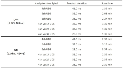
In this work, different navigator-free spiral acquisition schemes were investigated: (1) 4-shot conventional UDS with a readout duration of 41ms. (2) 5-shot conventional UDS with a readout duration of 32ms. (3) 6-shot conventional UDS with a readout duration of 28ms. (4) 4-shot variant 1# was designed based on the first strategy, $$$\alpha$$$=1.25, the readout duration is 32ms. According to the second strategy, (5) 4-shot variant 2# and (6) 4-shot variant 3# were derived from 5-shot / 6-shot conventional UDS, the readout duration is 32ms and 28ms, respectively.
Diffusion-weighted images were acquired using these spiral sampling patterns with the following scan parameters: FOV=220×220mm2, resolution=1.0×1.0×4.0mm3, acquisition matrix=220×220, b value=1000s/mm2, TE/TR=55/3000ms, 24 axial slices with a gap of 1mm. In the experiment 1: DWI with 3 orthogonal directions. Single-shot EPI DWI with resolution=2.0×2.0×4.0 mm3 at the same location was acquired as a reference, SENSE=2, PF=0.8, and TE/TR=64/3000ms. In the experiment 2: DTI with 12 diffusion directions. The other scan parameters were same as the experiment 1. SPIR technique was used to suppress fat signals. In addition, low resolution field maps acquired using multi-echo Cartesian sampling were used for off-resonance correction. The scan parameters were listed in Table 1.
Image reconstruction
The acquired multi-shot spiral diffusion-weighted images were off-line reconstructed using the POCS-ICE algorithm 4. Then these images were deblurred by conjugate phase off-resonance correction method 5. Colored FA maps were calculated using FSL toolbox.
Results and Discussion
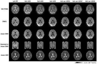
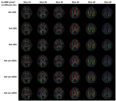
Figure 2 shows the T2-weighted images (b=0) and diffusion-weighted images (b=1000s/mm2) acquired by these different spiral acquisition schemes. Single-shot EPI DWI was shown as a reference. For conventional 4-shot UDS with a long readout duration of 41ms, there are residual blurring artifacts in the frontal lobe, where the static B0 gradient is steep6. In contrast, the T2W, single diffusion-weighted image, mean DWI acquired by conventional 5-shot and 6-shot UDS show fine anatomical details. As the number of shots increases, the blurring effects are alleviated. However, the scan time increases correspondingly. In this work, two acquisition strategies were proposed to reduce the readout duration of 4-shot diffusion imaging. The blurring effects were significantly reduced, and the image quality was improved. In addition, mean ADC maps were also shown here.Figure 3 shows the colored FA maps of six slices. The results show that fine DTI metrics can be obtained, which indicates that POCS-ICE algorithm can successfully solve motion-induced phase errors for these 4-shot spiral variants. Compared to conventional UDS sampling, there is no significant SNR degradation for these three variants because a lower acceleration factor of 1.25 was used.
In this work, we proposed two acquisition strategies to reduce the spiral readout duration, and then blurring effects can be controlled. POCS-ICE algorithm can successfully solve motion-induced phase errors and reconstruct DWI images simultaneously. High-resolution diffusion-weighted images can be acquired using a 4-shot navigator-free spiral acquisition.
Conclusion
In this study, two acquisition strategies were proposed to reduce spiral readout duration for alleviating blurring effects. Therefore, high-resolution DTI can be acquired using a 4-shot navigator-free spiral acquisition. In vivo results show that POCS-ICE algorithm has a powerful capability to solve the shot-to-shot phase errors and reconstruct DWI images simultaneously.Acknowledgements
No acknowledgement found.References
1. Liu C, Bammer R, Kim DH, Moseley ME. Self-navigated interleaved spiral (SNAILS): application to high-resolution diffusion tensor imaging. Magn Reson Med 2004;52(6):1388-1396.
2. Liu C, Moseley ME, Bammer R. Simultaneous phase correction and SENSE reconstruction for navigated multi-shot DWI with non-cartesian k-space sampling. Magn Reson Med 2005;54(6):1412-1422.
3. Li TQ, Kim DH, Moseley ME. High-resolution diffusion-weighted imaging with interleaved variable-density spiral acquisitions. J Magn Reson Imaging 2005;21(4):468-475.
4. Guo H, Ma X, Zhang Z, Zhang B, Yuan C, Huang F. POCS-enhanced inherent correction of motion-induced phase errors (POCS-ICE) for high-resolution multishot diffusion MRI. Magn Reson Med 2016;75(1):169-180.
5. Noll DC, Pauly JM, Meyer CH, Nishimura DG, Macovski A. Deblurring for non-2D Fourier transform magnetic resonance imaging. Magn Reson Med 1992;25(2):319-333.
6. Wilm BJ, Barmet C, Gross S, et al. Single-shot spiral imaging enabled by an expanded encoding model: Demonstration in diffusion MRI. Magn Reson Med 2017;77(1):83-91.
Figures


