1324
Multi-shot diffusion MRI of the human brain with motion-compensated oscillating gradients1Institute for Biomedical Engineering, ETH Zurich and University of Zurich, Zurich, Switzerland
Synopsis
High-resolution multi-shot acquisitions are commonly avoided in diffusion MRI because motion-related phase instability, which can result from diffusion encoding schemes with a nonzero first moment, often hampers image reconstruction. This issue can be circumvented through the use of motion-compensated diffusion gradient shapes derived from oscillating gradient spin-echo (OGSE) methodologies. The utility of this solution is demonstrated here for interleaved spiral scans performed using a high-performance gradient system. The robustness against motion of OGSE sequences provided a notable advantage compared to a standard diffusion sensitization sequence for the phase stability and subsequent quality of multi-shot acquisitions.
Introduction
Standard diffusion encoding schemes consisting of a pair of gradient pulses (i.e., pulsed gradient spin-echo, or PGSE) encode motion into the phase of MR signal due to the nonzero moments of the diffusion-sensitizing gradients. For multi-shot diffusion sequences, which rely on phase stability between shots, subject motion often produces sufficiently strong phase variation between shots, thereby resulting in artifacts in subsequent reconstructions.1,2 For this reason, most diffusion MRI methods are performed with single-shot readouts, like EPI or spiral, for which the resolution is limited to 2-3 mm with standard gradient systems.The use of multi-shot sequences capable of high resolution has been demonstrated with advanced correction methods,3,4 but such techniques are computationally challenging. Alternatively, motion-induced phase shifts can be reduced via motion compensation (i.e., elimination of first- and, possibly, higher-order moments) of the diffusion-sensitizing gradients, as demonstrated in single-shot cardiac diffusion imaging.5 Of particular interest are oscillating gradient spin-echo (OGSE) sequences; in their standard form, OGSE sequences are velocity-compensated (i.e., zero first-order moment) over the entire diffusion-sensitizing duration,6 as well as over each of the two oscillating gradient pulses. Compensation of higher-order moments may also be achieved through further modifications to OGSE sequences.
OGSE sequences, however, typically suffer relatively low diffusion sensitivity,6 rendering diffusion measurements vulnerable to noise. In this work, a high-performance gradient system7 is utilized to address this deficiency in order to investigate the feasibility of OGSE sequences in multi-shot acquisitions of the in-vivo human brain for the reduction of phase-induced reconstruction errors.
Methods
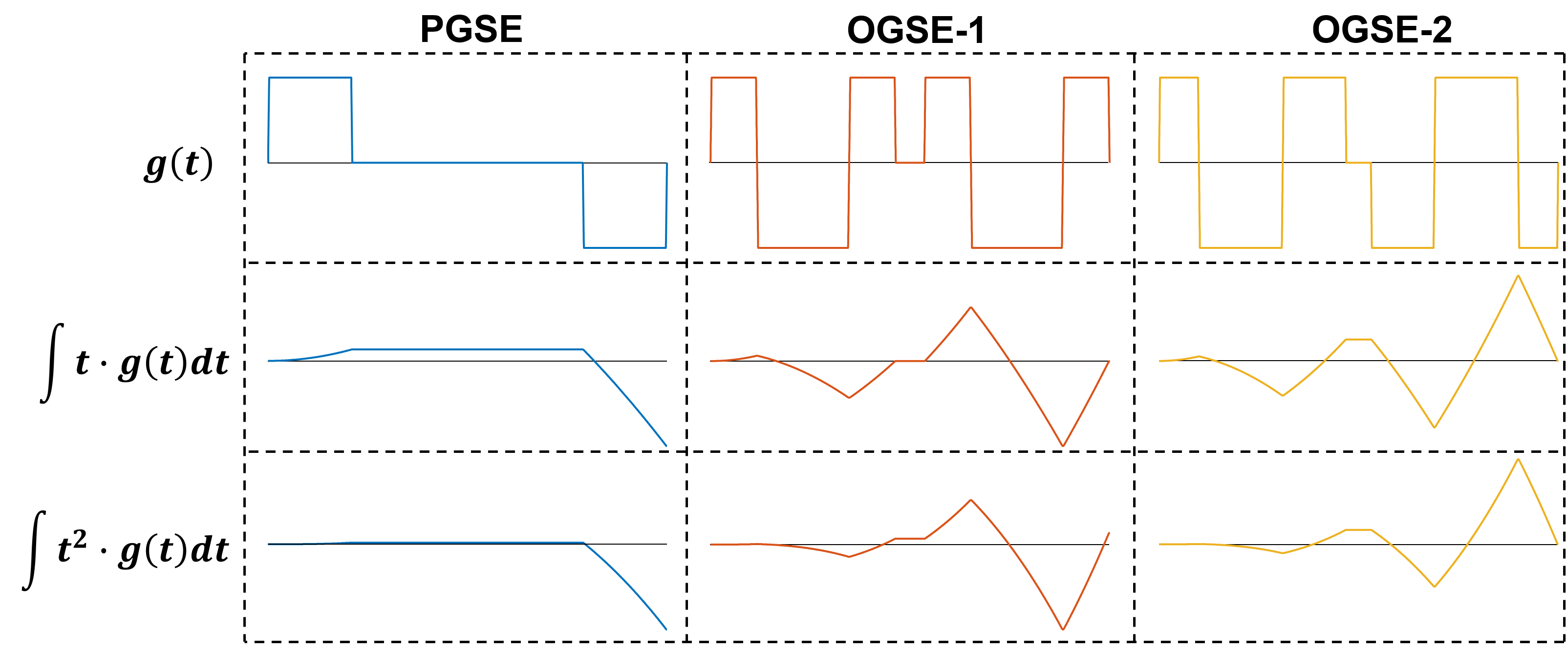
Scanning was performed with a 3T Philips Achieva system (Philips Healthcare, Best, the Netherlands) equipped with a high-performance gradient insert achieving gradient amplitudes up to 100 mT/m and slew rates up to 1200 mT/m/ms at 100% duty cycle.7 Two healthy adult volunteers were scanned with PGSE DWI and OGSE DWI sequences at b = 800 s/mm2 (10 slices, 3 mm slice thickness, 2 mm slice gap, TR/TE = 4000/75 ms, two b = 0 acquisitions, and 3 DWI directions, each aligned with a Cartesian coordinate axis). Two forms of oscillating gradient diffusion sensitization were employed: a commonly-used oscillating gradient shape at 33 Hz with apodized quarter-period trapezoidal lobes8 and a minor correction to external lobes9 (denoted OGSE-1), and a recently developed shape at 35 Hz providing increased diffusion sensitivity,10 further modified to eliminate velocity and acceleration sensitivity (denoted OGSE-2). Figure 1 provides further details on gradient shapes and moments. For all three diffusion weighting schemes, two readout variations were performed: single-shot spirals with 8 dynamics and three-shot spirals with 4 dynamics (in-plane resolution 2 mm and 1 mm, respectively). Readout trajectories were designed in-house to exploit the maximum gradient amplitudes and slew rates denoted above.Following scanning, the spiral readouts of each sequence were monitored using a field camera11 (Skope Magnetic Resonance Technologies, Zurich, Switzerland); the field probe data were fitted to a third-order spherical harmonic model of the magnetic field dynamics for each readout. Images were reconstructed using the resulting information, as well as off-resonance maps, in a higher-order algebraic reconstruction algorithm,12 and reconstructed images were smoothed with a Hamming filter.
Results
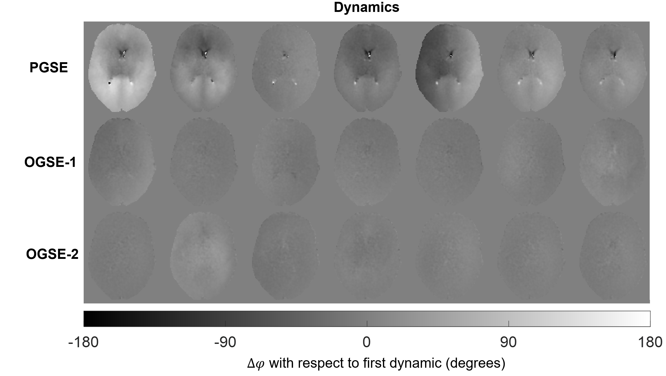
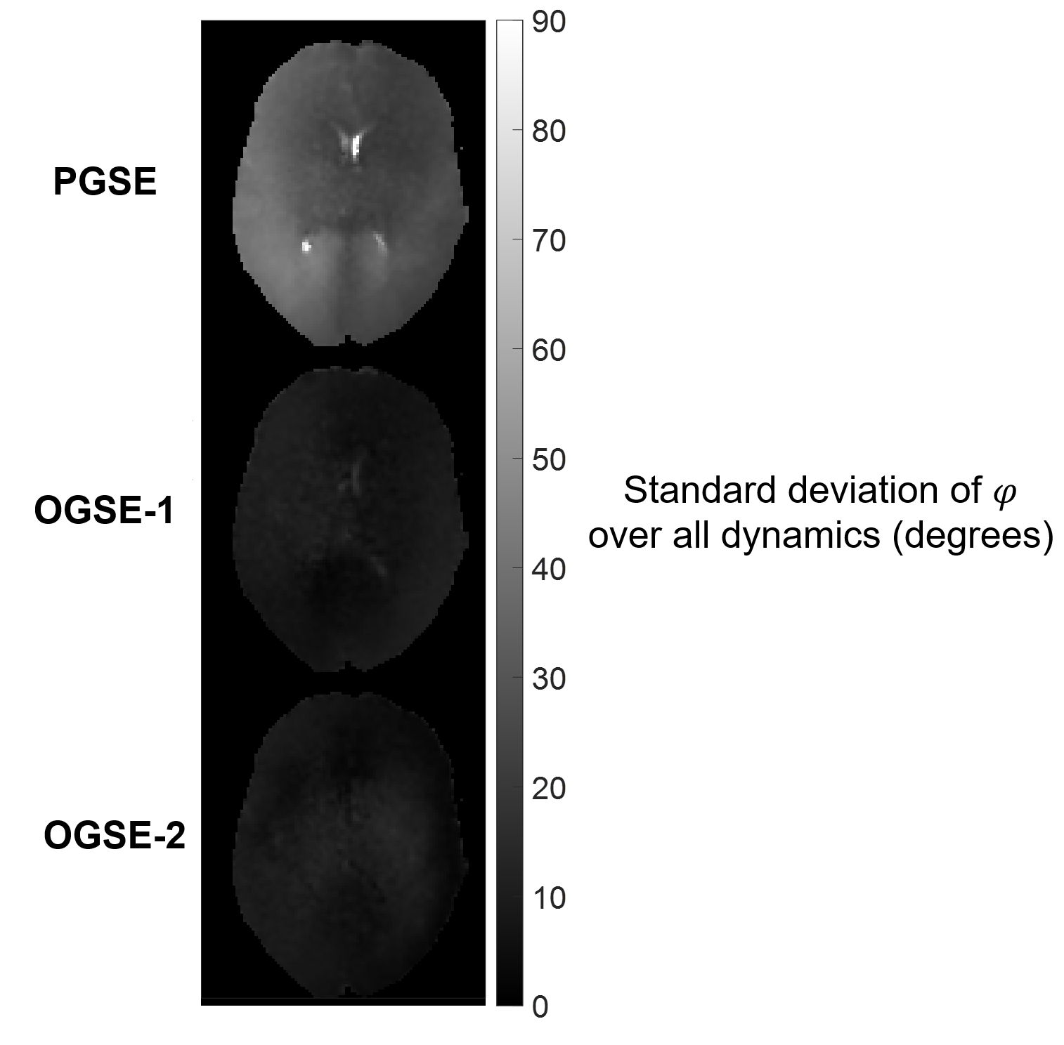
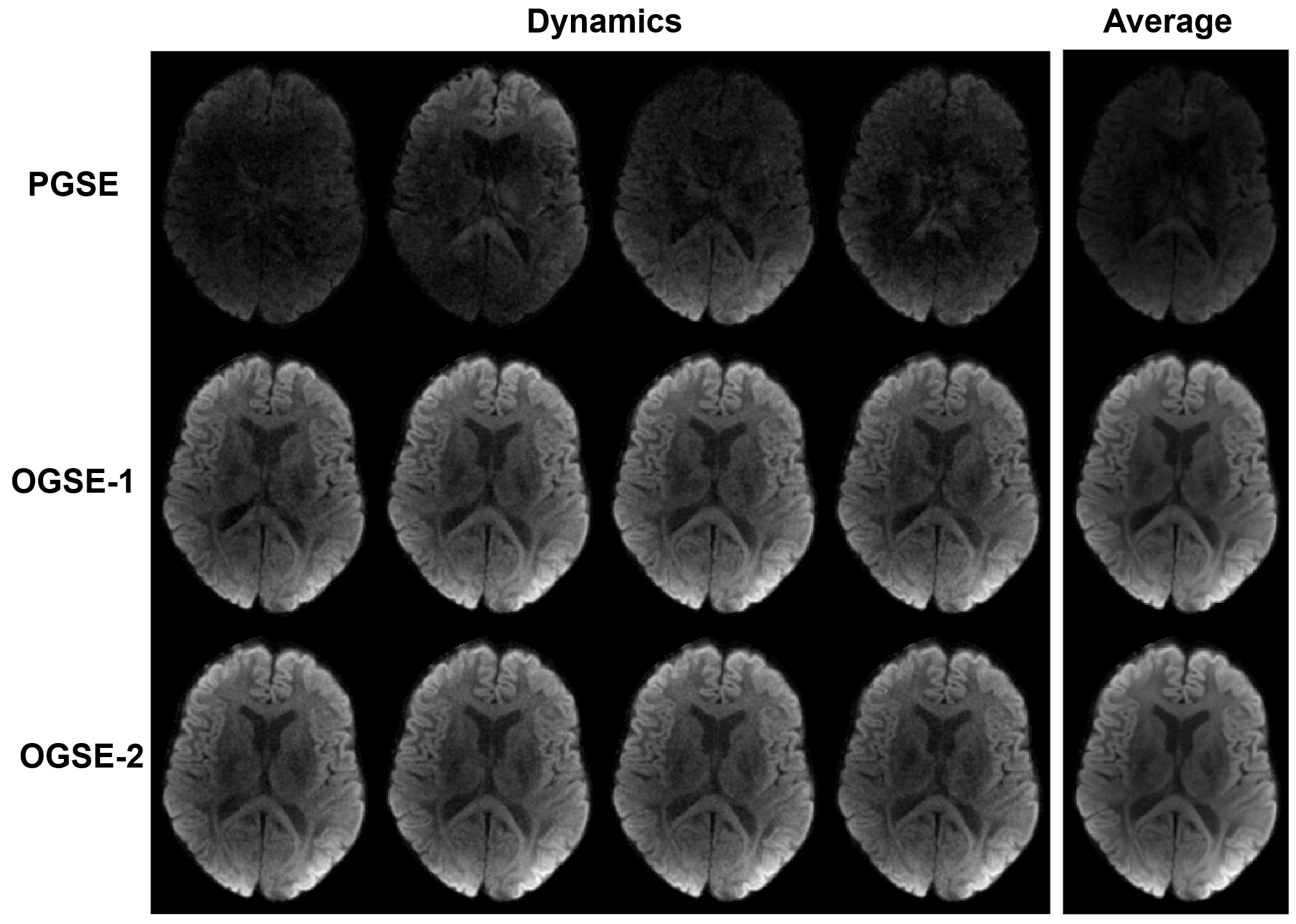
Figure 2 depicts the phase fluctuations of single-shot acquisitions over several dynamics for each method of diffusion sensitization. The phase variations of the OGSE acquisitions are substantially diminished with respect to those of the PGSE acquisition, as illustrated further in Figure 3, which contains maps the standard deviation of the phase variation for the slice shown in Figure 2. Figure 4 shows reconstructions of each dynamic of the multi-shot acquisitions for a single DWI direction (aligned with the x-axis), as well as the complex average of these images.Discussion
The utility of OGSE shapes is apparent based on the phase variability of repeated single-shot acquisitions: for the slice shown in Figure 3, the spatially averaged standard deviation of phase is 17.8° for the PGSE acquisition, and just 5.0° and 4.6° for OGSE-1 and OGSE-2, respectively. Phase variability was consistently lower for OGSE-2 than for OGSE-1 images (albeit by a small amount) across all the data, indicating a slight advantage of acceleration compensation over shorter interval velocity compensation. The relative lack of phase fluctuations for first- and second-order motion-compensated diffusion encoding notably benefitted multi-shot acquisitions; the OGSE images are practically artifact-free. On the contrary, the PGSE images (even after averaging) suffer severe artifacts, leaving the images diagnostically meaningless without further corrections. This improvement was observed for all diffusion directions and for both subjects, for both OGSE variants.The feasibility of the comparison among diffusion-weighting gradient shapes performed here was enabled by the gradient system utilized in this work. For the same OGSE shapes applied here, a modern clinical gradient system (e.g., amplitudes up to 80 mT/m and slew rates up to 200 mT/m/ms) could have reached a maximum b-value (considering both OGSE shapes) of about 550 s/mm2 in the same TE, which is a considerable reduction in sensitivity.
Conclusion
The first- and second-order motion compensation provided by OGSE sequences, performed with a high-performance gradient system, permitted high-resolution, interleaved diffusion measurements of the in-vivo human brain. With this implementation, typical problems of multi-shot acquisitions associated with motion sensitivity were avoided without the need for advanced computational methods.Acknowledgements
No acknowledgement found.References
1. Anderson AW, Gore JC. Analysis and correction of motion artifacts in diffusion weighted imaging. Magn Reson Med. 1994;32(3):379-387.
2. Norris DG. Implications of bulk motion for diffusion-weighted imaging experiments: Effects, mechanisms, and solutions. J Magn Reson Imaging. 2001;13(4):486-495.
3. Chen N-k, Guidon A, Chang H-C, Song AW. A robust multi-shot scan strategy for high-resolution diffusion weighted MRI enabled by multiplexed sensitivity-encoding (MUSE). Neuroimage. 2013;72:41-47.
4. Mani M, Jacob M, Kelley D, Magnotta V. Multi-shot sensitivity-encoded diffusion data recovery using structured low-rank matrix completion (MUSSELS). Magn Reson Med. 2017;78(2):494-507.
5. Stoeck CT, von Deuster C, Genet M, Atkinson D, Kozerke S. Second-order motion-compensated spin echo diffusion tensor imaging of the human heart. Magn Reson Med. 2016;75(4):1669-1676.
6. Van AT, Holdsworth SJ, Bammer R. In vivo investigation of restricted diffusion in the human brain with optimized oscillating diffusion gradient encoding. Magn Reson Med. 2014;71(1):83-94.
7. Weiger M, Overweg J, Rösler MB, et al. A high-performance gradient insert for rapid and short-T2 imaging at full duty cycle. Magn Reson Med. 2018;79(6):3256-3266.
8. Does MD, Parsons EC, Gore JC. Oscillating gradient measurements of water diffusion in normal and globally ischemic rat brain. Magn Reson Med. 2003;49(2):206-215.
9. Baron CA, Beaulieu C. Oscillating gradient spin-echo (OGSE) diffusion tensor imaging of the human brain. Magn Reson Med. 2014;72(3):726-736.
10. Hennel F, Michael ES, Pruessmann KP. Improved gradient waveforms for oscillating gradient spin-echo (OGSE) diffusion tensor imaging. NMR Biomed. 2020;e4434. DOI: 10.1002/nbm.4434.
11. Dietrich BE, Brunner DO, Wilm BJ, et al. A field camera for MR sequence monitoring and system analysis. Magn Reson Med. 2016;75(4):1831-1840.
12. Wilm BJ, Barmet C, Pavan M, Pruessmann KP. Higher order reconstruction for MRI in the presence of spatiotemporal field perturbations. Magn Reson Med. 2011;65(6):1690-1701.
Figures