1314
Noise reduction in diffusion tensor imaging of the brachial plexus using single-shot DW-EPI with Compressed SENSE1Department of Radiology, Chiba University Hospital, Chiba, Japan, 2Diagnostic Radiology and Radiation Oncology, Graduate School of Medicine, Chiba University, Chiba, Japan, 3Philips Japan, Tokyo, Japan
Synopsis
The brachial plexus is a difficult region to obtain high-quality DTI. In DTI with SENSE, noise-like artifacts appear in the center of the image, resulting in poor image quality. It has been reported that C-SENSE (EPICS), which combines parallel imaging and compressed sensing, can reduce parallel imaging-derived artifacts and may be a more robust method for quantitative imaging than SENSE. This study compared EPICS with SENSE and SENSE×2 (same conditions as SENSE with increased NEX) in the brachial plexus region, and EPICS was a robust imaging method producing better reproducibility of ADC and FA values than SENSE and SENSE×2.
Introduction
Diffusion tensor Imagine (DTI) is a method that uses multiple diffusion-weighted images (DWI) with different directions of motion probing gradient (MPG). Since this method can produce anisotropy of water molecules, it can image nerve fibers indirectly. Although it has been used mainly in the brain, it has recently begun to be applied for the evaluation of peripheral nerves such as the sciatic nerve and brachial plexus.1 The brachial plexus is an area that is easily affected by peripheral neuropathy , so that it is often the target of MRI. However, it is difficult to obtain DTI with good image quality due to the inhomogeneity of the magnetic field and motion artifacts. DWI is generally selected by combining single-shot EPI with parallel imaging (SENSE), a type of high-speed imaging method, to correct for magnetic field distortions. However, DWI with SENSE produces noise-like artifacts in the center of the image, resulting in degrading image quality.2,3 These artifacts are likely to affect the quantitative values such as apparent diffusion coefficient (ADC) and fractional anisotropy (FA). An increasing number of excitations (NEX) is a conventional method to reduce noise. Recently, a combination of parallel imaging and compressed sensing technique (Compressed SENSE, C-SENSE) has been developed to accelerate the acquisition time without increasing the image artifacts.4 EPI with C-SENSE (EPICS) would be an application of the C-SENSE framework for reducing the noise artifacts in single-shot EPI images.5,6 EPICS has a potential to become a more robust method of DTI for the brachial plexus than conventional SENSE. The purpose of this study was to compare EPICS, SENSE, and SENSE with increased NEX (SENSE×2) imaging in the brachial plexus region and to investigate the feasibility of EPICS.Methods
MethodsThe subjects were 17 healthy volunteers (median age 34, age-range 24-59, 15 men and 2 women). The imaging system was a 3.0T MRI (Ingenia 3T, Philips Healthcare). The brachial nerve area was imaged with EPICS, SENSE (SENSE combined with single-shot EPI), and SENSE×2 (same conditions as SENSE, with NEX set to 2). The same scan parameters were used for comparison : voxel size of 2*2*4mm, FOV of 350mm, 60slices, b-value of 0 and 800 mm2/s, TR of 10000ms, TE of 68ms, SENSE/C-SENSE acceleration factor of 3 . Regions of interest (ROIs) were set at 24 locations (Fig. 1) in the ganglion and nerve root of the C5 to Th1 nerves and the trunks and cords of the brachial plexus. The mean ADC and FA values in the ROIs were used for analyses. The coefficient of variation (CV) within the ROI was calculated as follows: CV= Standard Deviation (SD) of ADC or FA on each case / Average value of ADC or FA on each case.CV of ADC and FA were defined as CVADC and CVFA, respectively. These were used to evaluate the reproducibility of ADC and FA values.ADC, FA, CVADC , and CVFA were tested by Wilcoxon signed rank-sum test. Intraclass correlation coefficients (ICC) were calculated for ADC and FA values to evaluate reproducibility among three protocols.Results
Fig. 2 shows the results of ADC and CVADC.ADC values were higher in the order of EPICS, SENSE×2, and SENSE, and there was a significant difference between the protocols.CVADC values were lower in the order of EPICS, SENSE, and SENSE×2, and EPICS was significantly smaller than the other protocols (P<0.001 and P=0.006).Fig. 3 shows the results of FA and CVADC. FA was higher in the order of SENSE, EPICS, and SENSE×2, and there were significant differences. CVFA was lower in the order of EPICS, SENSE, and SENSE×2, and there were significant differences between SENSE×2 and the others (P<0.001). There were significant differences in FA value and CVFA when nerve roots alone were analyzed (P<0.001). Fig. 4 shows the results of ICC analysis. ICC for SENSE×2 was less consistent for EPICS and SENSE, respectively (0.22 and 0.19).Discussion
In EPICS, the CVADC was the smallest, which meant the reproducibility of quantitative values is higher than SENSE and SENSE×2. There was a significant difference in FA values between EPICS and SENSE in nerve roots alone. This difference may be due to the effect of the artifact in the center of the image that is characteristics of SENSE (Fig. 5). The FA values of SENSE×2 was not consistent with those of the other two protocols. The brachial plexus is a region that is strongly affected by respiration.SENSE×2 has a longer acquisition time than the other two protocols. The method of increasing NEX causes a motion shift during imaging. This may have had a significant effect on the quantitative values, especially the anisotropic FA. In the brachial nerve region, the background shows farther quantitative values from the nerves because the background structure barely produces signals on DWI. The values varied greatly depending on the size and position of the ROI (Fig. 5).EPICS produced high-quality images, and it was feasible to reduce artifacts from the background and to measure robust ADC and FA.Conclusion
DTI with EPICS was a robust imaging method with better reproducibility of ADC and FA values than SENSE and SENSE×2.Acknowledgements
No acknowledgement found.References
1. Ryckie G. , Alexander W, Irvin T et al. Diffusion tensor imaging of the roots of the brachial plexus:a systematic review and meta‑analysis of normative values. Clin Transl Imaging. 2020;8(6):419-431
2. Patricia N, Roland B, Caroline R et al. Parallel Imaging Artifacts in Body Magnetic Resonance Imaging. Can Assoc Radiol J. 2009;60: 91–98.
3. Yanasak NE, Michael J .MR imaging artifacts and parallel imaging techniques with calibration scanning: a new twist on old problems.Radiographics.2014;34:532-48.
4. Geerts-Ossevoort L, et al. Compressed SENSE Speed done right. Every time. The Netherlands: Philips Healthcare; 2018 Jan. Report No: 4522 99131821. https://www.philips.de/content/dam/b2bhc/de/resourcecatalog/landingpages/ingeniaelition/White_Paper_Compressed_SENSE-opt.pdf
5. Yoneyama M, Kosuke M, Johannes P et al. Noise Reduction in Prostate Single-Shot DW-EPI utilizing Compressed SENSE Framework. Proc. ISMRM. 2019:1634.
6. Kayoko A, Kazufumi S, Masami Y et al. Ultra-high b-value single-shot echo planar diffusion-weighted imaging with Compressed SENSE. Proc. ISMRM. 2020:4322.
Figures
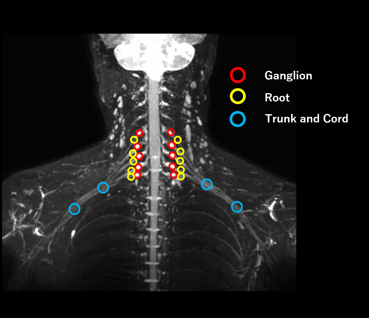
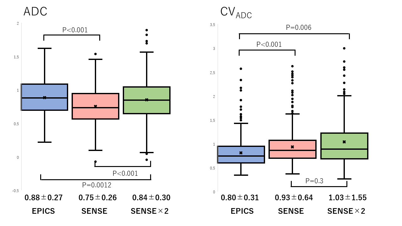
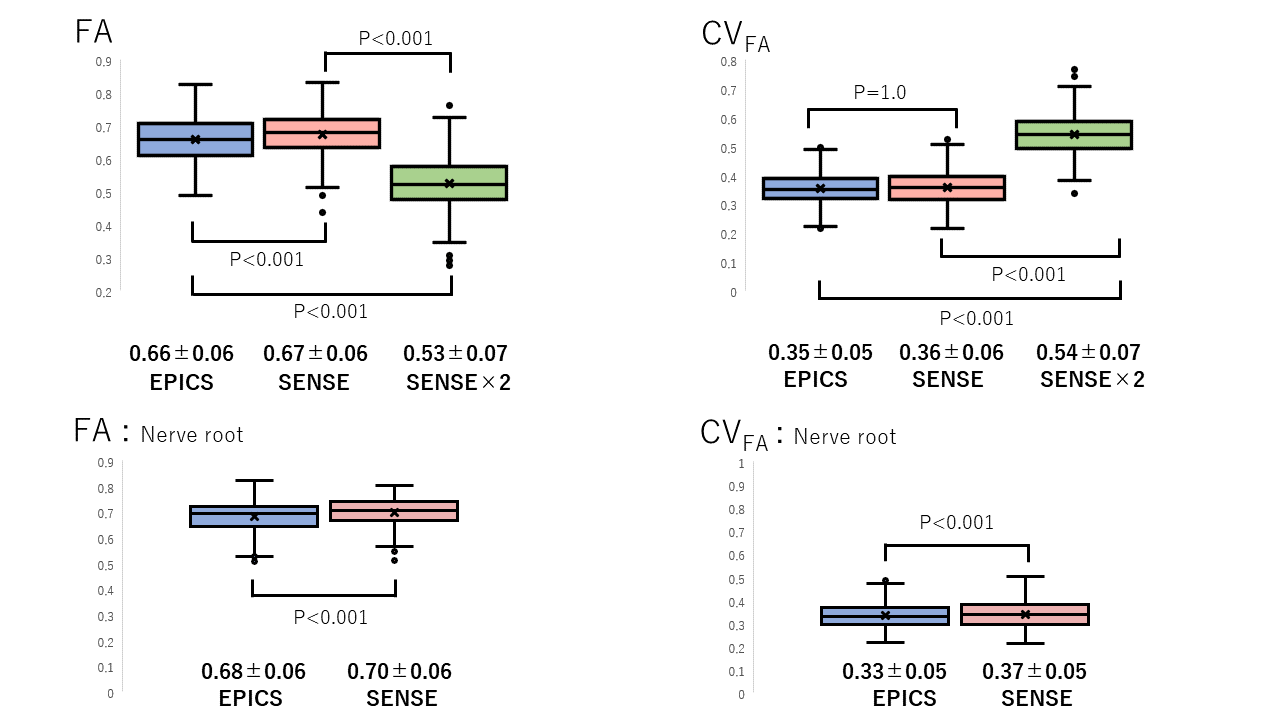
Figure 3.(Upper column) Fractional anisotropy (FA) and coefficient of variance (CVFA) of EPICS, SENSE, and SENSE×2. FA was significantly different among the three protocols, and CVFA was significantly smaller in EPICS and SENSE than in SENSE×2. (Lower column) Focusing on the nerve roots, FA in EPICS was significantly smaller and CVFA was smaller than SENSE.
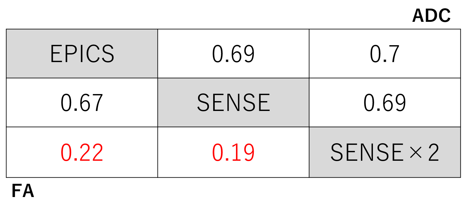
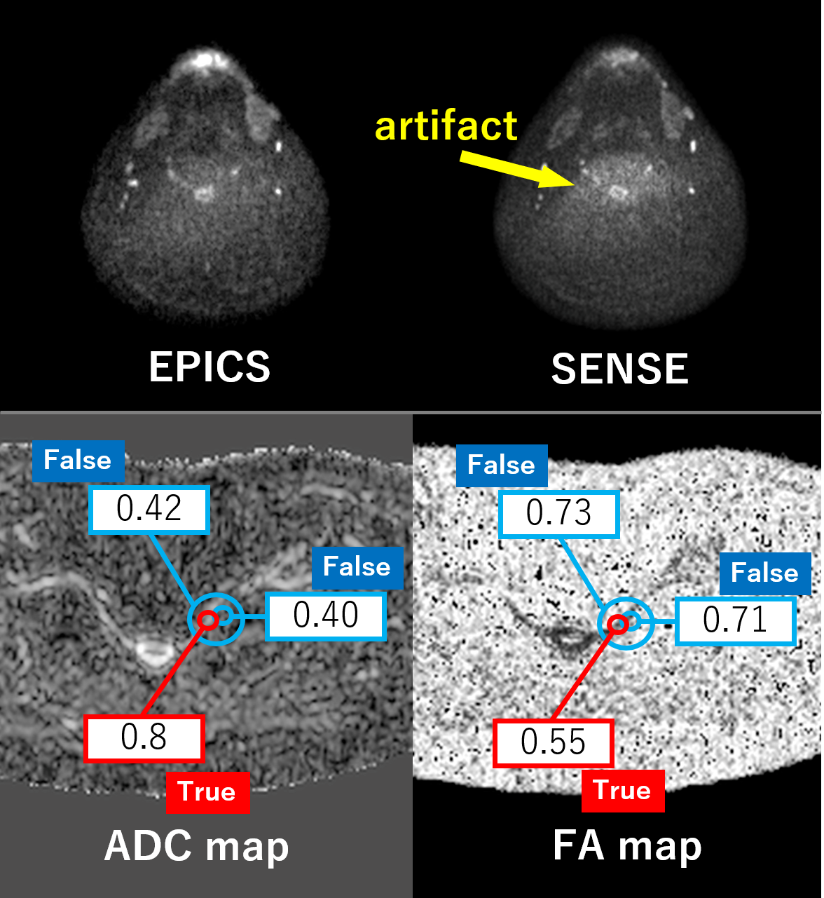
Figure 5. (Upper column) An example of noise-like artifacts in SENSE. The nerve root region is noisy, which may contribute to the reduced coefficients of variation in this region. (Lower column) There is a lot of noise in the area outside the nerve root. In the region of noise, ADC value is low and FA value is high. When the ROI is placed outside the nerve, the values are greatly shifted. It is important to draw ROIs on noise-free images.