1312
Whole-brain Diffusion Tensor Imaging Using Single-Shot Spiral Sampling1Center for Biomedical Imaging Research, Department of Biomedical Engineering, School of Medicine, Tsinghua University, Beijing, China, 2Center for Magnetic Resonance Research, Radiology, Medical School, University of Minnesota, Minneapolis, MN, United States
Synopsis
Single-shot acquisition techniques are commonly used to acquire diffusion weighted images. Single-shot spiral sampling allows shorter TE acquisition, thus provides higher SNR compared to EPI acquisition. However, spiral acquisition is sensitive to field inhomogeneity. Blurring effects degrade the quality of spiral images. In this study, compared to previous studies, a relatively large acceleration factor was used to reduce the spiral readout duration, and off-resonance correction was implemented for deblurring. The results show that the proposed single-shot spiral sampling can achieve whole-brain diffusion tensor imaging.
Introduction
Diffusion weighted imaging (DWI) is a very important imaging technique for clinical diagnosis and neuroscience research. Nowadays, DWI is generally performed using Cartesian EPI acquisition due to its high scan efficiency. Center-out spiral k-space sampling allows shorter TE acquisition, thus provides higher SNR compared to EPI acquisition 1-4. Single-shot imaging is the fastest and most efficient acquisition scheme. Currently, single-shot spiral technique has not been widely utilized in diffusion imaging. The major problem of single-shot spiral DWI is blurring and artifacts introduced by field inhomogeneity. In this work, compared to previous studies 1,3, a relatively large acceleration factor was used to reduce the spiral readout duration, and a robust off-resonance correction method was implemented for deblurring. In vivo results show that single-shot spiral sampling can achieve whole-brain diffusion tensor imaging.Methods
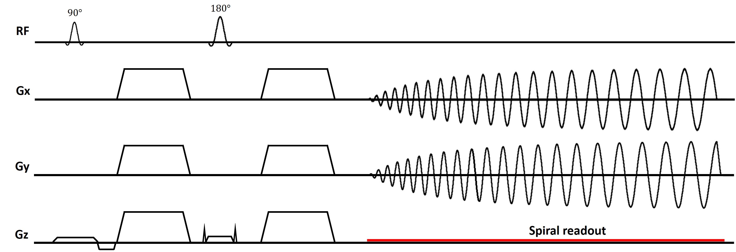
All spiral diffusion weighted imaging experiments were performed using a Stejskal-Tanner diffusion sequence (Figure 1) on a Ingenia 3.0T CX scanner (Philips Healthcare, Best, The Netherlands) using a 32-channel head coil. The gradient system was operated at a maximum gradient strength of 40 mT/m and with a slew rate of 200 mT/m/ms. Phantom studies were conducted to validate the imaging protocol and optimize the experiment design. Two healthy volunteers were recruited to participate in the following experiments, respectively.Experiment 1: FOV=220×220mm2, resolution=1.3×1.3×4.0mm3, acquisition matrix=168×168, b value=1000 s/mm2, 12 diffusion directions, TE/TR=55/3000ms, readout duration=26.0ms, SENSE=4. 24 axial slices with a gap of 1mm cover the whole brain. Single-shot EPI DTI with resolution=1.5*1.5*4.0 mm3 at the same location was acquired as a reference, SENSE=2, PF=0.75, and TE/TR=65/3000ms. In addition, T2W TSE and T2W Flair images were also acquired using conventional Cartesian sampling.
Experiment 2: FOV=220×220mm2, in-plane resolution=1.3×1.3mm2, b value=1000 s/mm2, 8 diffusion directions, TE/TR=55/3100ms, readout window=26.0ms, SENSE=4. 70 axial slices (no gap) with slice thickness of 2mm were used to cover the whole brain.
In all experiments, Spectral Presaturation with Inversion Recovery (SPIR) technique was used to suppress fat signals. In addition, low resolution B0 field maps acquired using multi-echo Cartesian sampling were used for deblurring. The single-shot spiral diffusion weighted images were off-line reconstructed, followed by conjugate phase off-resonance correction 5. Colored FA maps were calculated using FSL toolbox.
This study was approved by the Institutional Review Board and written informed consent was obtained from all the participants.
Results and Discussion
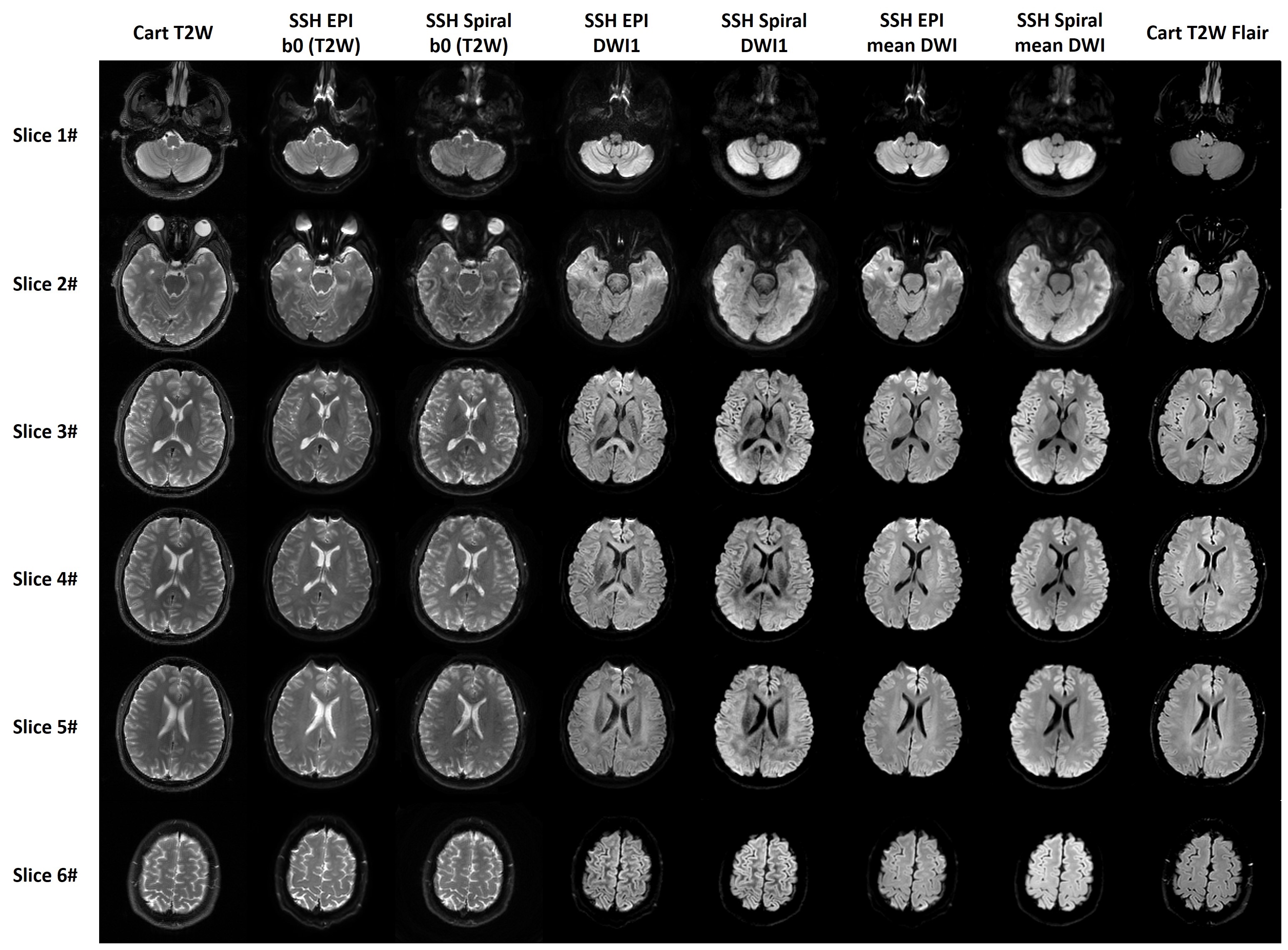
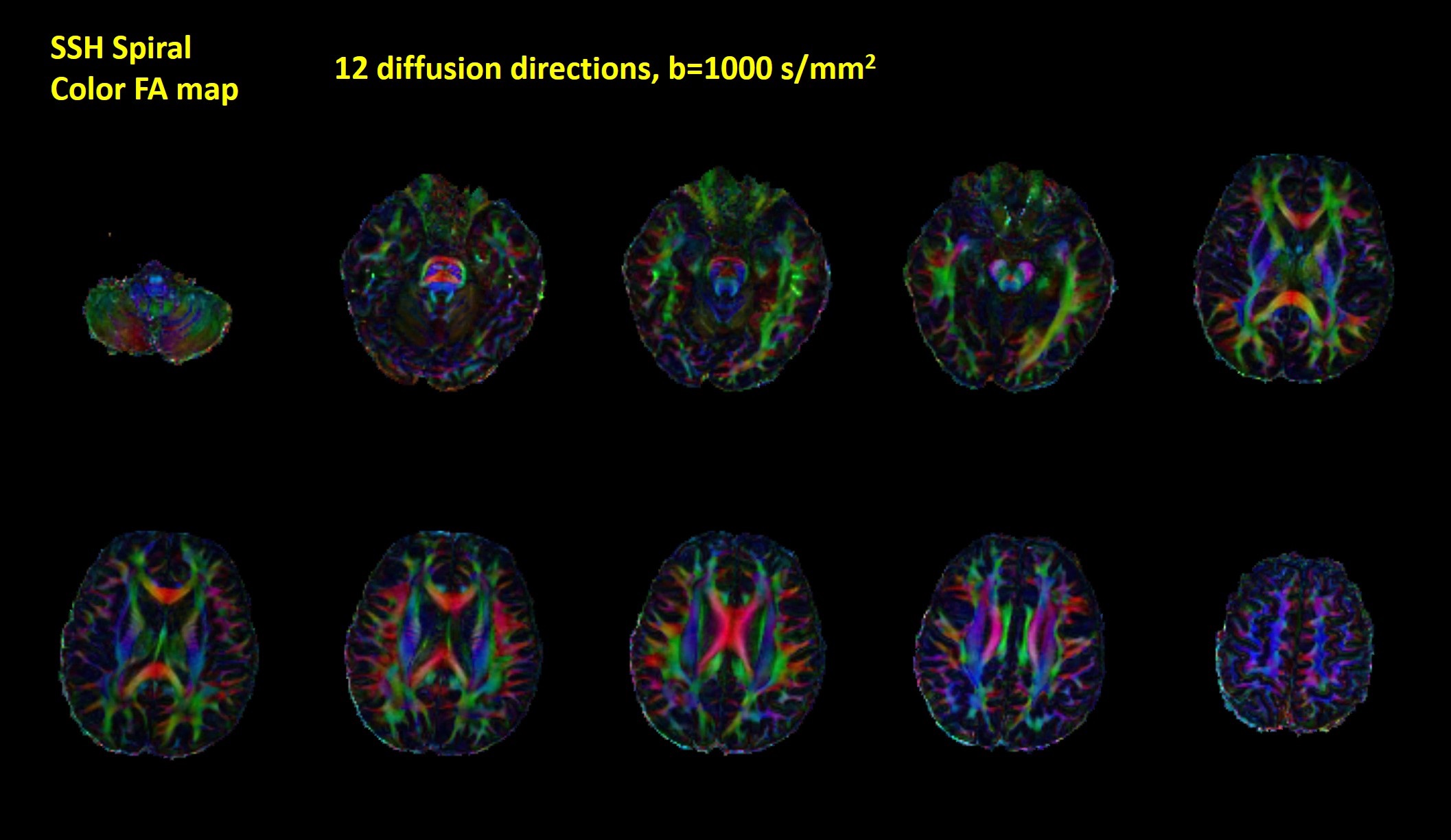
Figure 2 shows the single-shot spiral diffusion-weighted images. Image quality was improved after off-resonance correction. Single-shot EPI DWI, T2 weighted TSE and Flair images are also shown as reference. There are multiple hyper-intensities due to signal pile-up and noticeable distortions in the single-shot EPI images. In comparison, in the single-shot spiral images, slight residual blurring artifacts can be observed in the frontal lobe. Off-resonance related artifacts are visible near the sinuses and ear canal where the field inhomogeneity is severe. In general, fine anatomical details without distortions are shown in the single-shot spiral images, which have higher SNR, and better geometry fidelity than single-shot EPI images.Colored FA maps of ten representative slices covering the whole brain are shown in Figure 3. Accurate FA maps can be obtained in areas where field inhomogeneity is severe, such as in the brainstem. Based on the results, single-shot spiral diffusion imaging can provide accurate DTI metrics.
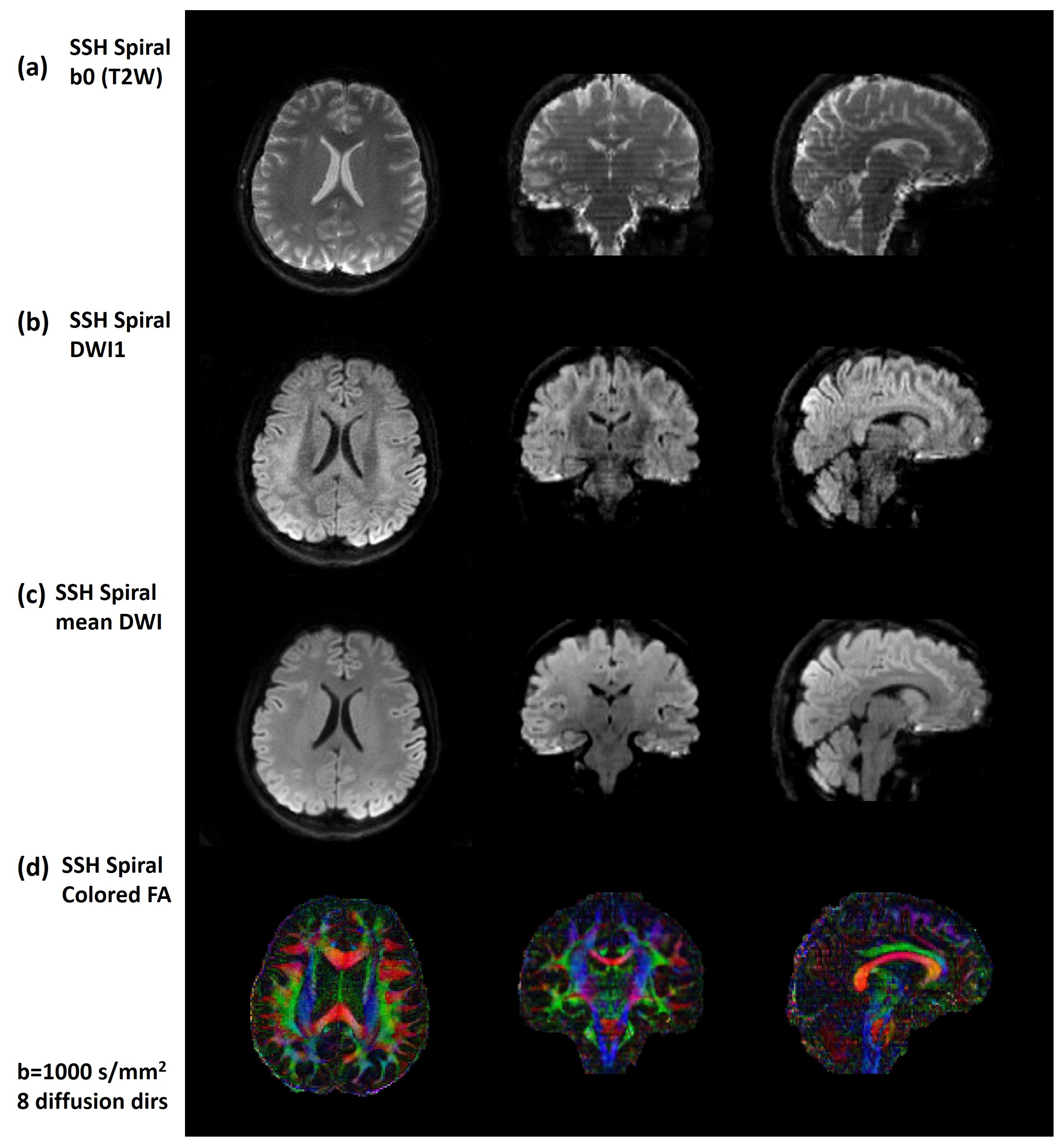
The reconstructed single-shot spiral diffusion images with an in-plane resolution of 1.3mm and slice thickness of 2.0mm are shown in Figure 4. Due to the high SNR efficiency of spiral sampling, whole-brain diffusion imaging with thin slice can be achieved using 2D spiral-out acquisition. After interpolation and reformation, axial, coronal and sagittal planes of b0 (T2W), single diffusion weighted image (DWI1), mean DWI with 1.3 mm isotropic resolution and colored FA maps are both shown here.
In this work, we found that spiral acquisition is sensitive to field inhomogeneity. Blurring effects degrade the quality of spiral images. To deal with this problem, on the one hand, a relatively large acceleration factor of 4 was used to reduce the spiral readout duration. On the other hand, accurate field maps are essential for off-resonance correction.
Conclusion
This study demonstrates that single-shot spiral sampling can achieve whole-brain diffusion tensor imaging with shorter TE and higher SNR. In addition, single-shot spiral DWI provides accurate DTI metrics, and has higher anatomical accuracy than single-shot EPI DWI.Acknowledgements
No acknowledgement found.References
1. Wilm BJ, et al. Diffusion Imaging with Very High Resolution and Very Short Echo Time. Proc ISMRM 2020; 0965.
2. Wilm BJ, et al. Single-Shot Spiral Imaging Enabled by an Expanded Encoding Model: Demonstration in Diffusion MRI. Magnetic Resonance in Medicine (2017) 77:83–91
3. Peter Börnert, et al. Single-shot Diffusion-weighted Spiral Imaging in the Brain on a Clinical Scanner. Proc ISMRM 2019; 0243.
4. Lee Y, et al. High-Resolution Diffusion MRI: In-Vivo Demonstration of the SNR Benefit of Single-Shot Spiral Acquisition vs. EPI. Proc ISMRM, 2019; 0767.
5. Noll DC, Pauly JM, Meyer CH, Nishimura DG, Macovski A. Deblurring for non-2D Fourier transform magnetic resonance imaging. Magn Reson Med 1992;25(2):319-333.
Figures