1291
3D Quantitative MRI of the Brain: Effects of B1 Inhomogeneity in 3D-QALAS1CMIV, Linköping University, Linköping, Sweden, 2Medical Radiation Physics, Linköping University, Linköping, Sweden, 3Siemens Healthcare AB, Malmö, Sweden
Synopsis
We have implemented the 3D QALAS sequence for a 3 T MR-scanner and validated the accuracy and evaluated the effect of B1+ inhomogeneities on the resulting R1 and R2 estimations. When using a small flip angle of 4° the determined R1 and R2 maps showed a high accuracy; however, for 10° there was a significant bias and dependence on B1+ inhomogeneities.
Introduction
Quantitative MRI (qMRI) provides objective tissue properties and can e.g. be used for various tissue classification [1] or for higher sensitivity of infiltrating tumor [2]. However, many robust qMRI techniques have been limited to 2D. In this project, the aims were to implement the 3D qMRI method QALAS (3D-QuAntification using an interleaved Look-locker Acquisition Sequence with T2 preparation pulse) [3], to validate the accuracy and to investigate the effect of B1 inhomogeneities.Materials and Methods
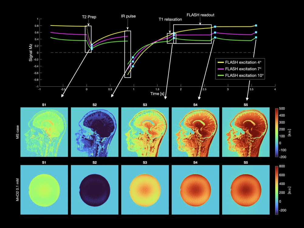
Implementation of QALASThe QALAS acquisition (cf. Figure 1) was implemented as a custom-built sequence, and experiments were performed on a 3 T MAGNETOM Prisma (Siemens Healthcare, Erlangen, Germany).
Phantoms and Patients
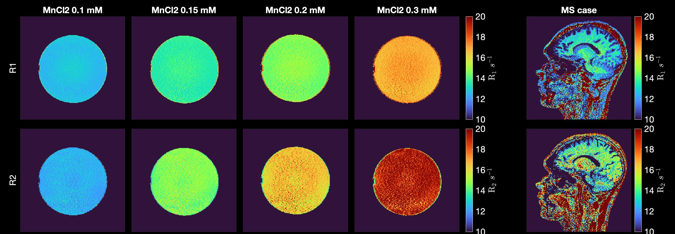
For the validation of accuracy and B1+ inhomogeneity, a set of four custom-made spherical FUNSTAR phantoms (Gold Standard Phantoms, London, UK) were used. The four different phantoms contained a range of MnCl2 concentrations of 0.10, 0.15, 0.20, and 0.30 mM. One MS patient (male, 27 y) was also examined using the implemented QALAS sequence.
MR experiments
For the phantom examinations, two types of QALAS data were acquired: one set with FLASH excitation flip angles of 4°or 10°, with an isotropic resolution of (1.2 mm)3, acquisition matrix 210×208×144, GRAPPA-factor 3 with 48 reference lines, resulting in a total acquisition time of 6 min. The QALAS examinations were scanned in SAG, COR, and TRA orientation for all four phantoms resulting in 12 volumes with 4° and 10°, respectively. For gold standard R1 quantification, a single-slice IR sequence with 10 IR times 35, 75, 100, 125, 250, 1000, 2000 to 3000 ms was used, with a total acquisition time of 25 min. And, for R2 estimation, a single-slice multi-echo sequence with TR 5 s, 32 echoes with TE from 10 to 320 ms in steps of 10 ms was used, resulting in a total time of 16 min. B1+ field maps were acquired using a B1-mapping sequence available on the scanner.
All data were post-processed using MATLAB 2020b (Mathworks inc., USA). The QALAS data was fitted using a least-squares fitting procedure, assuming mono-exponential relaxation, inversion pulse of 180°, and excitation pulse of either 4° or 10° in the FLASH readout. Gold standard R1 was post-processed using a grid fit method [4]. Gold standard R2 was estimated by least-squares fitting of a mono-exponential.
Results
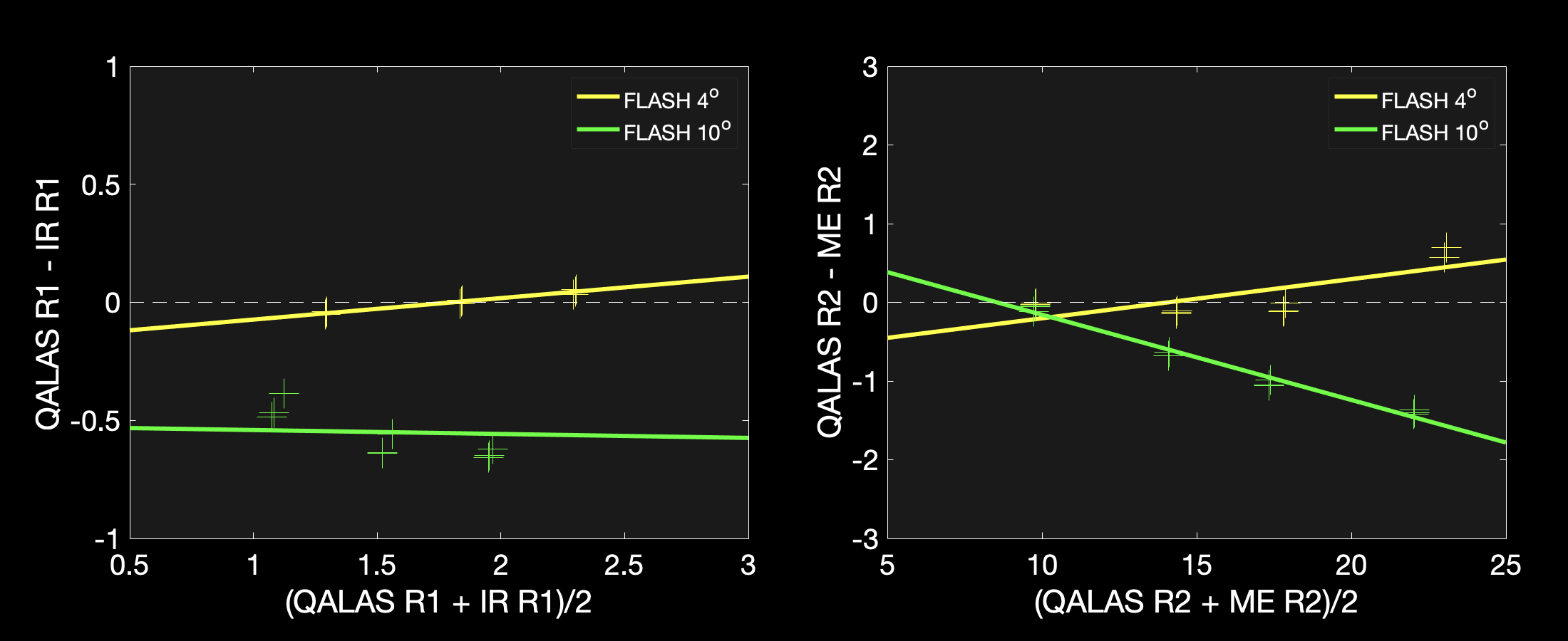
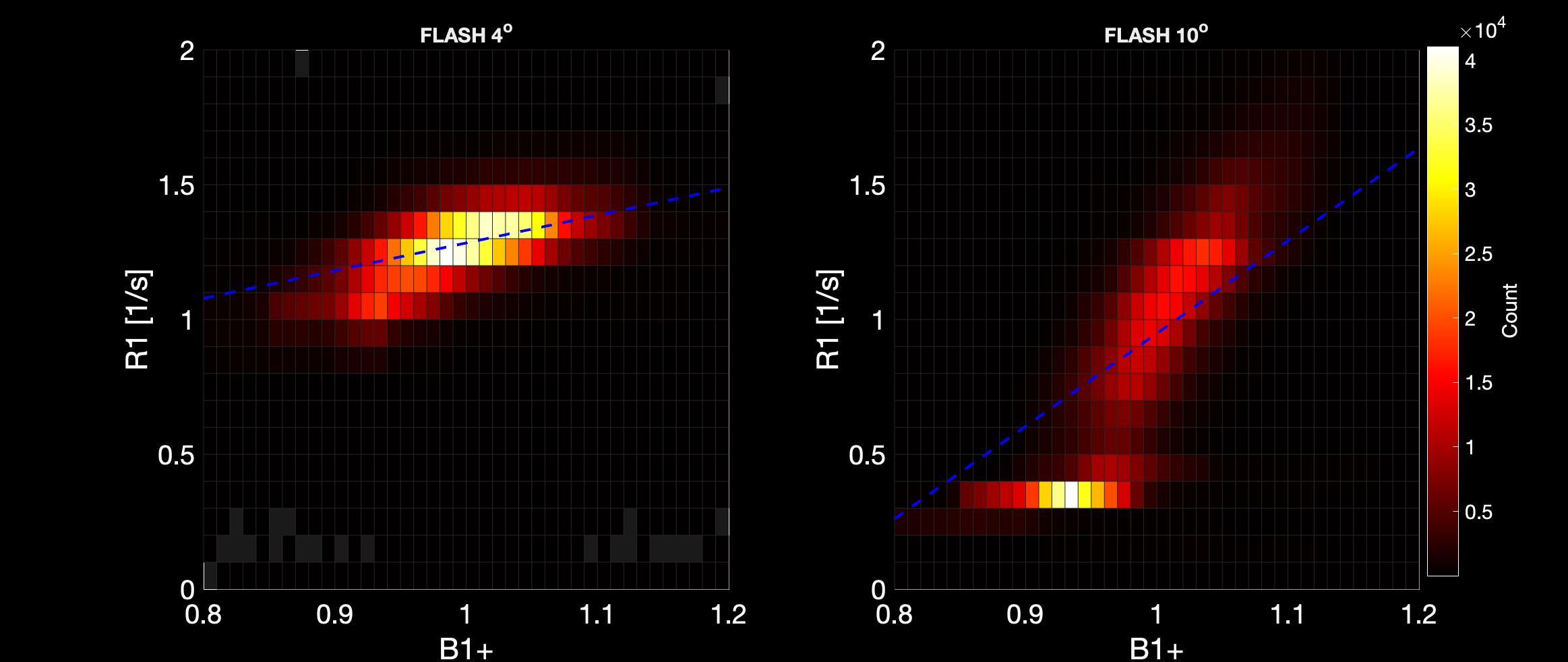
R1 and R2 maps of phantom examinations and one MS patient are shown in Figure 2. R1 and R2 estimated using the QALAS sequence with a flip angle of 4° showed high accuracy with a very small mean bias of 0.04 s-1 for R1 and 0.11 s-1 for R2 (Figure 3). In contrast, using a 10° excitation angle, R1-values were underestimated mean bias of –0.56 s-1 and R2-values were underestimated for high R2s and mean bias of –0.79 s-1. Moreover, the B1+ field and estimated R1 correlated for both 4° and 10°, and the effect was stronger for 10°, as depicted in the 2D histograms with linear regression lines in Figure 4°.Moreover, the B1+ field and estimated R1 correlated for both 4° and 10°, and the effect was stronger for 10°, as depicted in the 2D histograms with linear regression lines in Figure 4°.
Discussion and Conclusion
QALAS acquired with 4° excitation angle was very accurate compared to gold standard methods. However, a weak correlation to B1+ field was observed. That could be due to either non-ideal 180° inversions, or alternatively to different magnetization evolution during the FLASH readout due to non-ideal excitation pulses. Moreover, since there was an observed difference in B1+ dependence between 4° and 10°, it would be possible to derive the B1+ field, if the results obtained using both flip angles was combined.In conclusion, we observed a high accuracy of both R1 and R2 compared to gold standard measurements. However, we also see a small effect of B1+ inhomogeneities in the determined R1 maps.
Acknowledgements
No acknowledgement found.References
1. West, J., J.B. Warntjes, and P. Lundberg, Novel whole brain segmentation and volume estimation using quantitative MRI. Eur Radiol, 2012. 22(5): p. 998-1007.
2. Blystad, I., et al., Quantitative MRI using relaxometry in malignant gliomas detects contrast enhancement in peritumoral oedema. Sci Rep, 2020. 10(1): p. 17986.
3. Kvernby, S., et al., Simultaneous three-dimensional myocardial T1 and T2 mapping in one breath hold with 3D-QALAS. J Cardiovasc Magn Reson, 2014. 16: p. 102.
4. Barral, J.K., et al., A robust methodology for in vivo T1 mapping. Magn Reson Med, 2010. 64(4): p. 1057-67.
Figures