1275
Deep Learning Enables 60% Accelerated Volumetric Brain MRI While Preserving Quantitative Performance – A Prospective, Multicenter Trial1Neuroradiology, RadNet, Woodland Hills, CA, United States, 2Subtle Medical, Menlo Park, CA, United States, 3Cortechs.ai, San Diego, CA, United States, 4Stanford University, Stanford, CA, United States, 5RadNet, New York, NY, United States
Synopsis
In this prospective, multireader, multicenter study, we explore the impact of deep learning (DL) enhancement of 60% accelerated 3D T1 weighted brain MR image acquisitions. We found that the DL processed images demonstrated high volumetric quantification accuracy, matched clinical disease status predictability, and provided what readers perceived as superior image quality when compared with the longer standard-of-care exams, suggesting good generalizability, accuracy, and potential utility of DL enhancement in routine clinical settings. The results of this trial support the use of DL enhancement to shorten clinical MR brain examinations, even when additional quantitative tools such as volumetric analysis are applied.
PURPOSE
In this prospective, multicenter, multireader study, we evaluate the impact on both image quality and quantitative image analysis consistency (NeuroQuant™) of 60% accelerated volumetric MRI scans processed with a commercially available, vendor agnostic, DICOM-based, deep learning (DL) tool (SubtleMR™) compared to that of standard of care (SOC).MATERIALS AND METHODS
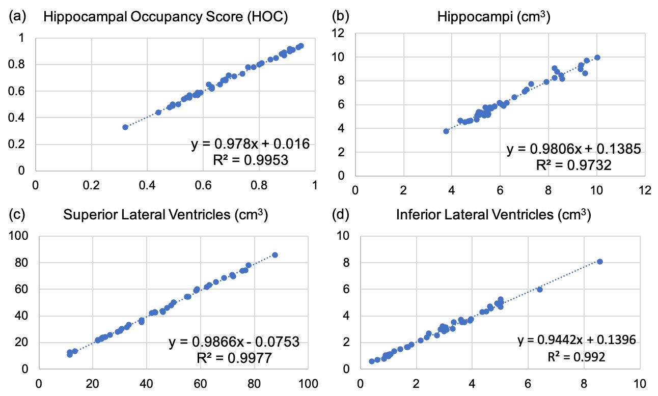
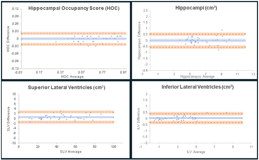
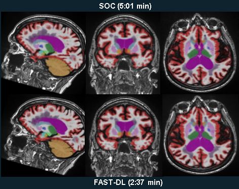
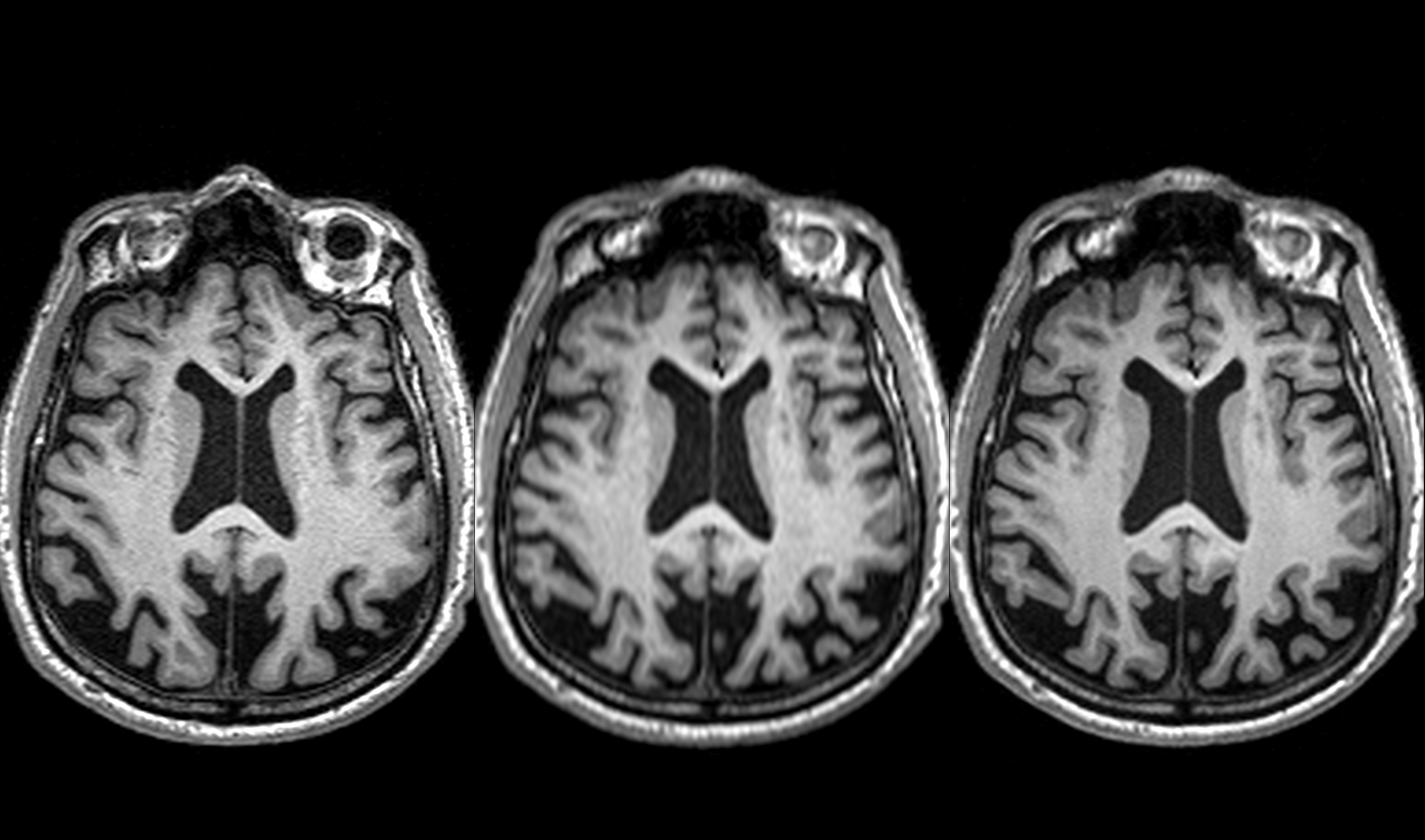
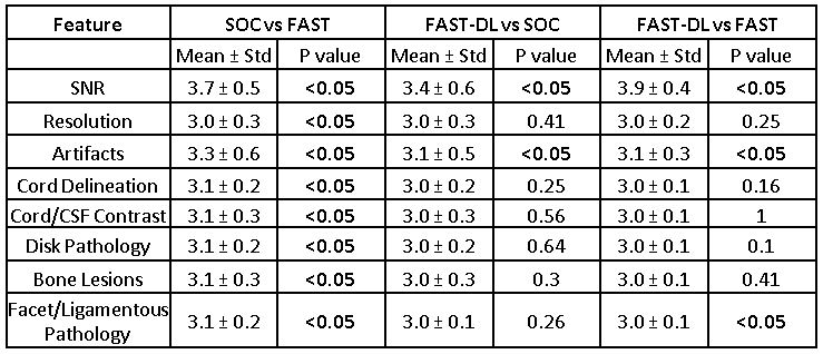
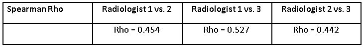
40 subjects underwent brain MRI exams on 6 scanners from 5 institutions. SOC and accelerated (FAST) datasets were acquired for each subject and FAST scans were enhanced with DL processing (FAST-DL). Both SOC and FAST-DL were subjected to NeuroQuant™ quantitative analysis and classified by a neuroradiologist into clinical disease categories. Concordance of SOC and FAST-DL biomarker measurements were assessed. To evaluate image quality, randomized, side-by-side, multiplanar datasets (360 series) were presented blinded to 2 neuroradiologists and rated for apparent signal-to-noise ratio (SNR), image sharpness, artifacts, lesion conspicuity, image contrast, and gray-white differentiation.RESULTS
FAST-DL was statistically superior to SOC for perceived quality across all imaging features despite a 60% scan time reduction. Both FAST-DL and SOC were superior to FAST for all features. There was no difference in quantitative volumetric biomarkers or clinical classification for SOC and FAST-DL datasets.CONCLUSION
DL reconstruction allows 60% scan time reduction while maintaining high volumetric quantification accuracy, consistent clinical classification, and what radiologists perceive as superior image quality when compared with SOC. This trial supports the reliability, efficiency, and utility of DL based enhancement for quantitative imaging. Shorter scan times may boost utilization of volumetric quantitative MRI in routine clinical settings.Acknowledgements
No acknowledgement found.References
1. Bash S. Eye on AI: Enhancing Neuroimaging with Artificial Intelligence. Appl Radiol. 2020; 49(1):20-21. https://appliedradiology.com/articles/enhancing-neuroimaging-with-artificial-intelligence
2. Tanenbaum LN, Bash S, Davis M. Appl Radiol (AR Connect Expert Discussions). AI: Clinical Applications. In: Proceedings of RSNA 2019, Chicago, IL. Dec 2020. https://www.appliedradiology.com/Communities/Artificial-Intelligence
3. Lunderwold AS, Lunderwold A. An overview of deep learning in medical imaging focusing on MRI. Elsevier 2019; 29:102-127. DOI: https://doi.org/10.1016/j.zemedi.2018.11.002
4. Tian C, Xu Y, Li Z, et. al. Attention-guided CNN for image denoising. Neural Netw 2020;124: 117-129. DOI: 10.1016/j.neunet.2019.12.0241
Figures