1274
Comprehensive Human Brain Imaging Enhancement with the Single-frequency Excitation Wideband MRI (SE-WMRI) Technique
Jordan Wang1, Po-Wei Cheng1, Ming-Jang Chiu2, Tzi-Dar Chiueh3, and Jyh-Horng Chen3
1Graduate institute of biomedical electronics and bioinformatics, National Taiwan University, Taipei, Taiwan, 2National Taiwan University Hospital, Taipei, Taiwan, 3Department of electrical engineering, National Taiwan University, Taipei, Taiwan
1Graduate institute of biomedical electronics and bioinformatics, National Taiwan University, Taipei, Taiwan, 2National Taiwan University Hospital, Taipei, Taiwan, 3Department of electrical engineering, National Taiwan University, Taipei, Taiwan
Synopsis
The Single-frequency Excitation Wideband MRI (SE-WMRI) technique was implemented on a Siemens PRISMA 3T human MRI system to demonstrate its multifaceted benefits for human brain imaging, including acceleration, SNR improvement, and spatial resolution enhancement. The accelerated Wideband brain results showed high consistency with standard scans, and the resolution enhanced Wideband images revealed more detailed brain structures details and clearer blood vessel due to reduced partial volume effect. Since SE-WMRI can enhance MR efficiency without any hardware upgrade, it’s highly cost-effective for medical institutions in pursuit of higher MR imaging quality.
Introduction
MR imaging efficiency is crucial for medical applications to provide sufficient medical information in a reasonable amount of time for adequate diagnoses. Such enhancement, in general, could be achieved by better system hardware or more efficient sequences. Since hardware upgrade is often costly, improving sequence efficiency with advanced MR technologies is a more practical option. SE-WMRI is one of those advanced MR acceleration techniques which achieves resolution and SNR enhancement without phased-array coils.1 This study will present multiple usages of SE-WMRI to demonstrate its multifaceted benefits for human brain imaging.Method
In this study, SE-WMRI is applied to the standard Gradient-Echo (GRE) sequence on a Siemens 3T Prisma MRI system. All images were acquired with a 16-channel head coil, using the GRE sequence and the newly designed SE-GRE sequence. All the data were processed and analyzed by MATLAB R2015A (The MathWorks, Inc.) programs.Four sets of brain images were acquired to demonstrate how SE-WMRI elevates imaging efficiency. For the first three image sets, TR/TE = 300/10 ms and slice thickness = 5 mm.
The first image set demonstrates the acceleration of SE-GRE under the same resolution (0.5x0.5 mm2). Since N = 4, the scan time was 11m34s for GRE and 5m47s for SE-GRE.
The second image set demonstrates the SNR enhancement of SE-GRE under the same scan time (5m47s) and resolution (0.5x0.5 mm2). For this image set, N = 2 for GRE and 4 for SE-GRE.
The third image set shows the resolution enhancement of SE-GRE under the same scan time (5m47s). The resolution is 1.0x1.0 mm2 for GRE and 0.5x0.5 mm2 for SE-GRE.
The fourth image set shows the z-axis resolution enhancement, namely slice thickness, under the same scan time. For GRE, TR/TE = 500/11.4ms, thickness = 4mm and slice number = 25; for SE-GRE, TR/TE = 1000/11.4ms, thickness = 2mm and slice number = 50. The duration for both scans is 9m36s.
Results
Due to temporal resolution enhancement, the image acquisition speed for Wideband is doubled under the same image resolution. Fig.1 compares GRE and the accelerated SE-GRE images with the derived signal-to-noise ratio (SNR) and structural similarity index (SSIM) values. The calculation results show that the two images have nearly the same SNR, and their SSIM is approximately 0.99, indicating high resemblance in terms of signal strength, contrast, and structural similarity. As shown in the zoomed-in images, the fine brain structures are highly comparable without noticeable differences. The result proved that even with a twofold acceleration, SE-GRE still provides nearly identical images with conventional GRE.The time-saving effect of SE-GRE can also be utilized to increase the number of averages and achieve a 1.4-time SNR improvement. As demonstrated in Fig.2, with lower SNR, the GRE image appears grainier, and the thinner brain vessels are blurry due to higher noise. On the contrary, the SE-GRE image shows smoother structures with more visible vessels.
SE-GRE can also be used to achieve double spatial resolution within the same scan time, as shown in Fig.3. The comparison between magnified GRE and SE-GRE images shows significant enhancement for the demarcation between gray and white matters, clarity of brain vessels, and sharpness of edges between tissues.
Another usage for spatial resolution enhancement is to reduce the slice thickness and enhance the z-axis resolution for a full brain MRI scan. Within the same scan time, the SE-GRE scan provides twice as many slices as the GRE scan, each with half the original slice thickness. Thus, fine details of brain structures can be better distinguished in different slices. Fig.4 presents one of the slices in the GRE full brain scan and the two corresponding slices in the SE-GRE scan. Since the partial volume effect reduced with thinner slices, the details of the cerebral arteries and the anterior lobe of the cerebrum became more apparent, as shown in the zoomed-in images. In summary, SE-GRE provides more medical information with enhanced spatial resolution within the same scan time, which will massively benefit disease diagnoses.
Conclusion
In this study, we presented various usages of SE-WMRI to demonstrate its versatility. The acceleration effect is flexibly traded-off against resolution enhancement or SNR improvement according to demand. Under the same resolution, SE-GRE massively accelerates imaging speed and provides images with high structural similarity and similar SNR as conventional GRE images. While the scan time remains constant, SE-GRE achieves double spatial resolution to reveal finer structural details. Since SE-WMRI is appliable on any existing MRI system without any hardware upgrade, this technique could be highly cost-effective for all medical institutes pursuing higher MRI throughput and better image quality. For future development, SE-WMRI will be applied to various sequences on different human MRI systems for more comprehensive implementation.Acknowledgements
No acknowledgement found.References
1. Wu EL, Huang YA, Chiueh TD, Chen JH. Single-frequency excitation wideband MRI (SE-WMRI). Med Phys 2015;42(7):4320-4328.Figures
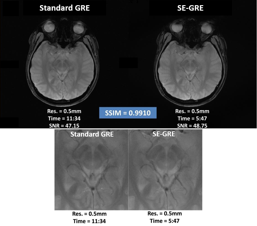
Fig. 1 Conventional GRE and 2-time accelerated SE-GRE image comparison under the same resolution with the zoomed-in comparison of
the center region. The two images have similar SNR, and their SSIM value is above 0.99, indicating an extremely high resemblance for image quality in terms of
signal strength, contrast, and structural
similarity.
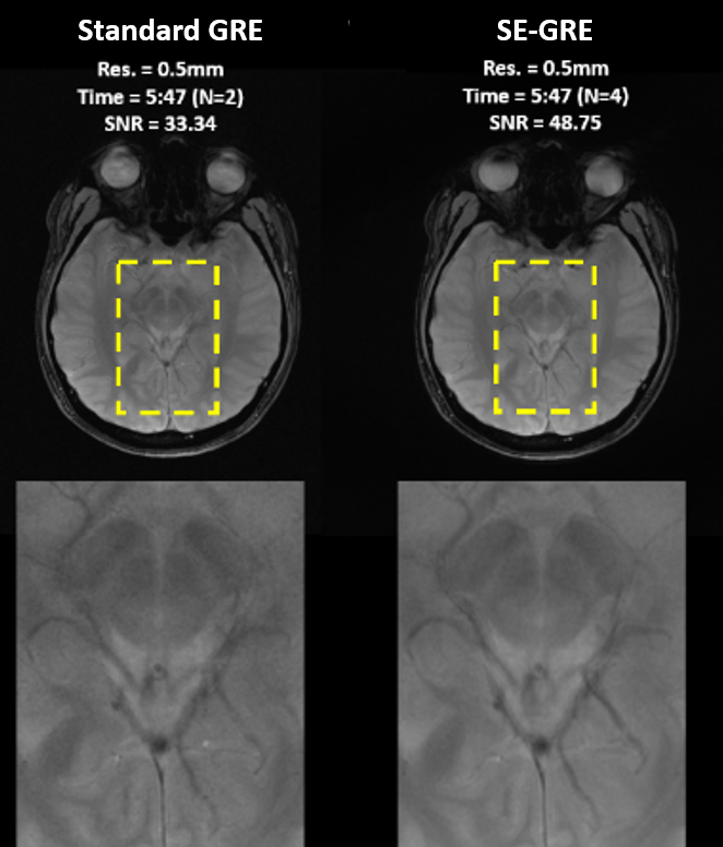
Fig. 2 Conventional GRE and 2-time average SE-GRE image comparison under the same resolution with the zoomed-in comparison. Due to the 1.4-time SNR increase, the SE-GRE image shows smoother structures and more distinct vessels.
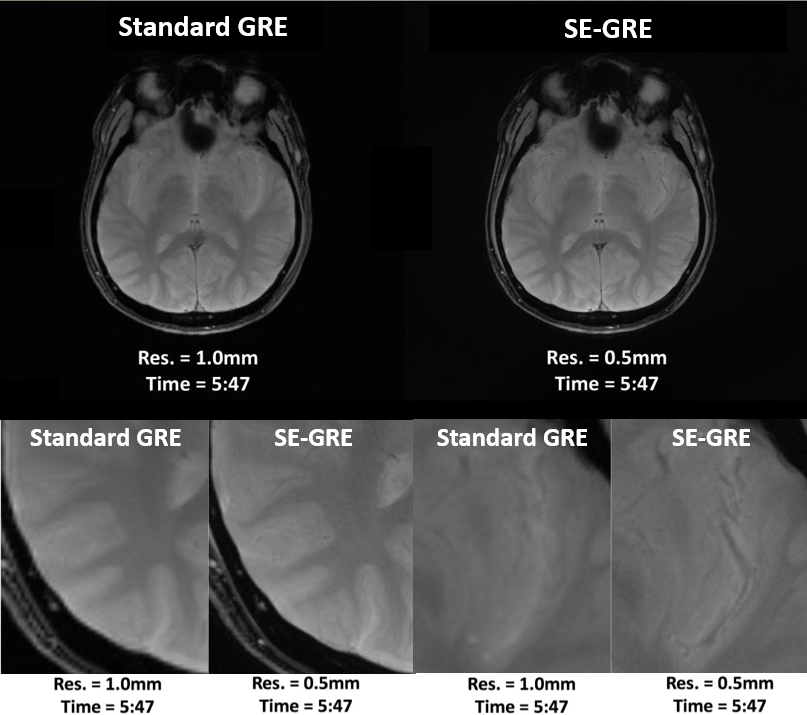
Fig.3 Standard GRE (Res. = 1 mm) and SE-GRE (Res. = 0.5 mm)
images acquired within the same scan time and the zoomed-in comparisons of the
bottom-left region and the top-right region. The comparison shows significant improvement for clarity of structural
details, visibility of vessels, and edge sharpness between different tissues.
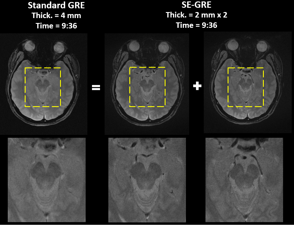
Fig.4 One of the slices for a traditional GRE full brain scan and the two corresponding slices in a 2x-thinner SE-GRE scan. Since the partial volume effect reduced with thinner slices, the details for cerebral arteries and other brain structures are more distinct.