1244
Simultaneous morphological and quantitative lumbar MRI with 3D isotropic high-resolution using MIXTURE T21Radiology, Eastern Chiba Medical Center, Chiba, Japan, 2Philips Japan, Tokyo, Japan
Synopsis
We propose a new sequence called MIXTURE (Multi-Interleaved X-prepared TSE with inTUitive RElaxometry). MIXTURE based is a 3D TSE that can set arbitrary echo times using the T2 preparation pulses, and enables several image contrasts (such as PDW, T2W) and T2 mapping by acquiring at least two echo time images. MIXTURE could simultaneously provide morphology, pathology and T2 quantitative lumbar imaging in one single scan. This sequence is very promising to evaluate the degeneration of nerve roots and intervertebral disc for 3D T2 mapping and improve the ability of diagnostic imaging around the lumbar for 3D multi contrast MRI.
PURPOSE
In previous studies, T2 mapping has been used for the diagnosis of lumbar intervertebral disc herniation and nerve root disorders1. Recently, an isotropic 3D T2 mapping technique of knee cartilage with a clinically feasible acquisition time has been developed. It’s allows for reformats in any plane and minimization of partial volume effects, which results in a more accurate T2 relaxation time quantification2,3. Therefore, it is possible to make a morphological diagnosis for 3D MRI and a quantitative diagnosis for T2 mapping simultaneously. However, clinical lumbar MRI requires multiple contrasts to diagnose various diseases. Thus, we propose a new sequence called MIXTURE (Multi-Interleaved X-prepared TSE with inTUitive RElaxometry). MIXTURE based is a 3D TSE that can set arbitrary echo times using the T2 preparation pulses, and enables several image contrasts (such as PDW, T2W) and T2 mapping by acquiring at least two echo time images. The purpose of this study was to evaluate the clinical feasibility of morphological and pathological MRI with two contrasts images and quantitative MRI with T2 mapping using MIXTURE.METHODS
All subjects were examined with 3.0T whole-body clinical system (Ingenia CX, Philips Healthcare). The study was approved by the local IRB, and written informed consent was obtained from all subjects. Scheme of the MIXTURE T2 sequence for 3D simultaneous morphological, pathological and T2-quantificagtion imaging is shown in Figure 1. T2 mapping was performed using T2-prepared 3D segmented turbo spin- echo (TSE) with variable refocusing pulse trains. Two images with different TE (TE = 0 and 50ms) were acquired with interleaved acquisition. In this study, since previous studies have reported the usefulness of 3D PDWI4, 1st echo time was set for PDWI (without any pre-pulses) and 2nd echo time was set for fat suppressed T2WI (by applying spectral fat suppression (SPAIR) and T2prep) [Fig.2].We compared T2 values between Multi-echo TSE and MIXTURE in sucrose phantom of various T2 values. Furthermore, the following evaluations were also performed on five healthy volunteers (22-39y, 1 female, 4 males) using MIXTURE. We compared T2 values of intervertebral disc and bilateral lumbar nerve roots at the level of L4-S1, and we evaluated ability of diagnostic imaging for multi contrast MRI. Imaging parameters for Multi-echo TSE were; Coronal, voxel size=1.0×1.0×2.0mm3, FOV=150×150mm2, 9 slices. band width=289.7Hz/px, TR=2500ms, TE=15, 30, 45, 60, 75, 90ms, and total acquisition time=5m34s. Imaging parameters for MIXTURE were; Coronal, voxel size=1.3×1.3×1.3mm3, FOV=350×350mm2, 220 slices. band width=1227.3Hz/px, fat suppression=SPAIR, TR=1240ms, TE=20ms, T2prep time=0, 50ms, CS-SENSE factor=4 and total acquisition time=7m30s.RESULTS AND DISCUSSION
T2 values of Multi-echo TSE and MIXTURE in the sucrose phantom showed a high correlation [Fig. 3A]. T2 values of healthy volunteers for nerve roots and intervertebral discs were comparable to previous studies [Fig. 3B]1,5. Clinical images taken by MIXTURE are shown [Fig. 4]. 3D PDWI contributed to the morphological evaluation of vertebrae, intervertebral disc,nerve roots and ligaments, and 3D FS T2WI contributed to qualitative diagnosis of inflammation, edema, bone contusion and tumor. For lumbar MRI where fine tissue is the object of observation, thin slice 3D images are useful. The major advantage of the MIXTURE set in iso voxel is that it can perform multi planer reconstruction (MPR) of PDWI, FS T2WI and T2 mapping.CONCLUSION
MIXTURE could simultaneously provide morphology, pathology and T2 quantitative lumbar imaging in one single scan. In addition, this sequence is very promising to evaluate the degeneration of nerve roots and intervertebral disc for 3D T2 mapping and improve the ability of diagnostic imaging around the lumbar for 3D multi contrast MRI.Acknowledgements
No acknowledgement found.References
1. Takashi Sato, et al. Diagnosis of lumbar radiculopathy using simultaneous MR neurography and apparent T2 mapping. J Clin Neurosci. 2020 Aug;78:339-346.
2. Roberto Colotti, et al. Isotropic three-dimensional T 2 mapping of knee cartilage: Development and validation. J Magn Reson Imaging. 2018 Feb;47(2):362-371.
3. Roberto Colotti, et al. Simultaneous fat-free isotropic 3D anatomical imaging and T 2 mapping of knee cartilage with lipid-insensitive binomial off-resonant RF excitation (LIBRE) pulses. J Magn Reson Imaging. 2019 May;49(5):1275-1284.
4. Naoko Kinoshita, et al. High contrast between lumbar nerve roots and surrounding structures using dual echo 3D turbo spin echo additional fusion images. Jpn J Radiol. 2018 Jun;36:472–476.
5. Izaya Ogon, et al. Analysis of chronic low back pain with magnetic resonance imaging T2 mapping of lumbar intervertebral disc. J Orthop Sci. 2015 Feb;20:295-301.
Figures
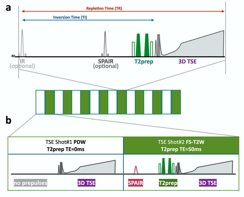
Fig.1 Scheme of the MIXTURE (Multi-Interleaved X-prepared tse with inTUitive RElaxometry).
(a) T2-mapping was performed using T2-prepared 3D segmented turbo spin- echo (TSE) with variable refocusing pulse trains. (b) Two images with different TE (TE = 0 and 50ms) were acquired with interleaved acquisition. To obtain the compatible contrasts with routine TSE images, TSE shot#1 did not apply any pre-pulses (as “PDW) and shot#2 applied both SPAIR and T2pre (as “fat-suppressed T2W”).
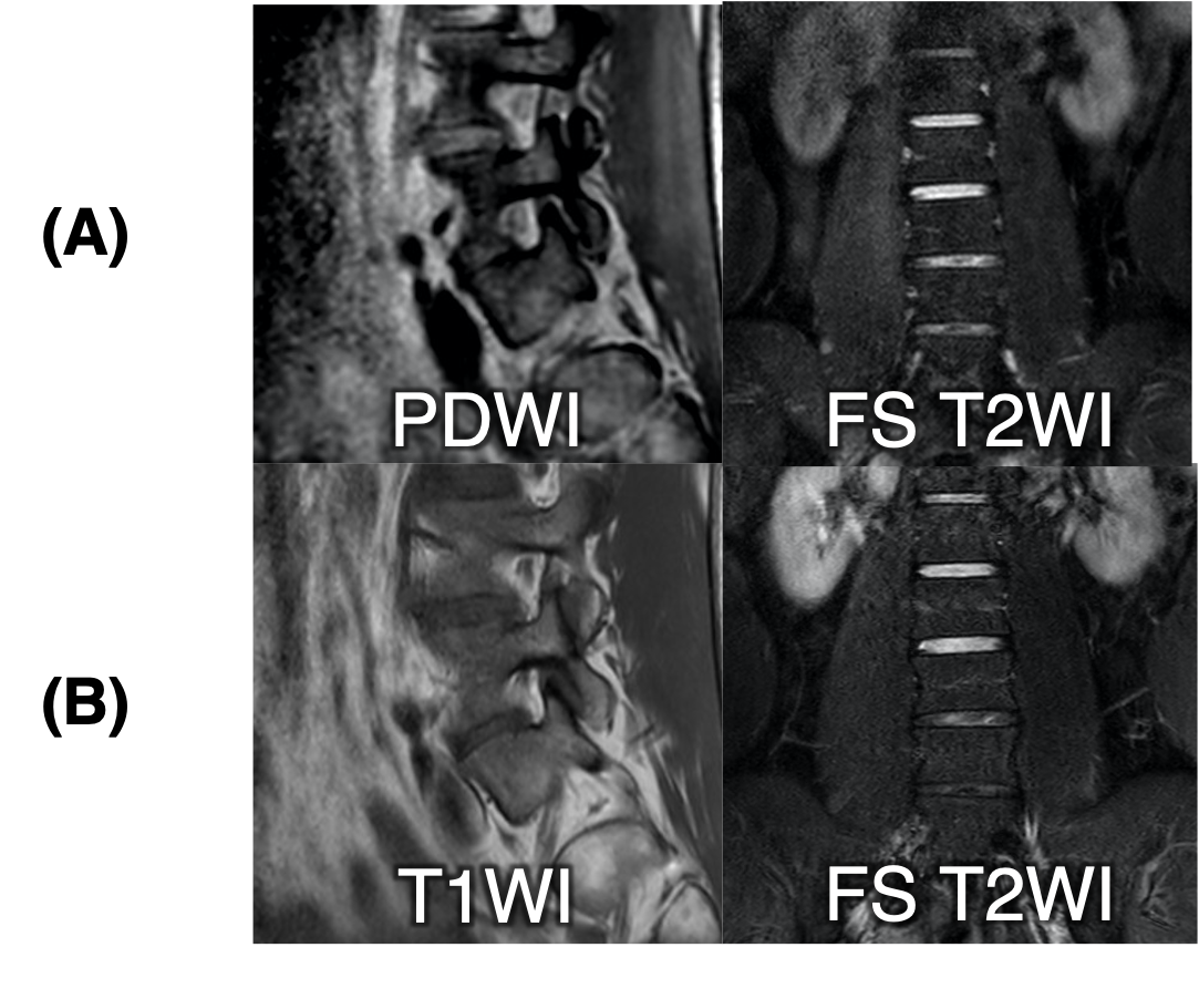
Fig.2 Comparison of MIXTURE and conventional methods (2D TSE and 3D TSE).
(A) MIXTURE, Multiple Interleaved X-prepared TSE with Universal Relaxation-mapping Extensions,(B) Conventional 2D TSE
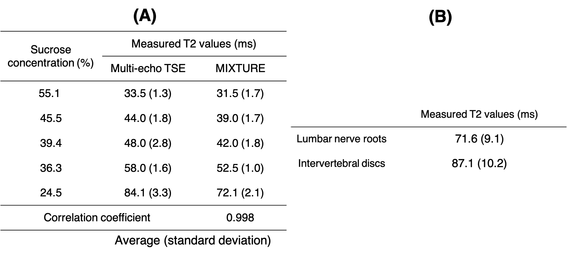
Fig.3 Measurement of T2 values in sucrose phantom (A) and in healthy volunteers using MIXTURE (B).
MIXTURE, (Multi-Interleaved X-prepared tse with inTUitive RElaxometry).
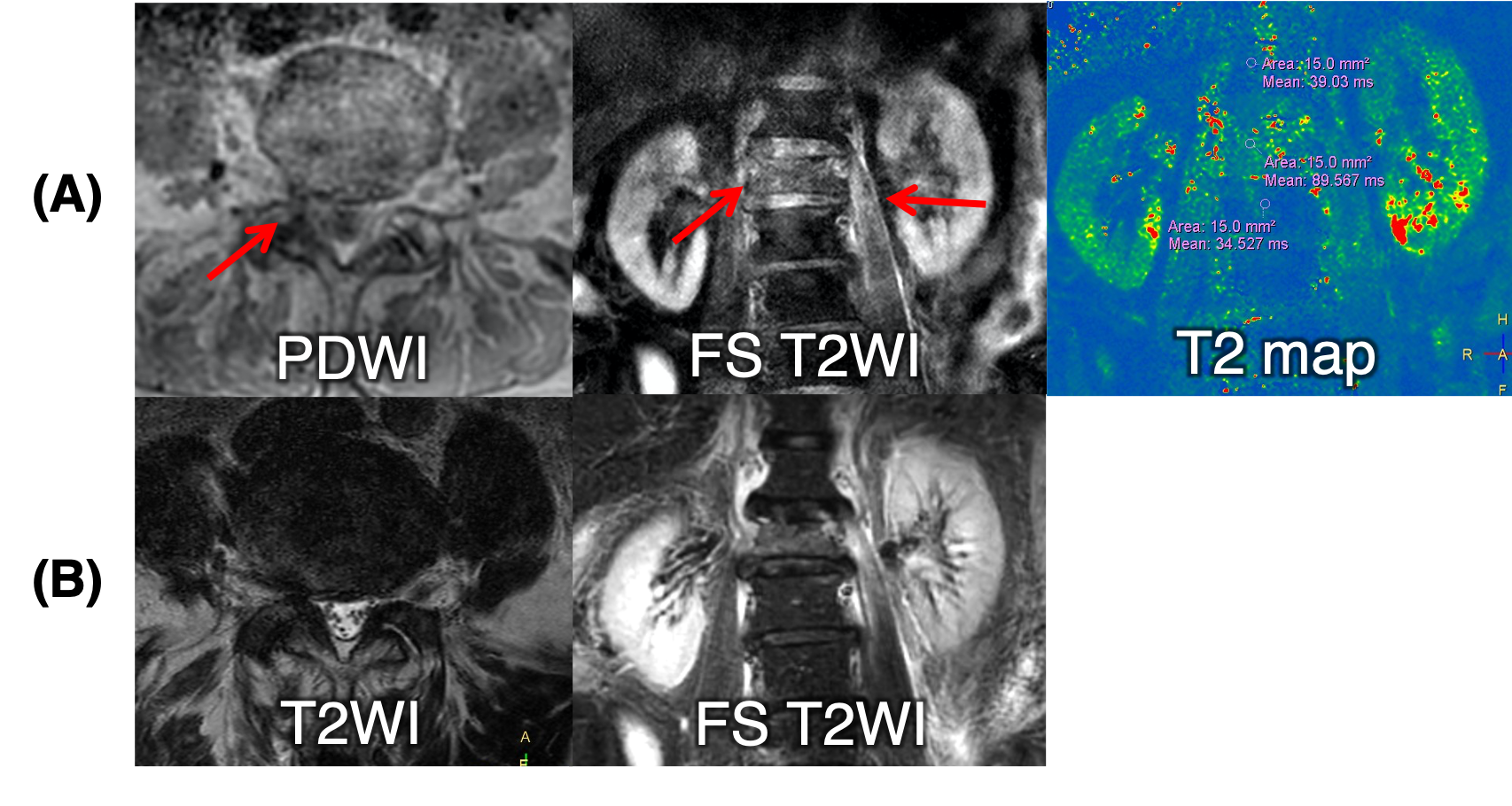
Fig.4 Clinical images with low back pain using MIXTURE.
(A) MIXTURE, Multiple Interleaved X-prepared TSE with Universal Relaxation-mapping Extensions,(B) Conventional 2D TSE