1236
Generation of co-registered multi-contrast MR images for carotid atherosclerosis evaluation based on a single SIMPLE sequence1Center for Biomedical Imaging Research, School of Medicine, Tsinghua University, Beijing, China, 2School of Biomedical Engineering and Imaging Sciences, King's College London, London, United Kingdom, 3Department of Radiology, University of Washington, Seattle, WA, United States
Synopsis
Carotid atherosclerosis is a leading cause of ischemic stroke worldwide. Multi-contrast magnetic resonance imaging (MRI) is an ideal non-invasive technique to assess carotid plaque. However, its clinical application is limited by long scan time and non-rigid registration between different contrasts. In this study, we generated a set of co-registered T1w, T2w, PDw images, and MR angiogram (MRA) in one scan based on the SIMPLE (SImultaneous T1 and T2 Mapping of carotid PLaquE) sequence. Preliminary experiments on patients validated the feasibility of the generated multi-contrast images for carotid plaque assessment and comparable performance with conventional sequences.
Introduction
Carotid atherosclerosis is a leading cause of stroke worldwide1. Luminal stenosis and plaque compositions are two key features of atherosclerosis2. A multi-contrast Magnetic Resonance (MR) imaging protocol, including time-of-flight (TOF), T1-weighted (T1w), T2-weighted (T2w), and proton density-weighted (PDw) images3, is typically used for plaque assessment, but it suffers from long acquisition time and mis-registration of different image contrasts.Recently, a technique named SIMPLE4 was proposed to achieve simultaneous T1 and T2 mapping of carotid plaque in one scan. In addition to the quantitative T1 and T2 maps, SIMPLE also has the potential for yielding multi-contrast weighted images, which would allow direct visual inspection of atherosclerosis in a way similar to the conventional multi-contrast sequences.
In this study, we aimed to generate a set of co-registered 3D high-resolution T1w, T2w, and PDw images, and MR angiogram (MRA) based on SIMPLE. Moreover, the generated images were used to assess atherosclerotic plaque, and the performance was compared with the conventional multi-contrast sequences.
Methods
SIMPLE Sequence and Data AcquisitionFigure 1 showed the SIMPLE sequence diagram and simulated signal evolutions of the vessel wall and blood. With the approval of institutional review board, 5 patients with carotid atherosclerosis (5 males, 62.2±3.9 years) were imaged on a 3T scanner (Achieva CX, Philips) with a dedicated 8-channel carotid coil, using SIMPLE sequence. Conventional multi-contrast sequences were also performed, including TOF, black-blood T1w-turbo spin echo (TSE), T2w-TSE, and magnetization prepared-rapid gradient echo acquisition (MPRAGE). Detailed scan parameters are shown in Figure 1C.
Multi-contrast MR Images Generation Strategies
Four different image contrasts were reconstructed using low-rank modeling and sparsity constraints (LRS)6 reconstruction method with a time window width (TW) of 254. Real-value representation of images, with the signal polarity preserved, was used to enhance the contrast between blood and other tissues.
MRA: Similar to SNAP7, an optimal inversion time (TI) of 390 ms, at which luminal signal was negative while background tissues were positive, was applied to maximize the contrast between vessel wall and blood. Then MRA was generated by using spokes with T2-prep of 25 ms and taking absolute values of the negative signals.
T1w: Using spokes with T2-prep of 0, T1w images were generated at a chosen TI=640 ms around the zero-crossing point of blood signal for good flow suppression.
T2w: Numerical simulation4 showed that short TI leads to less T1 relaxation effects. Limited by a TW of 25 used, this study adopted the minimum available TI of 140 ms. Spokes within the temporal window centered at the minimum TI and acquired with T2-prep of 50 ms were used for reconstructing T2w images.
PDw: Assuming that the chosen TI is long enough for near-complete T1 relaxation, spokes at the end of the shots without T2-prep pulses were used to obtain PDw reconstruction. Furthermore, the blood signal was suppressed by subtracting the phase of the black-blood T1W image from the PDw image.
Image Review and Statistical Analysis
All the generated images were reformatted into slices that matched with T1w-TSE sequence. Two trained reviewers used CASCADE8 to interpret the two datasets respectively. Quantitative morphological measurements (Table 1) were calculated from the two datasets respectively and the agreement was analyzed with intraclass correlation coefficient (ICC) at a 95% confidence interval (CI). The presence of plaque components, including lipid-rich necrotic core (LRNC), intraplaque hemorrhage (IPH), and calcification (CA) were identified at a slice-based level. Confusion matrix and Cohen’s kappa (κ) analysis were used to evaluate the agreement of plaque components identification between the two datasets.
Results
Multi-contrast images were successfully generated from SIMPLE on all 5 subjects. Figure 2 demonstrated a carotid plaque with IPH, in which both SIMPLE-T1w and T1w-TSE images showed hyperintensity. Maximum intensity projection (MIP) of SIMPLE-MRA demonstrated clear depictions of bilateral carotid arteries. For LRNC, the SIMPLE dataset showed iso-intensity T1W and hypointensity T2W and agreed with conventional multi-contrast images (Figure 3). A calcified plaque (Figure 4) presented hypointensity on all contrasts except SIMPLE-T2w, which might due to the negative blood signal on the real SIMPLE-T2w image. SIMPLE-T1w images provided a clearer visualization of CA than T1w-TSE.A total of seventy-four slices from the 5 subjects were analyzed. ICC of lumen area, wall area, maximum wall thickness, mean wall thickness, and NWI showed good agreement (ICC>0.94) between the two datasets (Table 1-1). For plaque components characterization, the SIMPLE-generated images showed good agreement with the conventional ones for identification of LRNC (κ=0.75) and excellent agreement for IPH (κ=0.96) and CA (κ=0.96, Table 1-2).
Discussion and Conclusion
In this study, a set of naturally registered multi-contrast images, including MRA, T1w, T2w, and PDw images, were generated based on a single SIMPLE sequence, and the acquisition time was less than conventional protocol (8 min vs 16 min). The preliminary experiment showed a good agreement between the SIMPLE-generated images with the conventional ones for both lumen and vessel wall delineation and plaque components identification. A larger study, with histology as the gold standard, for further comparison with conventional multi-contrast images, should be performed in the future. In conclusion, the feasibility of SIMPLE for generating multi-contrast MR images and simultaneous T1 and T2 mapping suggested its great potential in the comprehensive assessment of carotid atherosclerosis.Acknowledgements
None.References
1. Takaya, N., et al., Association between carotid plaque characteristics and subsequent ischemic cerebrovascular events - A prospective assessment with MRI - Initial results. Stroke, 2006. 37(3): p. 818-823.
2. Gupta, A., et al., Carotid Plaque MRI and Stroke Risk A Systematic Review and Meta-analysis. Stroke, 2013. 44(11): p. 3071-3077.
3. Cai, J.M., et al., Classification of human carotid atherosclerotic lesions with in vivo multicontrast magnetic resonance imaging. Circulation, 2002. 106(11): p. 1368-1373.
4. Qi, H., et al., Simultaneous T-1 and T-2 mapping of the carotid plaque (SIMPLE) with T-2 and inversion recovery prepared 3D radial imaging. Magnetic Resonance in Medicine, 2018. 80(6): p. 2598-2608.
5. Qiao, H., et al., Quantitative evaluation of carotid atherosclerotic vulnerable plaques using in vivo T1 mapping cardiovascular magnetic resonaonce: validation by histology. Journal of Cardiovascular Magnetic Resonance, 2020. 22(1).
6. Qi, H., et al., Highly Undersampled Kooshball Reconstruction with Low-rank Modeling and Sparsity Constraints for High-resolution T1 Mapping. International Society for Magnetic Resonance in Medicine, 2018. 0248.
7. Wang, J., et al., Simultaneous noncontrast angiography and intraPlaque hemorrhage (SNAP) imaging for carotid atherosclerotic disease evaluation. Magnetic Resonance in Medicine, 2013. 69(2): p. 337-345.
8. William, K., Magnetic resonance imaging of carotid atherosclerosis: plaque analysis. %J Topics in magnetic resonance imaging : TMRI. 2007. 5(18).
Figures
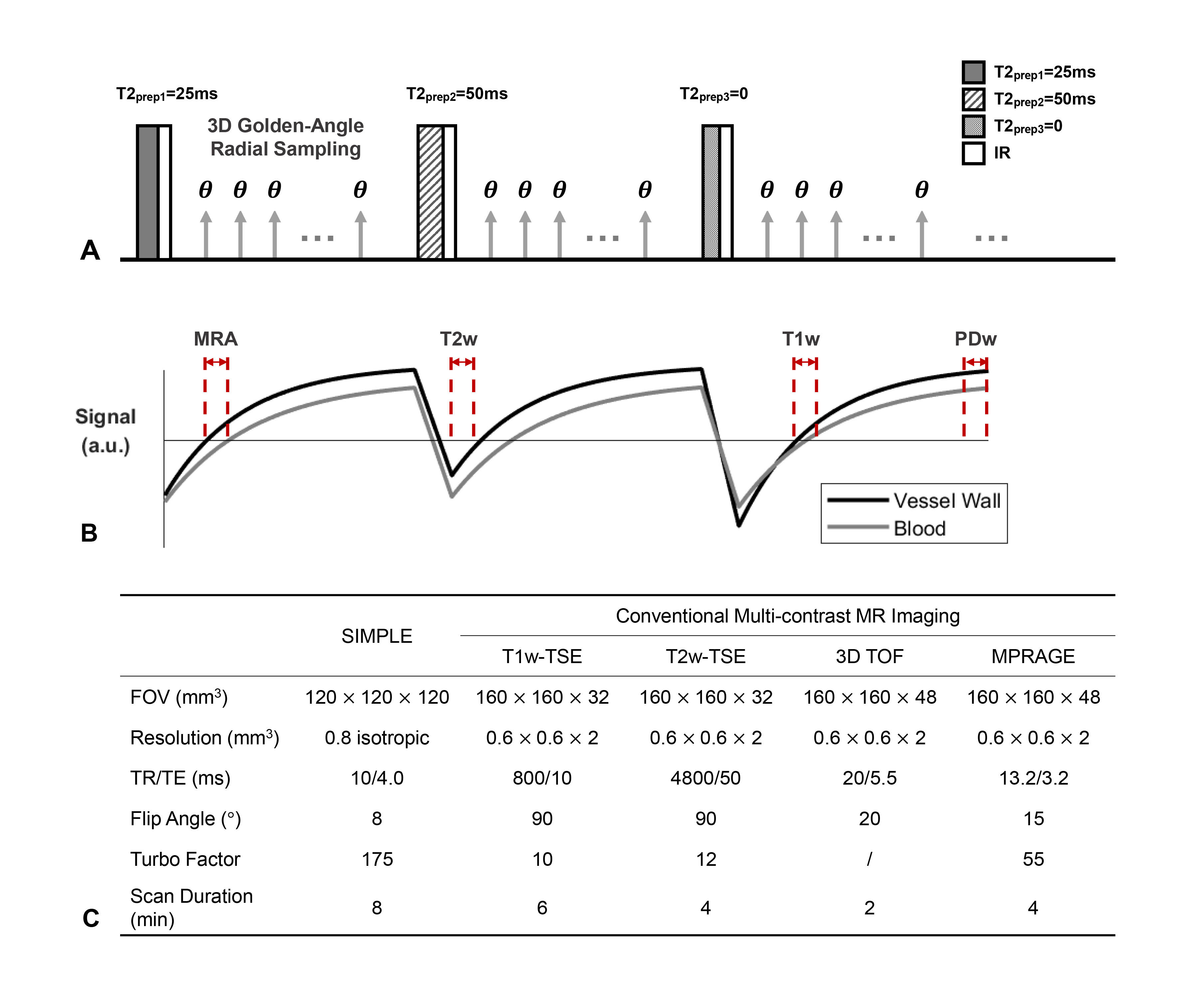
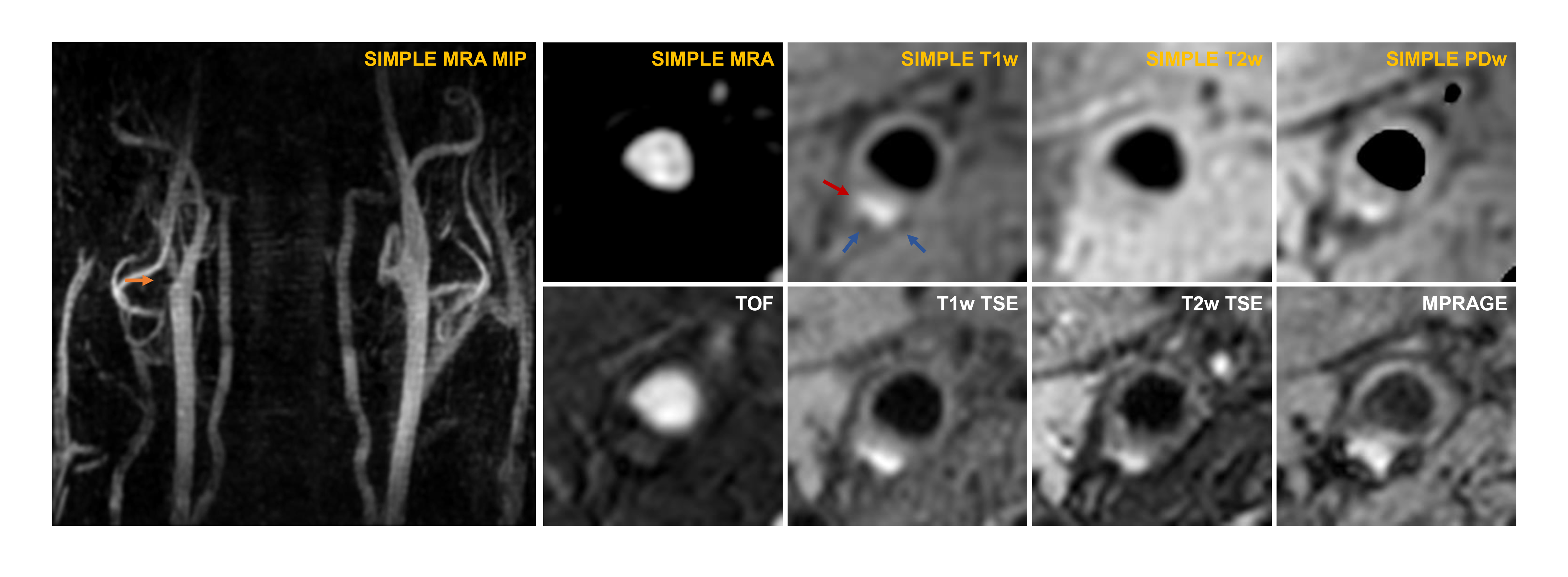
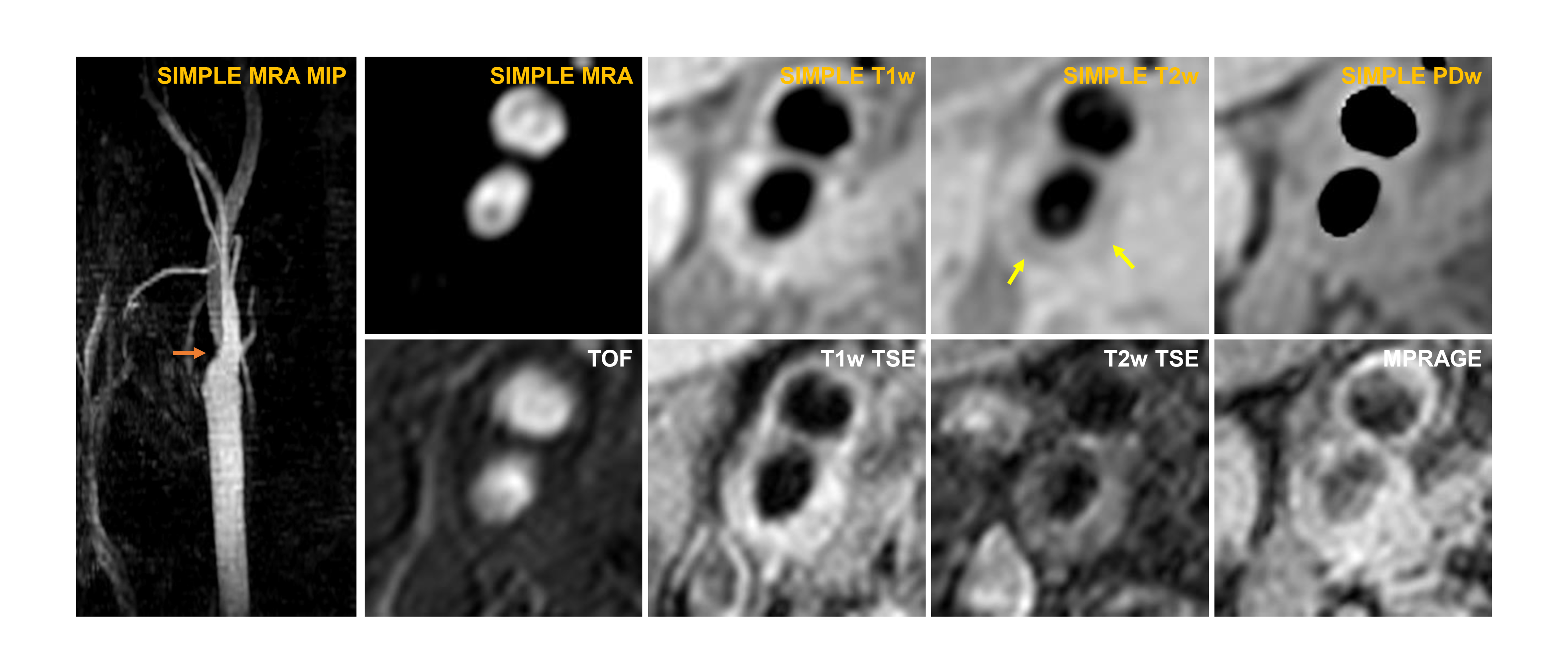
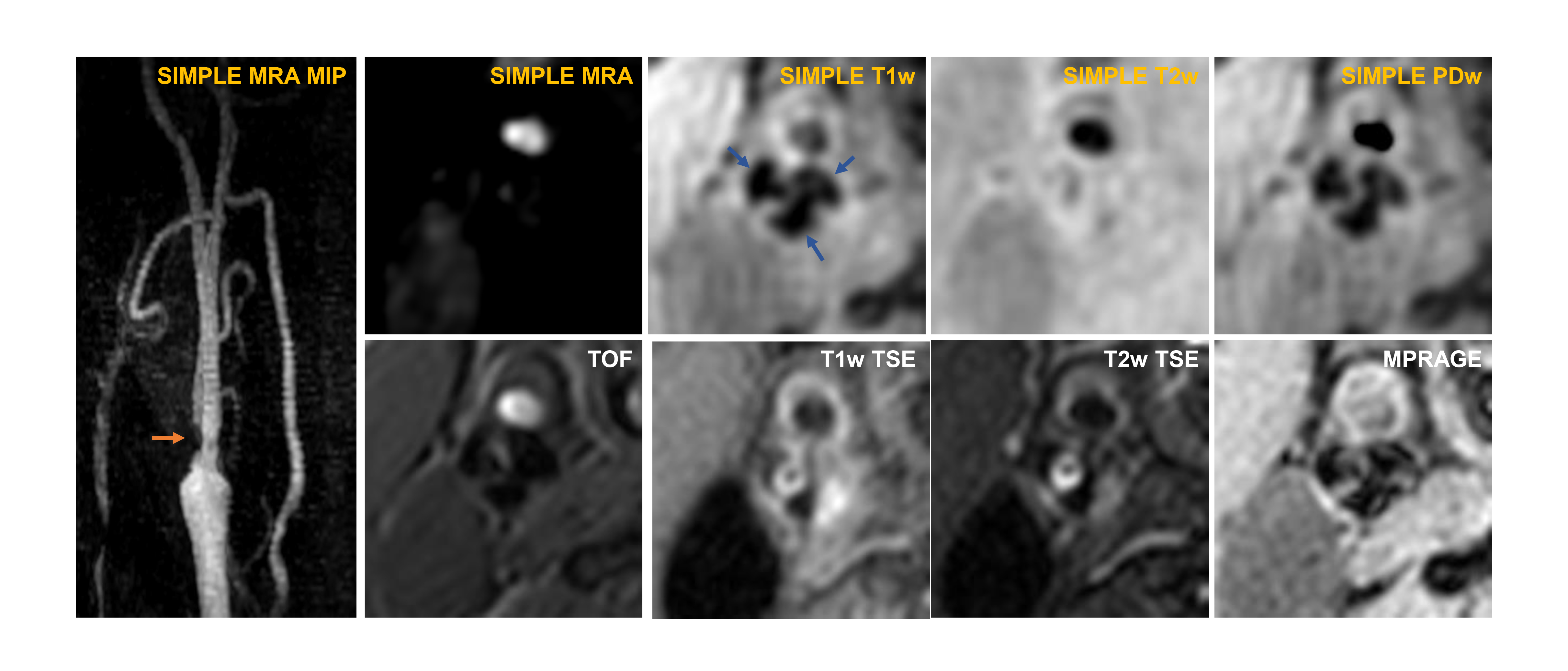
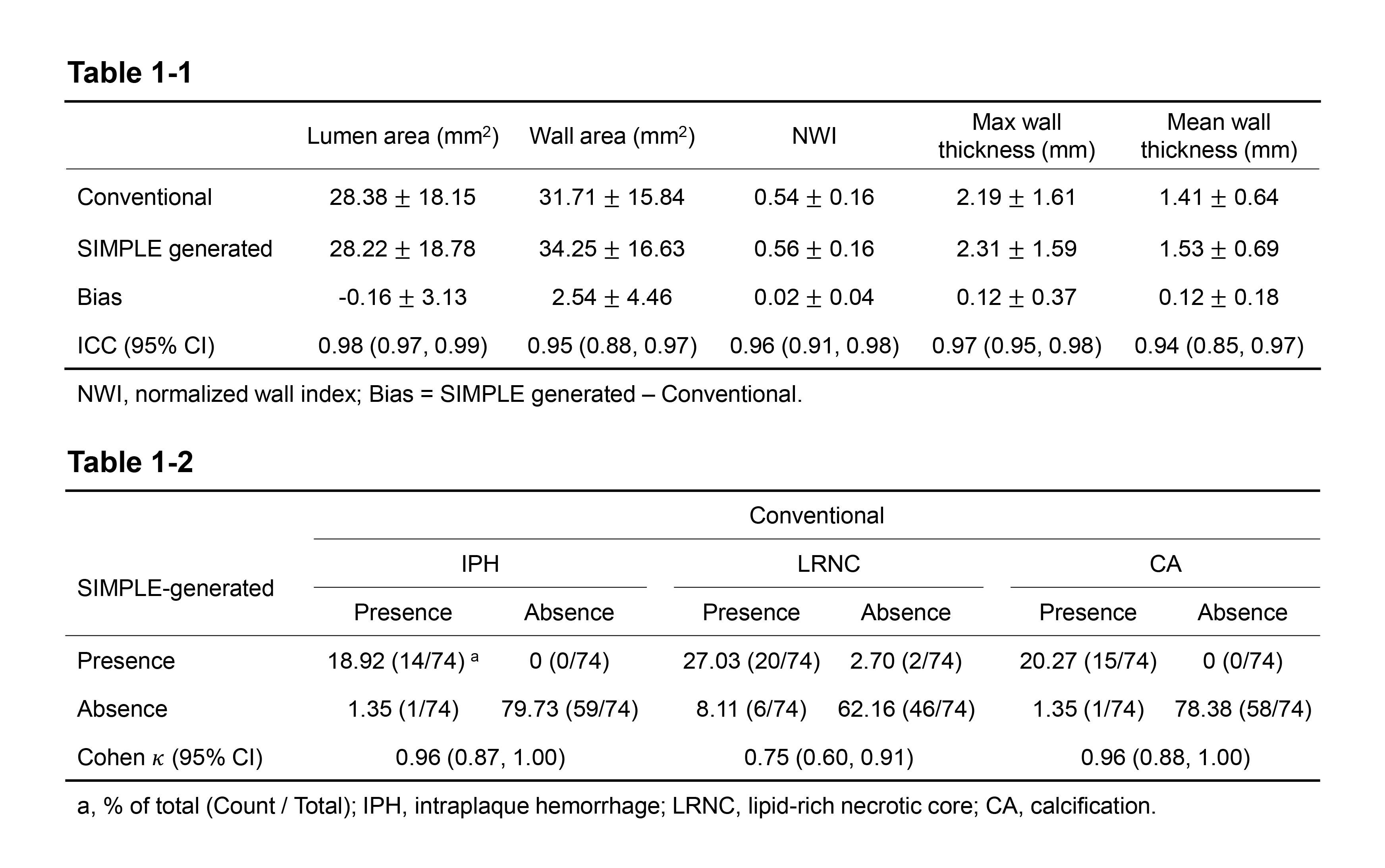
Table 1-1 Comparison of SIMPLE generated images and conventional multi-contrast MR images in measuring vascular morphological features.
Table 1-2 Agreement between SIMPLE generated images and conventional multi-contrast images in plaque components identification.