1231
PET-MR imaging of reactive macrophages in an orthotopic model of ovarian cancer
Catherine Foss1, Desmond Jacob1, Flonné Wildes1, and Marie-France Penet1
1The Russell H. Morgan Department of Radiology and Radiological Science, The Johns Hopkins University School of Medicine, Baltimore, MD, United States
1The Russell H. Morgan Department of Radiology and Radiological Science, The Johns Hopkins University School of Medicine, Baltimore, MD, United States
Synopsis
Major limitations of radiological evaluation of ovarian cancer arise from the inability to consistently detect small lesions. Here, we combine the anatomic imaging capabilities of MRI with the highly sensitive molecular imaging capabilities of PET in an orthotopic syngeneic model of ovarian cancer. [124I]iodo-DPA-713, a macrophage-specific imaging agent, is used to detect and stage ovarian cancer. Specific uptake of the radiotracer was observed within and proximal to the primary tumor at early stages, and in the peritoneal ascitic fluid and within metastases at later stage. These studies will provide insights into ovarian cancer progression that will complement emerging host-directed therapeutics.
Introduction
Ovarian cancer is a lethal gynecologic malignancy and the fifth leading cause of cancer-related deaths in women in the United States. Early detection is a critical unmet need, as patients have significantly improved prognosis when a tumor is discovered at an early stage. Disease staging for prognosis and treatment strategy is critical to ensure proper clinical management. Major limitations of radiological evaluation of ovarian cancer arise from the inability to consistently detect small lesions. The availability of multiplexed hybrid imaging systems, such as simultaneous PET-MRI scanners, presents unique opportunities to combine the strengths of anatomic, physiologic and molecular detection within a single exam for the purpose of early detection and staging of ovarian cancer.Tumor associated macrophages (TAMs) play a critical role in ovarian cancer, and consistently present a pro-tumoral M2-like profile, and promote tumorigenesis. TAMs are among several promising targets in host-directed therapies in cancer.Here, we combine the anatomic imaging capabilities of MRI with the highly sensitive molecular imaging capabilities of PET in an orthotopic syngeneic model of ovarian cancer to detect and stage ovarian cancer. [124I]iodo-DPA-713, a macrophage-specific imaging agent is used for defining host signatures. It is a low molecular weight probe that is selectively trapped within reactive phagocytes in both peripheral and central nervous system tissues, including TAMs, and exhibits very low background retention. [125I]Iodo-DPA-713 SPECT and a fluorescent analog (DPA-800) have been used to specifically image reactive TAMs associated with expanding pancreatic cancer xenografts as well as granulomas associated with Mycobacterium tuberculosis infection. PET-MR imaging allows visualization of ovaries and the peritoneal environment.Methods
Pieces of ID8-Defb29-VEGF tumor were orthotopically implanted onto the ovary of C57BL/6 mice. Subcutaneous ID8-Defb29-VEGF tumors were imaged for comparison, as well as sham control mice, for which surgery was performed without grafting the tumor piece. Bioluminescent imaging (BLI) was used to follow tumor progression, as these cancer cells constitutively express luciferin. Experiments were performed in mice presenting with different disease stages, from early stage when tumors are less than 3 mm with no metastases, to late stage with metastases and ascites present.For the PET imaging, mice were injected with ~525 µCi each of [124I]iodo-DPA-713 in 10% ethanol in PBS, pH 7.5 as a 100-200 µL intravenous bolus. PET-MR images were acquired 24h post injection on a Bruker 7T/30 MRI scanner with the three ring Bruker PET insert Si 198. Simultaneous 10 mins whole body static PET scan and a T2-weighted (T2w) 2D coronal scan were acquired for 2 mice placed side by side on the animal bed. 3D maximum likelihood expectation maximization (MLEM) iterative image reconstruction algorithm with a pixel size of 0.5 mm and 16 iterations was used for PET reconstruction. A 72 mm Tx/Rx PET optimized RF coil centered inside the PET detector was used for the MRI acquisition. The multislice TurboRARE T2w MRI scan had a FOV of 70mm x 60mm, 18 slices, 1mm slice thickness, TR/TE 3000/32 ms, matrix size of 280 x240 and 4 averages. The multislice T2w axial scans were acquired using a TurboRARE sequence with an FOV of 25mm x 60mm, 20 slices, 1mm slice thickness, TR/TE 2500/25 ms, matrix size of 100 x240 and 4 averages. Image fusion and analysis was done using PMOD.Results
BLI data were used to stratify the mice into different groups (Figure 1). Specific uptake of the radiotracer was observed within and proximal to the primary tumor at early stages (Figure 2). At later stages, uptake was observed in the peritoneal ascitic fluid and within metastases in the lungs, in addition to clear halos surrounding the primary tumor (Figure 3). Accumulation of [124I]iodo-DPA-713 in the tumor was confirmed by optical imaging after injection of DPA-713-IRDye800CW conjugate.(data not shown).Discussion:
[124I]iodo-DPA-713 can be used in ovarian cancer to image macrophages in primary tumor as well as metastases and ascitic fluid.Conclusion
These studies will provide insights into ovarian cancer progression that will complement emerging host-directed therapeutics destined for the clinic. They are clinically translatable and would facilitate non-invasive detection of a nursery of tumor supporting reactive macrophages.Acknowledgements
Pilot Funding from the Cancer Molecular and Functional Imaging Program of the SKCCC (NIH P30 CA006973) is gratefully acknowledged.References
No reference found.Figures
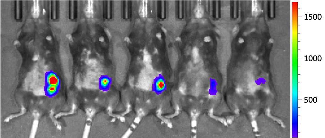
Bioluminescent images of 5 orthotopic ID8-Defb29-VEGF tumor bearing mice
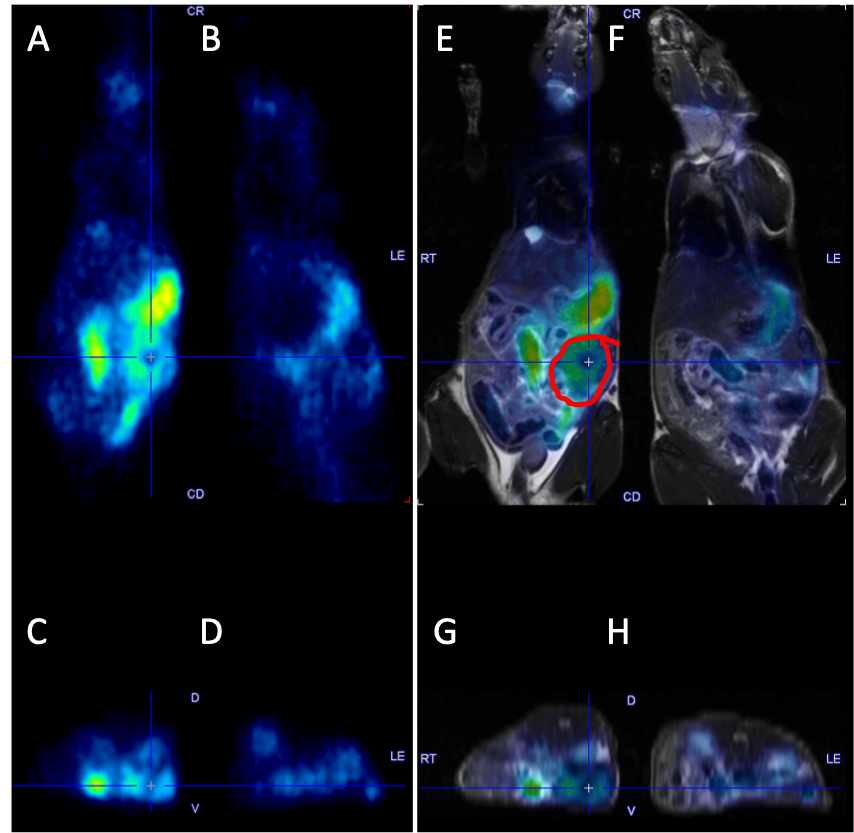
PET images of a tumor bearing mouse (A,C) and a sham mouse (B,D) with coronal (A,B) and corresponding axial slices (C,D). Corresponding fused PET-MR images (E-H).
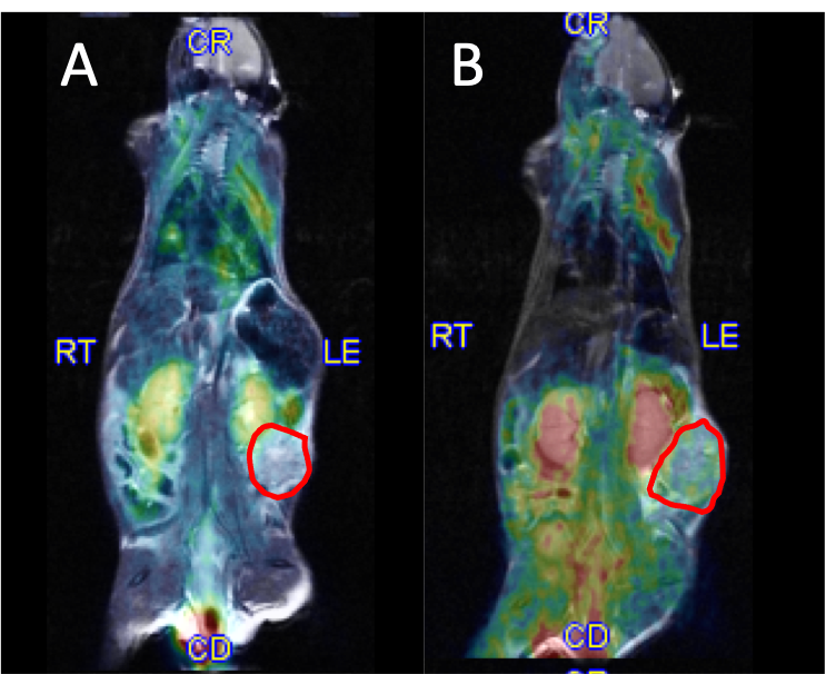
Fused PET-MR images of 2 tumor bearing mice with lung metastases (A) and ascitic fluid (B).