1181
Model-Based Iterative Reconstruction for Short-Axis Propeller EPI at 7T MRI1Radiologic Technology Department, Associated Medical Sciences, Chiang Mai University, Chinag Mai, Thailand, 2Department of Radiology, Mayo Clinic, Rochester, MN, USA, Rochester, MN, United States, 3Otto-von-Guericke University Magdeburg, Biomedical Magnetic Resonance, Magdeburg, Germany, 4National Nanotechnology Center (NANOTEC), National Science and Technology Development Agency (NSTDA), Bangkok, Thailand, 5Department of Radiology, Stanford University, Stanford, CA, United States
Synopsis
Short-Axis Propeller (SAP) EPI enables short echo spacing, thereby minimizing geometric distortion, while providing low resolution in the readout direction of single blades. These multiple low-resolution blades can be combined to create a final high-resolution image. However, off-resonance effect often results in blurring after blade combination. In this work, we extend a model-based framework for reconstructing SAP-EPI to minimize off-resonance induced blurring artifacts. Moreover, locally low rank (LLR) regularization is incorporated to estimate per-blade phase calibrations. As a result, the proposed technique enables high-resolution SAP-EPI images with minimizing blurring artifact and no need for phase calibration of different multi-blade directions.
INTRODUCTION
Short-Axis-Propeller (SAP) EPI enables short echo-spacing (ESP), thereby minimizing geometric distortion, while providing low resolution in the readout direction of single blades. Typically, multiple low-resolution blades can be combined to create a final high-resolution EPI image1-3. However, phase variations among blades and also off-resonance effects that result in blurring remain especially at high field (e.g., 7T). In this work, we extend a recent model-based framework4 for reconstructing SAP-EPI to minimize off-resonance induced blurring artifacts. Moreover, locally-low-rank (LLR) regularization5 is incorporated to estimate per-blade phase calibrations.METHODS
I. Discrete Signal Model for Single-Blade SAP-EPI: EPI uses high readout bandwidths such that $$$ \Delta{t}<<T$$$, where $$$\Delta{t}$$$ and $$$T$$$ are dwell time and echo-spacing, respectively - thus $$$\Delta{t}\approx{0}$$$ can be assumed, thereby off-resonance effects primarily manifest along the phase-encoding direction. With discretizing6, time-segmenting7, and polarity flipping, the single-blade SAP-EPI signal measured during readout $$$m\in [0\,M]$$$ of phase-encoding line $$$n\in [0\,N]$$$ can be modeled as: $$g_{c,\alpha}[m,n]=\sum_{l=0}^{L}Λ_{l}(n)\left\{\sum_{p=0}^{P-1}\sum_{q=0}^{Q-1}s_c[p,q]u[p,q]e^{-j(\triangle{w_0}[p,q][\curlyvee_l-\frac{N-1}{2}]T}e^{-j(k_{x,\alpha}[m]p+k_{y,\alpha}[n]q))}\right\}+\epsilon_{c,\alpha}[m,n]---(1)$$where $$$Λ_{l}$$$ is a spectral window, $$$\curlyvee_l$$$ denotes the center of segment $$$l$$$ and $$$p\in[0\,P]$$$ and $$$q\in[0\,Q]$$$ are pixel indices. $$$u$$$ is the complex-valued target image, $$$k_{x,\alpha}$$$ and $$$k_{y,\alpha}$$$ are the k-space coordinates in the readout and phase-encoding dimensions associated with blade angle $$$\alpha$$$. $$$s_c$$$ is the sensitivity profile for coil $$$c\in[0\,C-1]$$$, $$$\triangle{w_0}$$$ is the off-resonance, and $$$\epsilon$$$ is Gaussian noise. Defining $$$W_l=diag\left\{e^{-j(\triangle w_0[p,q][\curlyvee_l-\frac{N-1}{2}]T}\right\}$$$ and $$$S=[diag\left\{S_0\right\}\cdots{diag}\left\{S_{C-1}\right\}$$$, Eq. (1) abstracts to:
$$G_\alpha=\left(I{\otimes}\sum_{l=0}^{L}Λ_lF_\alpha{W_l}\right)Su_\alpha+\epsilon_\alpha=A_\alpha{u_\alpha}+\epsilon_\alpha---(2)$$
where $$$F_\alpha$$$ is the Fourier transform (or type-II NUFFT) and $$$\otimes$$$ is Kronecker’s product.
II. Joint Multi-blade Image Reconstruction with virtual-coil LLR: The virtual-coil LLR8,9 is performed to handle partial Fourier sampling. A set of target images ($$$u_\alpha$$$) are obtained by minimizing the following convex cost function.
$${\min_{\left\{u_1,...u_{N_\alpha}\right\}\in{\mathbb{C}}}}{\left\{\beta\sum_{b\in{\Psi}}\parallel{R_b}(\sum_{\alpha=1}^{N_\alpha}(u_\alpha\delta_\alpha^T+\overline{u_\alpha}\delta_{\alpha+N_\alpha}^T))\parallel_{*}+\sum_{\alpha=1}^{N_\alpha}\parallel{A_\alpha}{u_\alpha}-G_\alpha\parallel_2^2\right\}---(3)}$$
$$$\beta>0$$$ is a regularization parameter. $$$\delta$$$ is Kronecker delta. Operator $$$R_b$$$ extracts the $$$b^{th}\,(B^2\times2N_\alpha)$$$ blocks from the set $$$\Psi$$$ and reshapes each block into Casorati-matrix. Note that there are $$$2N_\alpha$$$ blades in total, including actual acquired blades and virtual blades with “$$$\bar{u}$$$ ” denoting complex-conjugation. The nuclear norm ($$$\parallel\cdot\parallel_*$$$) is the convex envelope of the rank functional. The optimization problem in (3) can be solved using fast composite splitting algorithm10 (FCSA), a generalization of the Fast Iterative Shrinkage Algorithm (FISTA) that allows independent management of penalties, which herein coincides with blockwise singular value thresholding (SVT) of the (conjugate) augmented Castorati matrices along with aggregation of nominal and virtual blade blocks.
III. Data Acquisition and Processing: In-vivo experiments were performed on a whole‐body 7T-MRI scanner (Siemens Healthcare, Erlangen, Germany) equipped with a gradient coil type SC72AB (70mT/m and 200 T/m/s) using a 32‐channel head-coil (Nova Medical, Wilmington, MA, USA). A healthy-volunteer was scanned after informed consent according to institutional review board-approved (IRB) protocol. six-echo SPGR and single-shot EPI were acquired using vendor-provided sequences. For ssEPI, the FOV was tilted eight times with different angles (22.5 degree each), resulting in SAP-EPI acquisition where readout is 64 samples, 256 phase-encoding steps, 6/8 partial Fourier, and SENSE-factor of 4. SPGR data were utilized for estimating the B0-field-map via fat-water separation toolbox11. In addition, coil sensitivity maps were estimated from their first echo data using the ESPIRiT technique12. The proposed MBIR was performed in Matlab using 20 iterations of FISTA with manually optimized $$$\beta\,(\beta=0.02)$$$, block-size 7⨯7, 2X-oversampled type-II NUFFT with a width $$$J=5$$$ Kaiser-Bessel kernel, and $$$L=N_{PE}$$$ time segments. Ramp sampling and odd-even phase correction was performed offline prior to MBIR.
RESULTS
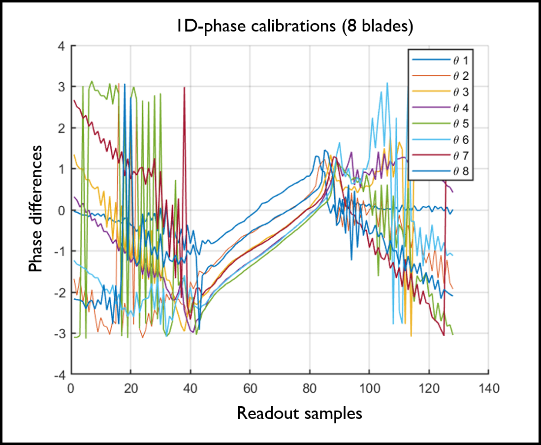
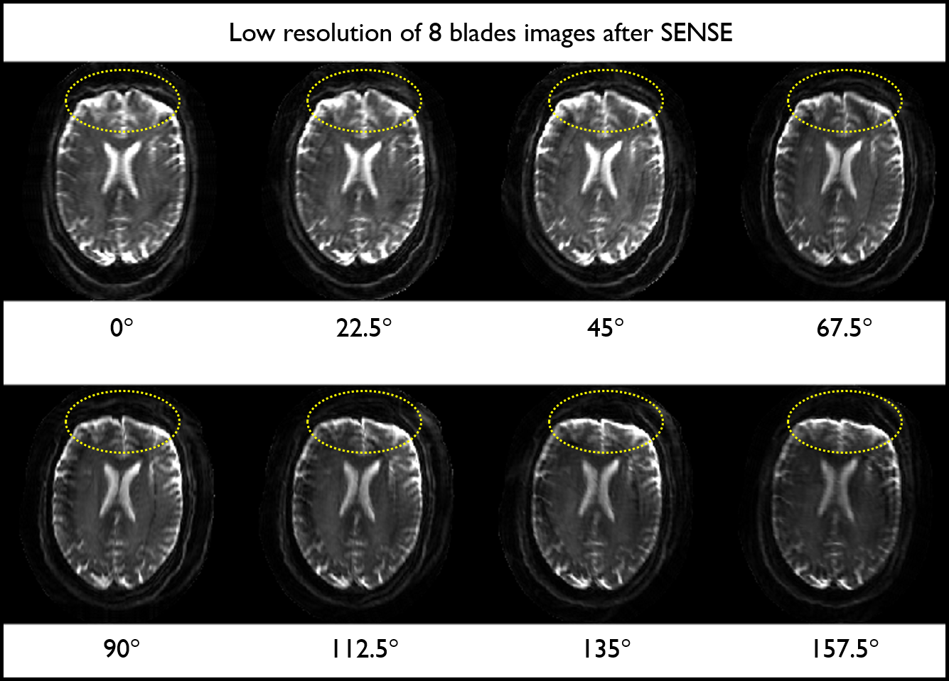
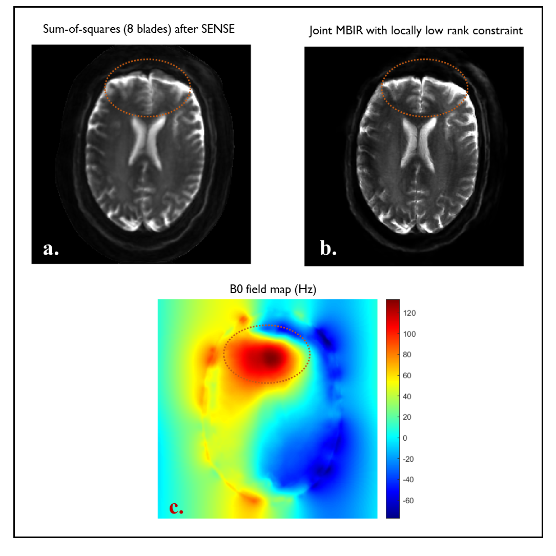
RESULTS: Fig. 1 shows 1D-phase differences between positive and negative readout echoes corresponding to the eight blades with different angles. After being fitted by magnitude weighted least squares, their slopes and intercepts are in the ranges of $$$[0.026\,0.031]$$$ and $$$[-2.54\,-1.62]$$$, respectively. Fig. 2a shows blade specific images obtained using standard SENSE reconstruction. These images look slightly blurry since only 64 samples along the readout were acquired and used to create 256x256 images after zero padding. The yellow circles highlight the geometric distortions that appear differently at different blade angles. The magnitude combination of eight blade images is shown in Fig. 3a, where the blurring artifact is highly visible, particularly in strong B0 field inhomogeneity regions highlighted by the red-circle (Fig. 3c), with up to 120 Hz. Fig. 3b shows that the proposed reconstruction can provide promising results – the blurring artifacts appear much reduced (red-circle) and the resolution is visibly improved.DISCUSSIONS
The proposed extended version of MBIR with LLR enables high-resolution SAP-EPI images with no need for phase calibration of different multi-blade directions. Typically, N/2-Nyquist ghosting in oblique-FOV-EPI is visible after standard 1D-phase correction due to cross-term gradient induced non-linear phase errors. However, in this study the 1D zeroth and first orders phase correction appears sufficient. This may be due to the use of short readout time, thus higher than first orders phase errors may be negligible. Highly accurate B0 field maps are crucial for the proposed scheme. Thus, highly effective technique in creating B0 maps, such as PSF13 may help to improve the image quality even further. It has been discussed how many shots/blades are sufficient for LLR. This point will be further investigated in future studies. Finally, the computational time is proportional to the number of blades, time segments and coil numbers. These issues may be handled by parallel-computing and coil compression14, respectively.Acknowledgements
This work was supported by NIH U01 EB024450 and SFB 1436 project Z02.References
1. Skare S, Newbould RD, Clayton DB, Bammer R. Propeller EPI in the other direction. Magn Reson Med. 2006; 55(6):1298‐1307.
2. Wen Q, Kodiweera C, Dale BM, Shivraman G, Wu YC. Rotating single-shot acquisition (RoSA) with composite reconstruction for fast high-resolution diffusion imaging. Magn Reson Med. 2018; 79(1):264‐275.
3. Chang HC, Chuang TC, Lin YR, Wang FN, Huang TY, Chung HW. Correction of geometric distortion in Propeller echo planar imaging using a modified reversed gradient approach. Quant Imaging Med Surg. 2013; 3(2): 73-81.
4. Yarach U , Bernstein MA, Huston J-III, In MH, Kang D, Shu Y, Gray EM, Meyer M, and Trzasko JD. Model-Based Single-Shot EPI Reconstruction with Sparsity Regularization. In Proc. Int. Soc. Magn. Reson. Med. 2019; pp. 0929.
5. Trzasko JD, Manduca A and Borisch E, Local versus global low-rank promotion in dynamic MRI series reconstruction, in Proc. Int. Soc. Magn. Reson. Med,, 2011, p. 4371.
6. Fessler J.A. Model‐based image reconstruction for MRI. IEEE Signal Process Mag 2010; 27:81–89. 7. Sutton B., Noll D., Fessler J. Fast, iterative image reconstruction for MRI in the presence of field inhomogeneities. IEEE Trans Med Imaging 2003; 22: 178–188.
8. Trzasko JD, Manduca A. Calibrationless parallel MRI using CLEAR. In Conference Record of the Forty Fifth Asilomar Conference on Signals, Systems and Computers (ASILOMAR), Pacific Grove, CA, 2011. pp. 75– 79.
9. Levine, E, Stevens, K, Beaulieu, C, Hargreaves, B. Accelerated three-dimensional multispectral MRI with robust principal component analysis for separation of on- and off-resonance signals. Magn Reson Med 2017; 79(3): 1495–1505.
10. Junzhou H, Shaoting Z, Hongsheng L, Dimitris M. Composite splitting algorithms for convex optimization, Computer Vision and Image Understanding 2011; 115(12): 1610-1622.
11. Hu H, Börnert P , Hernando D, et al. ISMRM workshop on fat–water separation: Insights, applications and progress in MRI. Magn Reson Med, vol. 68, no. 2, pp. 378–388, 2012.
12. Uecker M, Lai P, Murphy MJ, Virtue P, Elad M, Pauly JM, Vasanawala SS, Lustig M. ESPIRiT—an eigenvalue approach to autocalibrating parallel MRI: where SENSE meets GRAPPA. Magn Reson Med 2014; 71: 990– 1001.
13. In MH, Speck O. Highly accelerated PSF-mapping for EPI distortion correction with improved fidelity. Magn Reson Mater Phy 2012; 25: 183–192.
14. Zhang T, Pauly JM, Vasanawala SS, Lustig M. Coil compression for accelerated imaging with Cartesian sampling. Magn Reson Med 2013; 69: 571– 582.
Figures