1161
Simultaneous FLAIR T1W and T2W imaging using Temporal Harmonic Encoding1Department of Radiology, Mayo Clinic, Rochester, MN, United States
Synopsis
A temporal harmonic encoding scheme is proposed to improve T1W FLAIR imaging with TSE acquisition. The imaging method reduces blurring in T1W FLAIR and provides an extra T2W FLAIR contrast at the same time with no need of additional scan time.
Introduction
FLAIR imaging has been used in clinical exam to capture lesions in the brain without influence by the CSF1. The sequence typically adopts a turbo spin echo (TSE) acquisition with low-high phase encoding order for T1W contrast. As a result, the T2 decay with the echo trains usually introduces blurring along phase encoding direction. Parallel imaging was one method to reduce T2 blurring but balancing between artifact, SNR and scan time requires further consideration2,3. Over the past few years, imaging acceleration strategies involved with temporal dimension and/or additional non-spatial dimensions have been exploited to render images with better quality without sacrificing scan time4-7. The present work, Temporal Harmonic Encoding, adopted one of the shear grid spatial-temporal trajectory in the TSE acquisition to suppress T2 blurring effect. Meanwhile, the harmonic signals decomposed in the reconstruction could reveal additional T2W contrast for reference.Materials and Methods
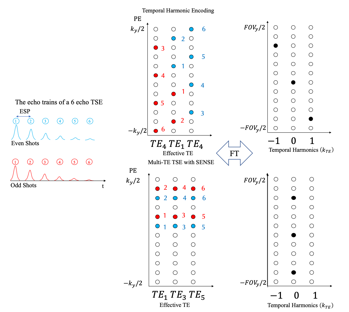
Fig 1. illustrates an example of Temporal Harmonic Encoding sampling strategy in conjunction with a SENSE accelerated multi-TE imaging at TSE factor = 6 and their point spread functions. The Temporal Harmonic Encoding factor $$$R_T$$$, and the acceleration factor of SENSE $$$R_S$$$ are both 3. Note that Temporal Harmonic Encoding only rearranges k-space filling pattern, the total scan time for Temporal Harmonic Encoding will be identical to the original fully sampled TSE sequence. The reconstruction of Temporal Harmonic Encoding data can be described as $$ min_{\rho_y, k_{TE} } |s_{k_y, {TE}} - EC\rho_{y,k{TE}}|^2_2-\lambda^2 |M^{-1}_{y,{TE}} \rho_{y,k_{TE}}|^2_2$$, where $$$ s_{k_y, {TE}}$$$nis the acquired data in the k-effective TE space, $$$rho_{y,k{TE}}$$$ is the signal in the Fourier Transformed space, E is the encoding matrix including a 2D Fourier Transform operation and the sampling mask, C is the coil sensitivity and M is the reference signal for regularization weighted by $$$\lambda$$$. The self-reference M is employed as in the Ref 4. The DC ($$$k_{TE} = 0 $$$)of reconstructed image is regarded as the equivalent contrast to the T1W FLAIR. The other harmonic components along $$$x-k_{TE}$$$ domain will be used to generate T2W contrast.Three health volunteers were recruited in this study following informed consent. The experiments were conducted on a 3.0 T scanner (Elition, Philips Healthcare, Best, Netherland) using a SPIR fat-sat T1W FLAIR TSE with ETL = 12, ESP = 8ms, TE1 = 7ms, TI = 1350ms, and TR = 3500ms. The spatial resolution of the images is $$$1\times1 mm^2$$$ with slice gap 2mm for a 10 slice scan. Three different k-space arrangements were tested: (1) Fully sampled k-space with low-high order, (2) Multi-TE SENSE with $$$R_S = 3$$$ , and (3) Temporal Harmonic Encoding with $$$ R_T = 3 $$$. A fitting procedure is performed to the Multi-TE SENSE reconstruction to extract T1W FLAIR and the T2 map for reference.
Results
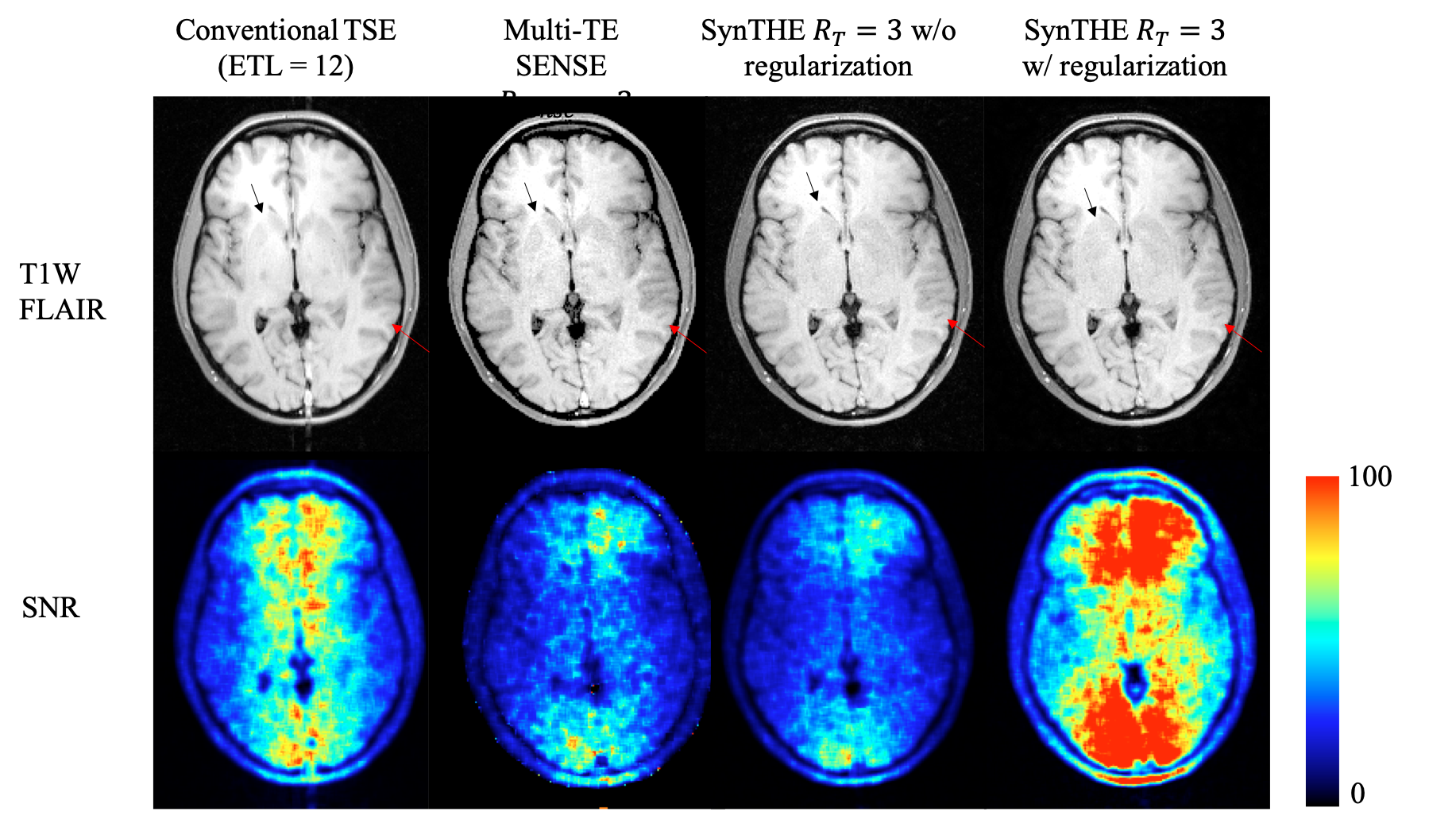
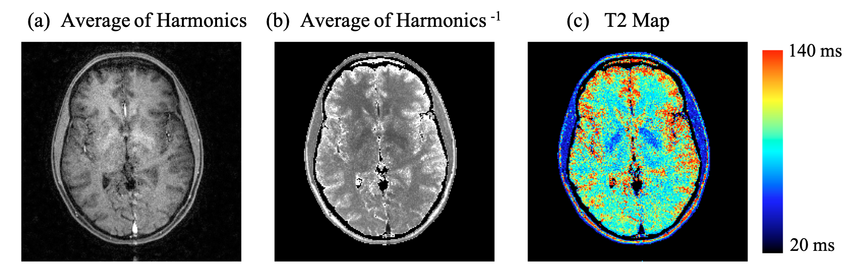
Fig 2. compares the images with equivalent contrast to T1W FLAIR. Blurring due to T2 decay can be found in the conventional scan while the edge becomes sharper in the multi-TE SENSE and Temporal Harmonic Encoding reconstructions. Meanwhile the contrast in Temporal Harmonic Encoding is comparable to the original scan in spite of some contrast loss in the center of the image. The SNR of Temporal Harmonic Encoding is also comparable to the multi-TE SENSE. The regularization in the Temporal Harmonic Encoding scheme could help SNR restored to a better level.Fig 3 shows calculated images from the other two harmonic components in Temporal Harmonic Encoding reconstruction. The information in these harmonic components reflects the T2 weighting in the parenchyma. In comparison with the T2 map calculated from the multi-TE SENSE, the T2W Temporal Harmonic Encoding image demonstrates similar contrast with better SNR.
Discussion
Minimizing T2 blurring in a TSE sequence has been a critical subject. The Temporal Harmonic Encoding method presented in this work combines the ideas of changing the sampling function in conjunction with sensitivity encoding to decode harmonic components along the echo trains. The DC component of Temporal Harmonic Encoding is suggested to resemble the primary contrast in the original sequence with sharper edges but a bit lower SNR due to the decoding process. This SNR penalty is equivalent to the g-factor in the parallel imaging reconstruction while additional regularization information has potential to reduce the noise amplification for improving image quality as shown in Fig 2. The harmonic components in Temporal Harmonic Encoding unveil the fast changing signal with TE. Although the dominant signal change is T2 decay, flow is another source causing signal reduction leading to high magnitude in the harmonic components found in the vessels in Fig 3(a). As far as the artifact is concern, the DC components, T1W FLAIR, is more robust due to the shear grid sampling pattern. However, the artifact related to inaccurate sensitivity map and motion will appear in the harmonic components and T2W image. Therefore, robust sensitivity estimation and motion control will be critical when applying the proposed method into practice.Conclusion
We have proposed a Temporal Harmonic Encoding method for acquiring T1W FLAIR image. The results suggest that it could reduce T2 blurring in a TSE image and provide additional T2W information without lengthening the scan time.Acknowledgements
This work was funded in part by Philips Healthcare.References
1. Greenspan, S.L., et al., FLAIR and HASTE imaging in neurologic diseases. Magn Reson Imaging Clin N Am, 1998. 6(1): p. 53-65.
2. Gabr, R.E., et al., Optimal combination of FLAIR and T2-weighted MRI for improved lesion contrast in multiple sclerosis. J Magn Reson Imaging, 2016. 44(5): p. 1293-1300.
3. Xiao, Z., et al., Comparison of parallel MRI reconstruction methods for accelerated 3D fast spin-echo imaging. Magn Reson Med, 2008. 60(3): p. 650-60.
4. Chao, T.C., et al., Fast diffusion imaging with high angular resolution. Magn Reson Med, 2017. 77(2): p. 696-706.
5. Madore, B., G.H. Glover, and N.J. Pelc, Unaliasing by fourier-encoding the overlaps using the temporal dimension (UNFOLD), applied to cardiac imaging and fMRI. Magn Reson Med, 1999. 42(5): p. 813-28.
6. Breuer, F.A., et al., Controlled aliasing in volumetric parallel imaging (2D CAIPIRINHA). Magn Reson Med, 2006. 55(3): p. 549-56.
7. Tsao, J., P. Boesiger, and K.P. Pruessmann, k-t BLAST and k-t SENSE: dynamic MRI with high frame rate exploiting spatiotemporal correlations. Magn Reson Med, 2003. 50(5): p. 1031-42.
Figures