1156
non-Cartesian Parallel imaging and compressed sensing adapted for accelerating hybrid trajectory PETRA1Siemens Shenzhen Magnetic Resonance Ltd., Shen Zhen, China
Synopsis
PETRA sequence, with ultra-short TE and ultra-low acoustic noise, has wide applications such as dental imaging, lung imaging, bone imaging, pediatric imaging as well as the routine anatomical imaging. However, the long scanning time and the vulnerability to motion hampered its usage. In this study, the combining non-Cartesian parallel imaging and Compressed sensing technique is adapted for the hybrid trajectory PETRA sequence. The image quality is clinically adequate for 2-5 folds acceleration.
INTRODUCTION
PETRA1 is an ultra-short TE based, hybrid trajectory pulse sequence with 3D radial acquisition in the outer k-space and Cartesian single points in the central k-space. With the advantage of ultra-short TE and ultra-low acoustic noise, it has wide applications such as dental imaging, lung imaging, bone imaging, pediatric imaging as well as the routine anatomical imaging. However, it usually takes a long scanning time (typically over 6min) for getting a high-resolution image with PETRA because of the huge number of radial views needed. In this abstract, the combining non-Cartesian parallel imaging (PI) and Compressed sensing (CS)2,3 is adapted for accelerating the hybrid trajectory PETRA imaging.METHODS
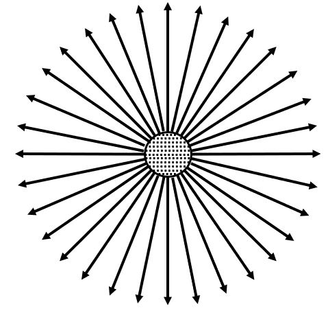
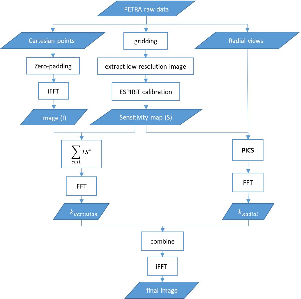
The acquisition scheme of PETRA consists of two parts: the 3D radial acquired spokes which cover most of the k-space except the central part, and the Cartesian single points in the k-space center. Figure 1 shows the 2D projection of the PETRA k-space trajectory. Since the acquisition of the Cartesian points only cost a little time, the total acquisition time is almost proportional to the number of radial views. For acceleration, only the radial views are under-sampled, while the Cartesian points in the k-space center are always fully sampled. The two parts have to be processed separately because it is not possible to apply the PICS algorithm directly on the data with hybrid trajectory. The flowchart of the image reconstruction is shown in Figure 2. Detailed steps are described as below:Step 1: Perform gridding to the PETRA raw data and then remove the high frequency part to get the low-resolution images. Perform an ESPIRiT4 calibration on the images for sensitivity maps.
Step 2: Extract the Cartesian points from the PETRA raw data. For each coil, the Cartesian points are expanded to the same size as the sensitivity map via zero-padding. Perform iFFT on the datasets to get multi-coil images. Then multiply the images with the conjugate of the sensitivity maps. Sum the result images to produce a coil-combined image. Then perform FFT to get the coil-combined k-space $$$k_{Cartesian}$$$.
Step 3: Extract the Radial views from the PETRA raw data. Perform a PICS reconstruction on the radial data with the sensitivity maps from Step 1. A coil-combined image was generated as the output of the reconstruction. Then perform FFT to get the coil-combined k-space $$$k_{Radial}$$$.
Step 4: Replace the center region of $$$k_{Radial}$$$ with the corresponding data from $$$k_{Cartesian}$$$. The new combined k-space data was then transformed to the final image by an iFFT.
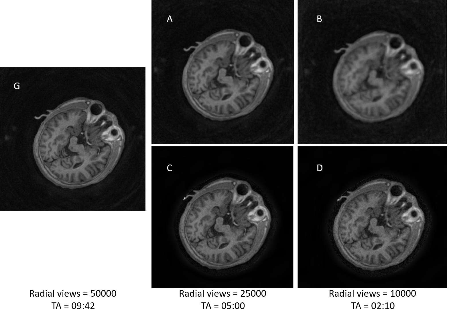
One dataset of 3D T1w PETRA was collected on a 1.5T MRI system (MAGNETOM Amira, Siemens Healthineers) with the following parameters: Resolution = 256*256*256; Turbo factor = 200; Number of radial views = 50000; Total scan time (TA) = 09:42. Two under-sampled datasets were generated by uniformly under-sampling the original raw data to Radial views = 25000, 10000, respectively.
RESULTS
Figure 3 shows one slice of the reconstructed images with different number of radial views by using a standard nuFFT reconstruction (A, B) and PICS reconstruction (C, D). The fully sampled image G is for comparison.A, B: Image are seriously blurring, especially under high acceleration factor.C, D: Image contrast and spatial resolution are well kept, with only slight SNR drop under high acceleration factor. The image quality is clinical adequate with 5-fold acceleration.DISSCUSION AND CONCLUSION
Parallel imaging and CS are effective tools for acceleration of MR scanning. For a hybrid trajectory sequence like PETRA, separate processing of the dataset is proved to be feasible: the outer k-space could be highly under-sampled to get a high acceleration factor, while keep the k-space center fully sampled for sensitivity maps calculation. With the proposed method, PETRA could be 2-5 times faster with only little reduction of image quality.Acknowledgements
The authors would like to thank Jinqiang He for his kind help with the working environment set up.References
1 David M. Grodzki, Peter M. Jakob and Bjoern Heismann, Ultrashort Echo Time Imaging Using Pointwise Encoding Time Reduction With Radial Acquisition (PETRA). Magnetic Resonance in Medicine 67:510–518 (2012).
2 Martin Uecker, Patrick Virtue, Frank Ong, Mark J. Murphy, Marcus T. Alley, Shreyas S. Vasanawala, and Michael Lustig. Software Toolbox and Programming Library for Compressed Sensing and Parallel Imaging. ISMRM Workshop on Data Sampling and Image Reconstruction, Sedona 2013
3 Martin Uecker, Frank Ong, Jonathan I Tamir, Dara Bahri, Patrick Virtue, Joseph Y Cheng, Tao Zhang, and Michael Lustig. Berkeley Advanced Reconstruction Toolbox. Annual Meeting ISMRM, Toronto 2015, In Proc. Intl. Soc. Mag. Reson. Med. 23:2486
4 Martin Uecker, Peng Lai, Mark J. Murphy, Patrick Virtue, Michael Elad, John M. Pauly, Shreyas S. Vasanawala, and Michael Lustig. ESPIRiT - An Eigenvalue Approach to Autocalibrating Parallel MRI: Where SENSE meets GRAPPA. Magnetic Resonance in Medicine, 71:990-1001 (2014)
Figures