1151
Evaluation of CEST-mDixon imaging for breast malignancy characterization and staging: correlation with histopathology1Radiology, University of Texas Southwestern Medical Center, Dallas, TX, United States, 2Philips Healthcare, Gainesville, FL, United States, 3Philips Research, Hamburg, Germany, 4Advanced Imaging Research Center, University of Texas Southwestern Medical Center, Dallas, TX, United States, 5Pathology, University of Texas Southwestern Medical Center, Dallas, TX, United States, 6Cancer Systems Imaging, University of Texas MD Anderson Cancer Center, Houston, TX, United States
Synopsis
CEST-mDixon imaging utilizing hydroxyl and amine pools could aid breast tumor diagnosis, as shown in previous preliminary studies. In the current study, CEST-mDixon is evaluated in a breast cancer patient cohort using improved saturation and acquisition parameters. The results agree with previous findings demonstrating moderate linear correlation of CEST effects at 1 ppm and 2 ppm with Ki-67. In addition, we observe a linear correlation with the percentage of cells positive for nuclear expression of the progesterone receptor (PR), the first such observation reported. Overall, the study confirms the potential for CEST-mDixon for characterizing breast tumor aggressiveness non-invasively.
Introduction
CEST-MRI is emerging as a powerful tool in assessment and characterization of tumor aggressiveness and treatment response1,2. Recent studies have demonstrated the application of CEST to breast malignancies and its potential to provide metabolic information to aid tumor characterization3-4. Previous work from our group demonstrated that CEST-mDixon is a promising MR technique for breast cancer characterization1. Hence, it is important to have an optimized protocol for diagnostic accuracy and clinical applicability. The objectives of this study are (i) to evaluate the CEST-mDixon acquisition and post-processing protocols with enhanced saturation of 2 s employing alternating parallel transmission5, and (ii) correlate CEST effect, as quantified using MTRasym for amide (3.5 ppm), amine (2 ppm), and hydroxyl (1 ppm) pools, with expression of histopathological markers of aggressiveness (estrogen receptor (ER), progesterone receptor (PR) , Her-2 and Ki-67) in the corresponding tumor tissue.Methods
For this IRB approved study, 8 female patients with suspicious breast lesions on diagnostic imaging (BIRADs 4 or 5) were recruited and scanned prior to biopsy, with written informed consent. The recruitment is ongoing with the targeted cohort of N>30. All 8 cases had confirmed malignancies with 7 Invasive Ductal Carcinoma (IDC) and 1 Mucinous Carcinoma (MC). The MC case was excluded from the analysis below (Fig.2 and 3), because of the different tumor type. The scans were performed on a 3T scanner (Ingenia, Philips, NL) using a 16-channel bilateral breast coil. CEST images were acquired using a 2D multi-slice multi-shot T1-weighted TFE sequence with 3-point multi-echo Dixon acquisition (TR/TE1/ΔTE=5.2/1.59/1.0 ms). The CEST saturation consisted of 40 hyperbolic secant shaped pulses, 50 ms each, B1rms =1.2 μT, for a total saturation length of 2 s, enabled by alternated parallel transmission5, increased from 0.5 s in a previous study1. 33 points in the Z-spectrum from -6 ppm to 6 ppm were acquired. Other imaging parameters included centric ordering, voxel size=2x2x5 mm3, FA=10°, 3 slices. Further B1rms power levels (0.7 and 1.4 mT) were acquired but not yet included in the evaluation. After the MRI study, the patients underwent routine clinical biopsies and histopathological analysis was performed.MTRasym maps for 1 ppm, 2 ppm, and 3.5 ppm were calculated. The region-of-interests (ROIs) were drawn under the guidance of a fellowship‐trained radiologist (>5 years of experience with breast imaging). ROIs were selected based on the water‐only images while referring to high‐resolution fast gradient-echo (eTHRIVE) images. Averaged MTRasym ROI-values were correlated with histopathology results. The immuno-histopathological expression scores of PR, and Ki-67 as well as the percentage of cells positive for nuclear expression of PR were obtained from the clinical biopsy reports for the correlation analysis.
Results
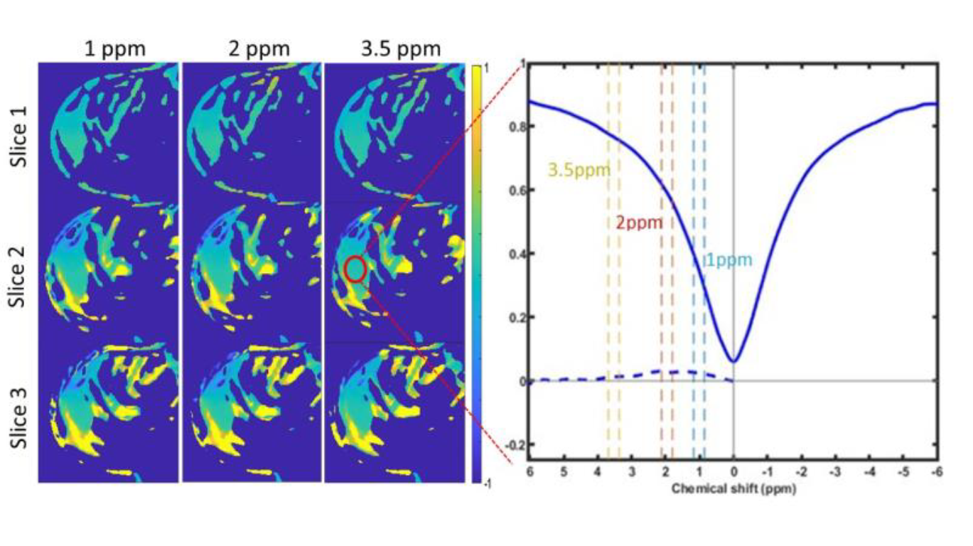
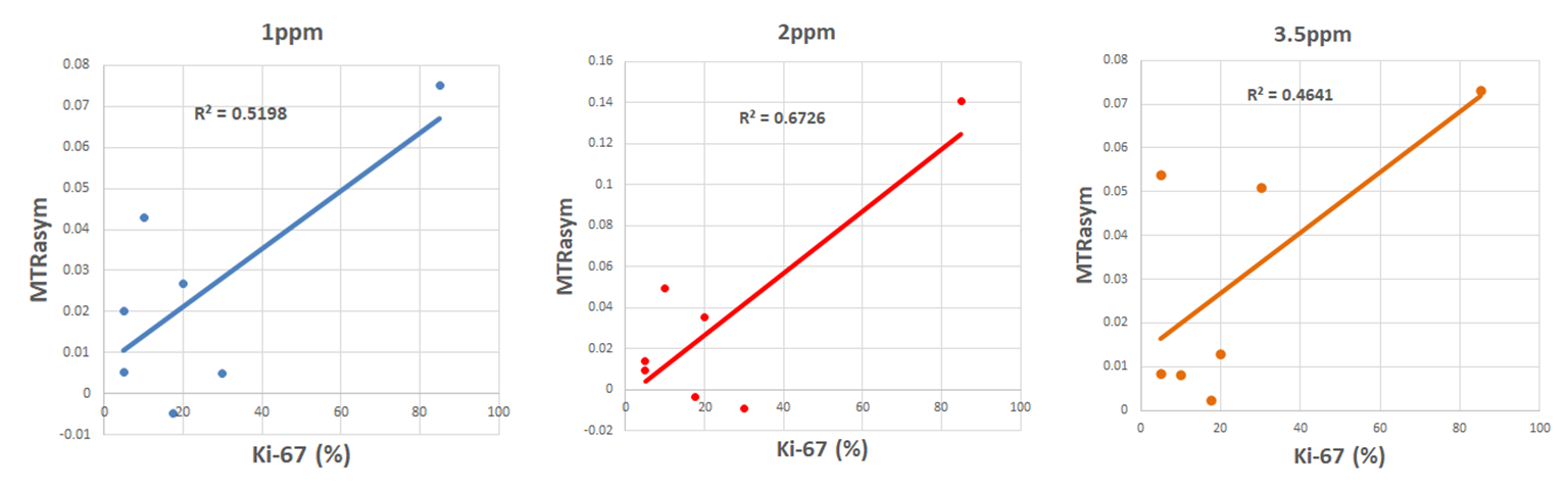
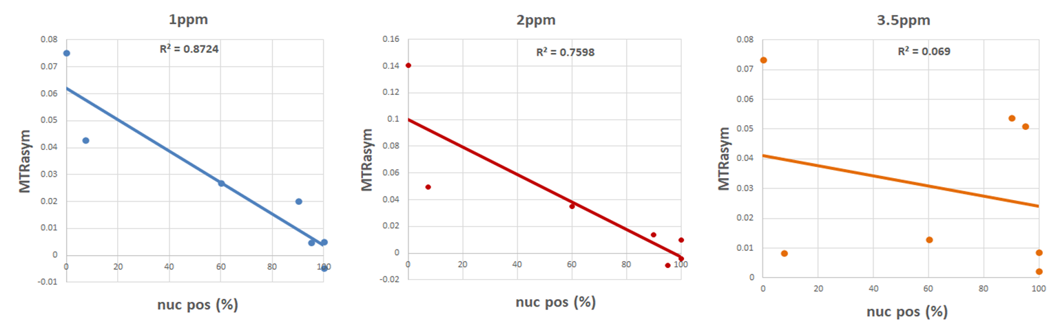
Fig. 1 demonstrates a typical CEST maps and a Z-spectrum from a suspicious lesion in a patient obtained with the CEST-mDixon protocol. Analysis of the 7 IDC patients shows moderate linear correlation with Ki-67 for MTRasym for 1 ppm and 2 ppm (R2=0.52 and R2=0.67, respectively, Figure 2). In addition, there is a strong correlation of MTRasym for 1 ppm and 2 ppm with percentage of cells positive for nuclear expression of PR (R2=0.87 and R2=0.76, respectively, Figure 3).Discussion
The observed correlations with Ki-67 status are overall consistent with our previous observations1, although longer saturation times were employed here. The correlation with the PR positive percentage of cells, while preliminary, is novel and to the best of our knowledge, had not been reported before. The correlations are observed at 1 ppm and 2 ppm, which are typically assigned to hydroxyl and amine chemical shifts, respectively. We are currently investigating the nature of this correlation and relevance to breast cancer aggressiveness in detail, and one area of interest are the alterations in choline metabolism. No correlations were observed for MTRasym (3.5 ppm), associated with amides (Figures 2 and 3), similar to previous observations.While MTRasym (3.5 ppm) has been optimized for neuro oncology in many studies, the CEST-mDixon optimization in breast cancer and for hydroxyls and amines is ongoing. Analysis of the further B1rms power levels will be performed with the goal of elucidating saturation parameters that provide best correlation with histopathology. Further markers like the estrogen receptor (ER) and Her-2 are available and will be included in the analysis.
Some slight shifts of the Z-spectral minimum after B0 correction with Dixon-type B0 maps were observed in partly fat containing areas. One potential source might be an influence of the direct saturation on the lipid spectra6, which needs further investigation.
Conclusion
The results of this study indicate that with different saturation parameters, we were able to reproduce previously reported results of CEST effect correlation with breast tumor aggressiveness, specifically a moderate correlation of MTRasym at 1 ppm and 2 ppm with Ki-67 index. For the first time, we observe a good correlation , in particular for hydroxyl-CEST (1 ppm), with percentage of cells positive for nuclear expression of PR. Work is in progress to further optimize CEST-mDixon and determine most robust and accurate analysis for breast lesion characterization.Acknowledgements
The work is supported by the CPRIT RP180031 grant.References
1) Zhang S, Seiler S, Wang X, et al. CEST‐Dixon for human breast lesion characterization at 3 T: A preliminary study. Magnetic resonance in medicine. 2018 Sep;80(3):895-903.
2) Jones CK, Schlosser MJ, Van Zijl PC, et al. Amide proton transfer imaging of human brain tumors at 3T. Magnetic Resonance in Medicine: An Official Journal of the International Society for Magnetic Resonance in Medicine. 2006 Sep;56(3):585-92.
3) Dula AN, Arlinghaus LR, Dortch RD, et al. Amide proton transfer imaging of the breast at 3 T: establishing reproducibility and possible feasibility assessing chemotherapy response. Magnetic resonance in medicine. 2013 Jul;70(1):216-24.
4) Song X, Airan RD, Arifin DR, et al. Label-free in vivo molecular imaging of underglycosylated mucin-1 expression in tumour cells. Nature communications. 2015 Mar 27;6(1):1-7.
5) Keupp J, Baltes C, Harvey PR, et al. Parallel RF transmission based MRI technique for highly sensitive detection of amide proton transfer in the human brain at 3T. InProc Int Soc Magn Reson Med 2011 May 7 (Vol. 19, p. 710).
6) Zhao Y, Yan X, Zhang Z, et al, Self‐adapting multi‐peak water‐fat reconstruction for the removal of lipid artifacts in chemical exchange saturation transfer (CEST) imaging. Magnetic resonance in medicine. 2019 Nov;82(5):1700-12.
Figures