1144
B-value derivation for diffusion-weighted Double-Echo Steady-State (dwDESS) sequences1Philips Research Europe, Hamburg, Germany, 2Philips Healthcare, Hamburg, Germany
Synopsis
To quantify diffusion weighting and to calculate the diagnostically valuable “Apparent Diffusion Coefficient” (ADC) in MR-based “Diffusion-Weighted Imaging” (DWI), a standardized b-value is typically used. To avoid typical shortcomings of standard DWI, diffusion-weighted double-echo-steady-state (dwDESS) sequences were proposed, which don’t show geometrical distortions. Balanced bipolar dwDESS, in particular, provides high SNR and is robust against motion. However, a proper definition of the b-value equivalent to standard DWI is not yet well addressed. This study investigated an estimation for effective b-values in dwDESS based on approximation of the underlying signal model, which was verified in phantom and human measurements.
Introduction
Standard Diffusion-Weighted Imaging (DWI) frequently suffers from geometric distortions, severely restricting diagnostic value, e.g. in localization of breast cancer lesions. To overcome this problem, DWI based on Double-Echo-Steady-State (DESS) sequences was proposed (so-called dwDESS1,2). However, it is yet unknown how to define a corresponding b-value, which would enable a quantitative comparison of dwDESS with other types of DWI. This study investigates the signal model previously developed1,2 and derives an effective b-value for a bipolar version of dwDESS.3Theory
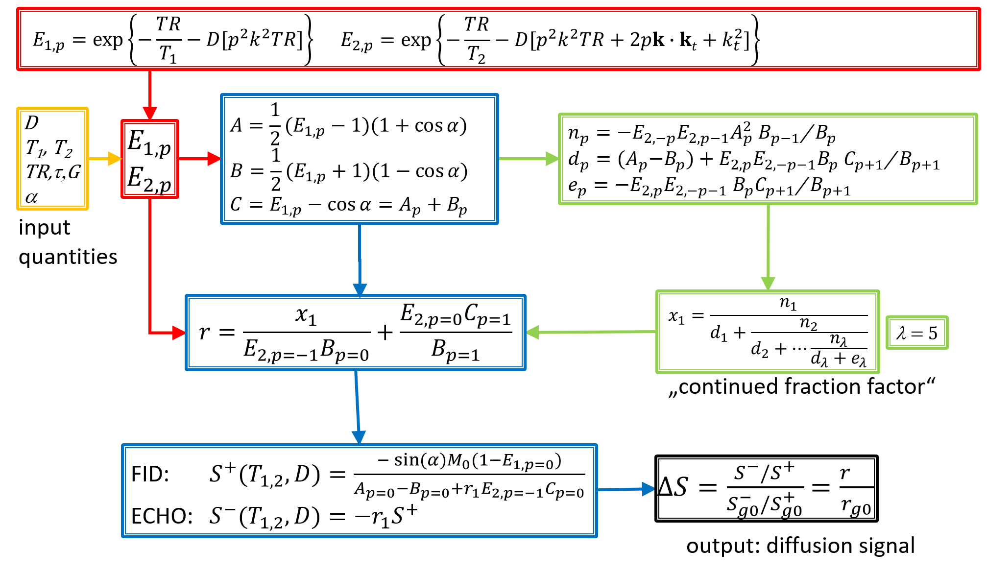
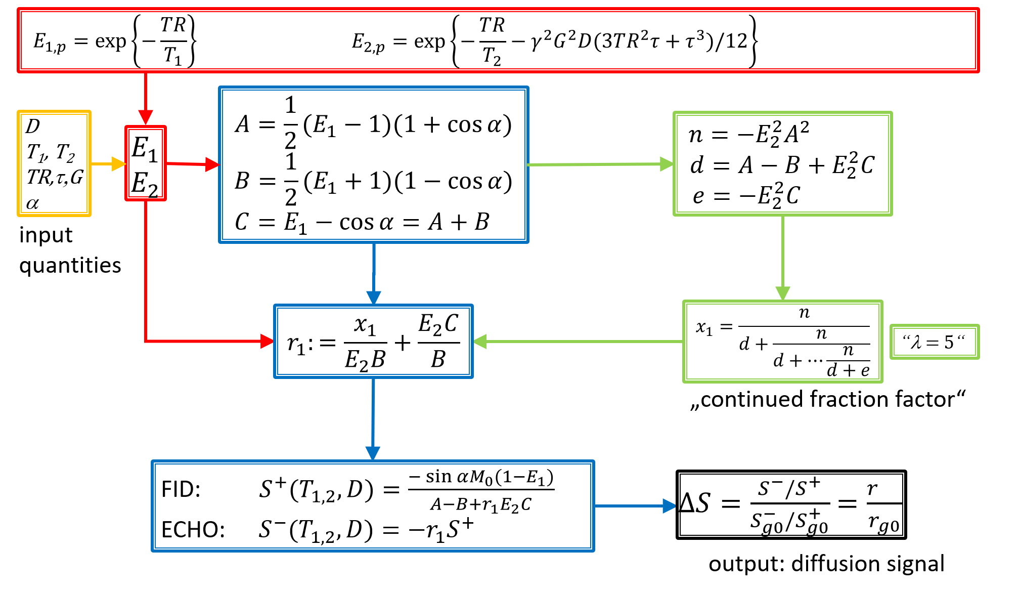
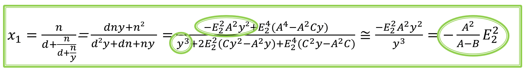
The general signal model of dwDESS in the framework of configuration theory1 is summarized in Fig.1. Based on predefined input quantities (yellow) belonging to tissue (T1,T2, diffusivity D) and sequence design (repetition time TR, diffusion gradient duration τ, diffusion gradient amplitude G, flip angle α), the exponential factors E1 and E2 are defined (red). These factors lead to several intermediate variables (blue, green), which allow calculation of signal amplitude of FID S+ measured shortly after a given RF pulse and echo S− measured shortly before next RF pulse. A double normalization (echo normalized by FID, ratio normalized by fixed gradient amplitude G=g0) yields the desired diffusion signal ΔS (black). Figure 2 summarizes the scenario for a bipolar diffusion gradient placed symmetrically within TR, consisting of two lobes each with duration τ/2 and opposite polarity.3 Due to this symmetry, E1,E2, and derived quantities simplify (particularly, mode number p completely cancels out), and so does the continued fraction factor x1, which now can be approximated by first order terms of its numerator and denominator (Fig.3). Ideally, x1 comprises an infinite number of levels λ, but usually converges after λ~5.1,2 However, first order terms of numerator/denominator don't change with λ. Thus using these first order terms to approximate x1, diffusion signal ΔS can be calculated analytically, yielding a mono-exponential function. The exponent b’ of this function readsEq.(1): b'=γ2G2(3TR2τ+τ3)/12
and can be used as effective b-value for dwDESS. For the limit of τ−>TR, Eq.(1) reads
Eq.(2): b’=γ²G²τ³/3
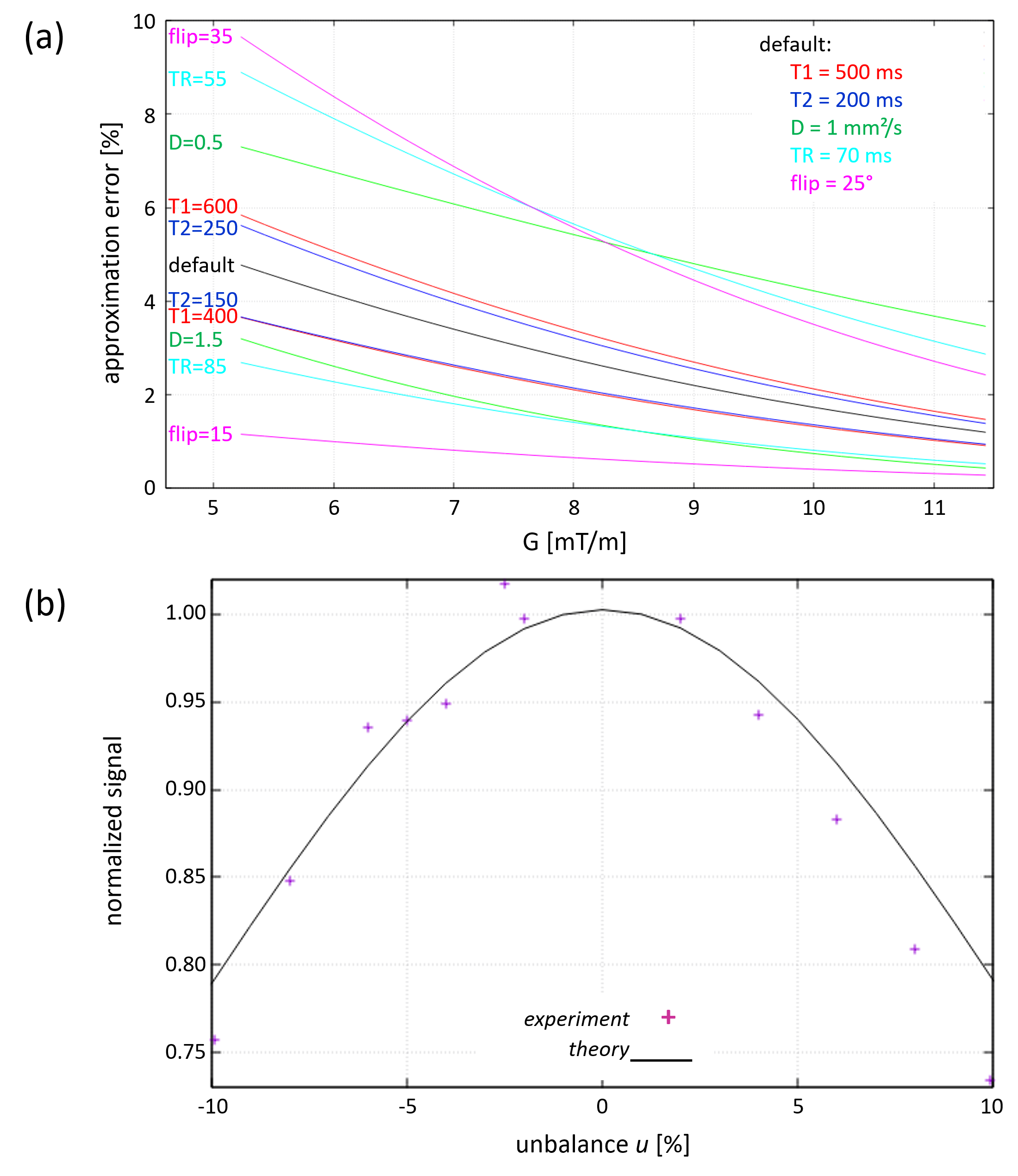
and is thus proportional to the textbook definition of b, related to standard DWI sequences. The error introduced by the approximation of x1 depends on the input variables (G,T1,T2,D,TR,α) and is typically below 10% (Fig.4a).
Furthermore, the investigated bipolar version of dwDESS3 requires a small unbalance u between the two diffusion gradient lobes to suppress adverse banding artefacts,4 i.e., two different lobe durations δ and ε
Eq.(3a): δ=τ(0.5+u)
Eq.(3b): ε=τ(0.5-u)
In practice, u can be as small as a few percent to sufficiently suppress banding,3 thus terms in E1 and E2 depending on p are negligible compared to terms not depending on p. The above discussed approximation of b’ now yields (with t≡TR/2-τ/2)
Eq.(4): b'=γ2G2[t2(δ+ε)+t(δ2+ε2)+(δ3+ε3)/3+2tδε+δ2ε+δε2]
reducing to Eq.(1) for u=0. Figure 4b shows the error introduced by u.
Methods
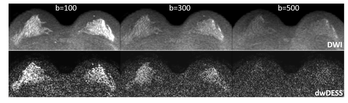
A female volunteer was measured after informed written consent obtained according to local Institutional Review Board (1.5T Ingenia, Philips Healthcare, Netherlands). Two sequences were applied, EPI-DWI (acquired voxel 1.8x1.8x3.0mm³, TR/TE=997/62ms, EPI factor=87, NSA=16 yielding roughly 20s per b-value and slice) and dwDESS (acquired voxel 1.8x1.8x2.0mm³, TR/TE1/TE2=54/5.4/48.4ms, NSA=2 yielding roughly 5s per b-value and slice). For both sequences, (effective) b-values of 100/300/500s/mm² were applied. To suppress banding artefacts, u=2.5% was chosen. The impact of u on the dwDESS signal was verified by applying the dwDESS sequences to a doped water phantom using same parameters as for in vivo, but varying u between -10% and +10%.Results
Phantom measurements confirmed the signal reduction caused by u as expected from theory (Fig.4b). Volunteer measurements show a high agreement between DWI and dwDESS using above derived b’ (Fig.5). The resulting ADC averaged over glandular area was (1.4±0.2)x10-3mm²/s for EPI-DWI and (1.3±0.4)x10-3mm²/s for dwDESS.Discussion
The estimation of ADC in the framework of dwDESS was previously performed by fitting the signal model to the measured data,5 thus circumventing the explicit determination of an effective b. This study derived an explicit estimation of an effective b, thus enabling also the comparison of diffusion-weighted images between different sequences, different measurements, and different scanners. Noteworthy that a consistent b-value definition is limited to the specifically analyzed bipolar dwDESS sequence, as diffusion weights strongly depend on other parameters (relaxation) in unipolar dwDESS.The estimation of b’ involves two assumptions: (1) approximation of x1 by first order terms, (2) unbalance u only partially integrated into signal model. However, the validity of these assumptions was confirmed by theoretical simulations as well as diffusion contrast found experimentally in vivo.
Conclusion
Estimation of effective b-values in bipolar dwDESS is possible and in good agreement with the conventional b-value of standard DWI.Acknowledgements
The authors would like to thank Oliver Bieri and Christian Stehning for their valuable support.References
1. Freed DE et al., Steady-state free precession experiments and exact treatment of diffusion in a uniform gradient. J Chem Phys 2001; 119: 4249
2. Bieri O et al., Quantitative In Vivo Diffusion Imaging of Cartilage Using Double Echo Steady-State Free Precession, Magn Reson Med. 2012; 68: 720
3. Keupp J et al., Simultaneous acquisition of diffusion weighted images and conductivity maps using a balanced double echo steady state (DESS) sequence. ISMRM 2020; 28: 955
4. Granlund KL et al., High-resolution, three-dimensional diffusion-weighted breast imaging using DESS. Magn Reson Imag. 2014; 32: 330
5. Staroswiecki E et al., Simultaneous Estimation of T2 and ADC in Human Articular Cartilage In Vivo with a Modified 3D DESS Sequence at 3T. Magn Reson Med. 2012; 67: 1086
Figures