1140
Combination of IVIM with DCE-MRI in diagnosis and prognostic evaluation of breast cancer1Department of Magnetic Resonance, LanZhou University Second Hospital, Lanzhou, China, 2Philips Healthcare, Xi’an, China
Synopsis
Intravoxel incoherent motion (IVIM) combined with Dynamic Contrast Enhanced MRI (DCE-MRI) are meaningful MRI techniques applied to breast cancer. This study used DCE-derived parameters (voiume translocation constant, Ktrans and rate constant, Kep) and IVIM-derived parameters (diffusion coefficient, D and perfusion fraction, f) to perform correlation analysis with prognosis of breast cancer indexes (ER, PR, her-2, Ki-67). The preliminary results showed that there were correlations between Ktrans, Kep, D and prognostic factors of breast cancer. Our research may provide more important clinical evidence for the treatment and prognosis of breast cancer.
Introduction
Breast cancer is the most common malignancy among females with high heterogeneous characterized by variant biological features. Its diagnosis, treatment and prognosis are affected by many factors, including the size and shape of breast cancer, pathological grade, receptor expression (ER, PR, her-2), the status of axillary lymph node metastasis, and the antigen KI-67 (Ki-67) index[1]. As a non-invasive and non-radiation method, MRI can not only evaluate the lesions from the macroscopic point of view of gross morphology, but also provide tumor micro-molecular level information through a variety of functional imaging methods (including DCE-MRI and IVIM) [2]. The prognosis of the tumor is closely related to the microscopic characteristics of the tumor[3-5] . Previous studies focused on the correlation between the microcosmic characteristics and prognosis factors, but very little was known on the multiparameter (including DCE-MRI, IVIM and prognosis factors) application[6,7]. Therefore, in this study, we compared the application of DCE-MRI and IVIM in the differential diagnosis of benign and malignant breast lesions, and analyzed the correlation between various parameters and prognostic factors of breast cancer.Methods
A total of 30 female patients with breast lesions were selected for breast MR imaging, and of these,14 patients were excluded for the following reasons: 1) failure to obtain clear immunohistochemistry and pathological results after scanning (8 patients); 2) missed scanning sequence or showed poor lesion visibility on DCE and IVIM parameter maps for analysis (6 patients). Finally, a total of 16 patients were included in the study. Sixteen patients with breast lesions (6 benign lesions and 10 malignant lesions) were recruited from the LanZhou University Second Hospital and underwent T1WI, T2WI, DCE-MRI and IVIM-MRI with a 3.0T scanner (Ingenia CX, Philips Healthcare, the Netherlands). The DCE-MRI related parameters including Ktrans, Kep were extracted by IntelliSpace Portal workstation. IVIM related D, and f values were obtained by MITK-Diffusion software (https://www.mikt.org/wiki/Downloads). Then, these multiple parameters were compared between the benign and malignant groups and between groups with different expression levels of prognostic factors. The software SPSS 22.0 was used for the statistical analysis. The measurement data using mean ± standard deviation (x±s); The independent-sample t-test, χ2 test exact probability analysis were used to compare the differences of parameters. The receiver operating characteristic (ROC) curve was used to evaluate the diagnostic efficacy of different parameters. The Pearson correlation coefficient was used to analyze the correlation. For multi-index combined diagnosis, we constructed a binomial logistic regression model to estimate the corresponding performance.Results
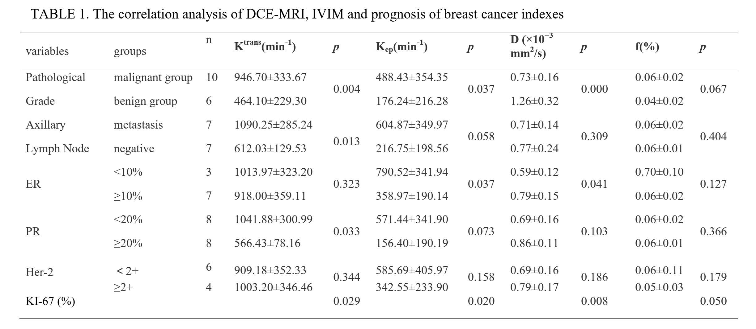
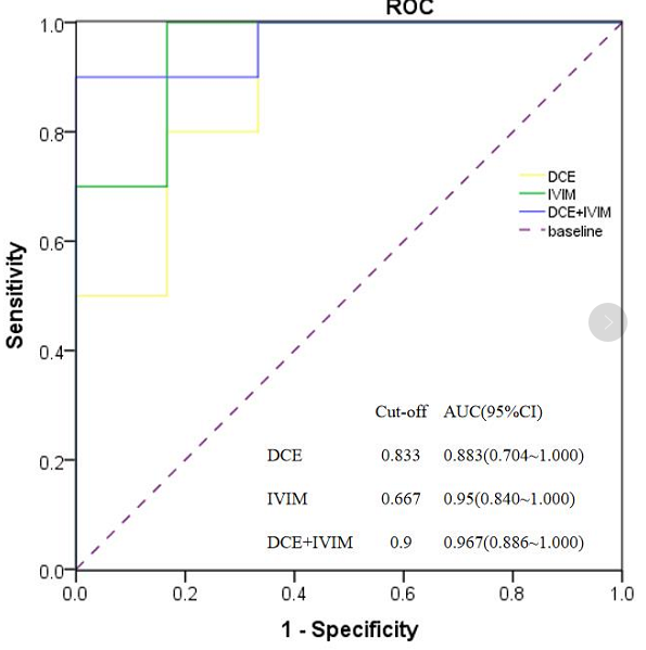
The D value of the malignant group were significantly lower than those of the benign group. However, Ktrans, Kep and f values were significantly higher than those of the benign group (p < 0.05). The areas under the curve (AUCs) of DCE (Ktrans + Kep), IVIM(D + f), DCE + IVIM were 0.883, 0.950, 0.967 respectively, FIGURE 1. The D value showed a high correlation with the pathological grade (|r| = 0.763,p<0.05); The f value showed no significant correlation with prognosis factors. The Ktrans showed a moderate correlation with the pathological grade, PR, Ki-67 and the status of axillary lymph node metastasis (|r| = 0.639, 0.601, 0.482, 0.692, p<0.05). The Kep showed a moderate correlation with the pathological grade, ER, Ki-67 (|r| = 0.460, 0.588, 0.516, p<0.05), TABLE 1.Discussion
DCE can quantify the contrast exchange between blood vessels and tissue spaces, and provide detailed information about the blood flow, microvascular and capillary permeability of the lesions. Ktrans reflects the penetration rate of contrast medium from intravascular to extracellular vascular space, which is determined by tissue blood volume, endothelial cell surface area and permeability, while Kep also reflects the rate from opposite direction in equilibrium state[8]. In this study, the values of Ktrans and Kep in malignant group were significantly higher than those in benign group. The Ktrans value showed a moderate correlation with the pathological grade, PR, Ki-67. The Ki-67 value increased, the greater the possibility of axillary lymph node metastasis, and the vascular permeability and blood perfusion at the microscopic level increased, which was consistent with previous reports. Ki-67 can induce the production of vascular endothelial growth factor (VEGF), which can increase tumor vascular perfusion. ER, PR was negatively correlated with Ktrans and Kep, possibly because ER downregulates the expression of vascular endothelial growth factor and thus inhibits tumor angiogenesis. PR changed the endocrine microvascular environment of the tumor. IVIM can reflect both tissue cell diffusivity and microcapillary perfusion. The D value showed a high correlation with the pathological grade, Ki-67 and ER. Because the higher the degree of malignant tumor, the faster the cell proliferation, the higher the value of Ki-67. Therefore, the D value are reduced due to the limited diffusion.Conclusion
This research showed that IVIM and DCE-MRI can be used in the differential diagnosis of benign and malignant breast lesions. DCE-derived Ktrans, Kep and IVIM-derived D and f are associated with prognostic factors of breast cancer. These results suggest that our method may provide complementary information to breast cancer imaging biomarkers, which potentially leading to earlier diagnosis and treatment of breast cancer.Acknowledgements
No acknowledgement found.
References
[1]. Meng Nan, Wang Xue-Jia, Sun Jing et al. Comparative Study of Amide Proton Transfer-Weighted Imaging and Intravoxel Incoherent Motion Imaging in Breast Cancer Diagnosis and Evaluation.J Magn Reson Imaging, 2020, 52: 1175-1186.
[2]. Tudorica Alina, Oh Karen Y, Chui Stephen Y-C et al. Early Prediction and Evaluation of Breast Cancer Response to Neoadjuvant Chemotherapy Using Quantitative DCE-MRI. Transl Oncol, 2016, 9: 8-17.
[3]. El Khouli Riham H,Macura Katarzyna J,Kamel Ihab R et al. 3-T dynamic contrast-enhanced MRI of the breast: pharmacokinetic parameters versus conventional kinetic curve analysis.AJR Am J Roentgenol, 2011, 197: 1498-505.
[4]. Huang Wei, Li Xin, Chen Yiyi et al. Variations of dynamic contrast-enhanced magnetic resonance imaging in evaluation of breast cancer therapy response: a multicenter data analysis challenge.Transl Oncol, 2014, 7: 153-66.
[5]. Huang Wei, Chen Yiyi, Fedorov Andriy et al. The Impact of Arterial Input Function Determination Variations on Prostate Dynamic Contrast-Enhanced Magnetic Resonance Imaging Pharmacokinetic Modeling: A Multicenter Data Analysis Challenge.Tomography, 2016, 2: 56-66.
[6].Cho Gene Young, Moy Linda, Kim Sungheon G et al. Evaluation of breast cancer using intravoxel incoherent motion (IVIM) histogram analysis: comparison with malignant status, histological subtype, and molecular prognostic factors. Eur Radiol, 2016, 26: 2547-58.
[7]. Iima Mami, Kataoka Masako, Kanao Shotaro et al. Intravoxel Incoherent Motion and Quantitative Non-Gaussian Diffusion MR Imaging: Evaluation of the Diagnostic and Prognostic Value of Several Markers of Malignant and Benign Breast Lesions.Radiology, 2018, 287: 432-441.
[8]. Pinker Katja, Helbich Thomas H, Morris Elizabeth A, The potential of multiparametric MRI of the breast.Br J Radiol, 2017, 90: 20160715.
Figures